. 2019 Nov;37(11):2476-2485.
doi: 10.1002/jor.24397. Epub 2019 Jul 12.

Non-Invasive Ultrasound Quantification of Scar Tissue Volume Identifies Early Functional Changes During Tendon Healing

Affiliations

Non-Invasive Ultrasound Quantification of Scar Tissue Volume Identifies Early Functional Changes During Tendon Healing

Jessica E Ackerman et al. J Orthop Res. 2019 Nov.

Abstract

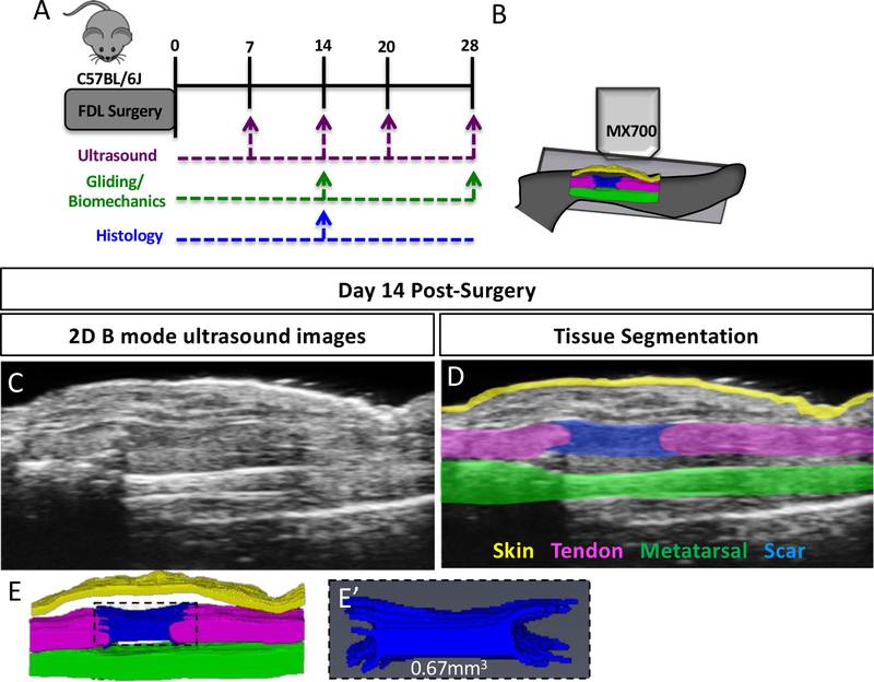
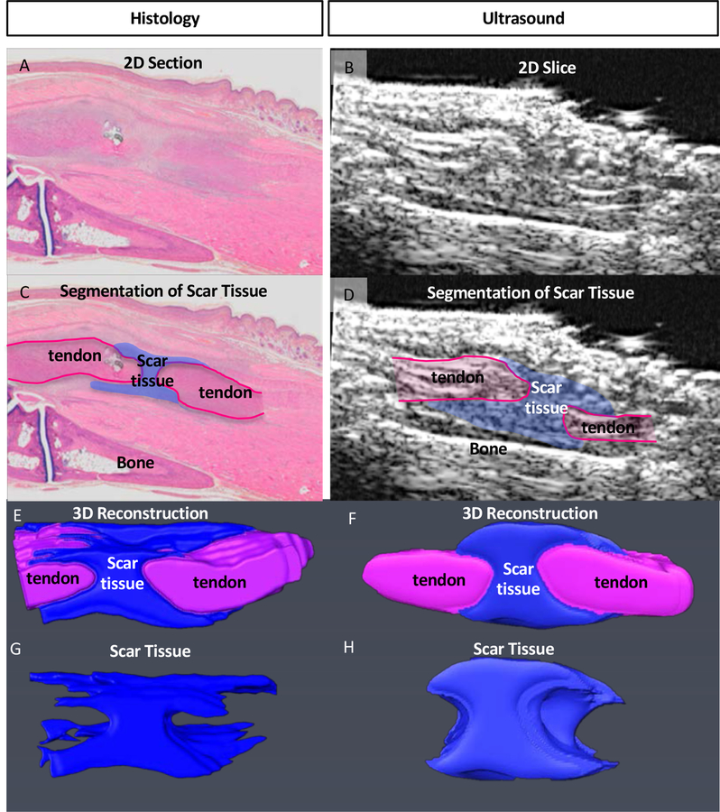
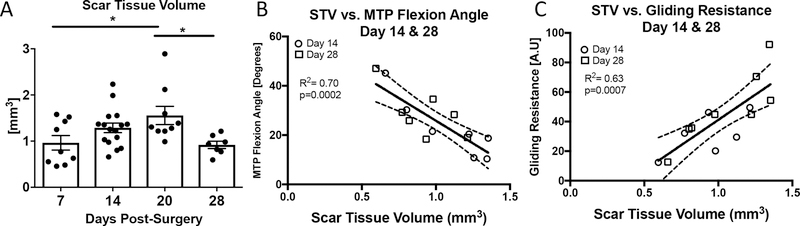
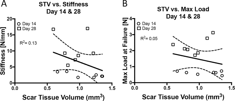
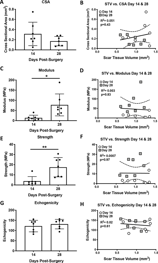
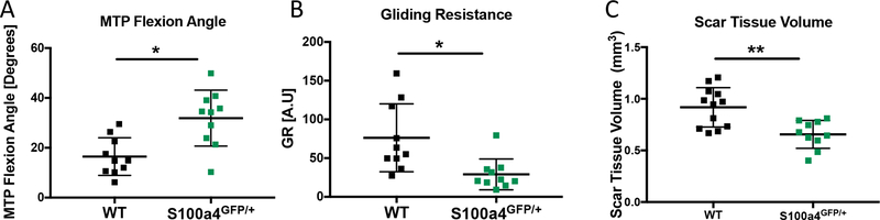
Tendon injuries are very common and disrupt the transmission of forces from muscle to bone, leading to impaired function and quality of life. Successful restoration of tendon function after injury is a challenging clinical problem due to the pathological, scar-mediated manner in which the tendons heal. Currently, there are no standard treatments to modulate scar tissue formation and improve tendon healing. A major limitation to the identification of therapeutic candidates has been the reliance on terminal endpoint metrics of healing in pre-clinical studies, which require a large number of animals and result in destruction of the tissue. To address this limitation, we have identified quantification of scar tissue volume (STV) from ultrasound (US) imaging as a longitudinal, non-invasive metric of tendon healing. STV was strongly correlated with established endpoint metrics of gliding function including gliding resistance and metatarsophalangeal (MTP) flexion angle. However, no associations were observed between STV and structural or material properties. To define the sensitivity of STV to identify differences between functionally discrete tendon healing phenotypes, we utilized S100a4 haploinsufficient mice (S100a4GFP/+ ), which heal with improved gliding function relative to wild-type (WT) littermates. A significant decrease in STV was observed in S100a4GFP/+ repairs, relative to WT at day 14. Taken together, these data suggest US quantification of STV as a means to facilitate the rapid screening of biological and pharmacological interventions to improve tendon healing, and identify promising therapeutic targets, in an efficient, cost-effective manner. © 2019 Orthopaedic Research Society. Published by Wiley Periodicals, Inc. J Orthop Res 37:2476-2485, 2019.

Keywords: mouse model; range of motion; scar tissue; tendon healing; ultrasound.


Figures

Figure 1.
Figure 1.. Quantification of Scar Tissue Volume from ultrasound images.
(A) Experimental design for longitudinal ultrasound assessment of tendon healing at 7, 14, 20- and 28-days post-surgery, followed by assessment of gliding function at day 28. An additional cohort of animals underwent ultrasound imaging only at day 14, followed by either histological analysis, or assessment of gliding function. (B) Schematic of ultrasound setup showing sagittal plane of imaging. (C) 2D B Mode ultrasound image of a healing tendon at day 14 post-surgery. (D) Segmentation of skin (yellow), metatarsal (green), FDL tendon (pink) and scar tissue (blue) at day 14 post-surgery. (E) 3D Reconstruction of segmented tissues at day 14 post-surgery. (E’) 3D reconstruction and volumetric quantification of STV at day 14 post-surgery.
Figure 2.
Figure 2.. Validation of Scar Tissue Segmentation using histology.
(A & B) 2-Dimensional sagittal Histological (A) and US (B) image sections from the same specimen. (C & D) Segmentation of scar tissue (blue) and tendon (pink) from (C) histology, and (D) US images. (E & F) Following segmentation of all 2D images containing scar tissue from (E) histology, and (F) US, the segmented slices were reconstructed in 3D, and (G & H) scar tissue was volumetrically quantified.
Figure 3.
Figure 3.. Scar Tissue Volume is correlated with changes in gliding function.
(A) Scar Tissue Volume was quantified longitudinally at 7, 14, 20, and 28 days post-surgery. Peak STV was observed at day 20. (*) indicates p<0.05. (B-C) Linear regression analyses of STV and (B) MTP Flexion Angle, (C) Gliding Resistance at 14 and 28 days. White circles represent day 14 and grey squares represent day 28.
Figure 4.
Figure 4.. Scar Tissue Volume is not correlated with tensile mechanical properties.
Linear regression analyses of STV and (A) Stiffness, (B) Max load at failure, (C) Energy to maximum load, (D) Yield load at 14, and 28 days post-surgery. White circles represent day 14 and grey squares represent day 28.
Figure 5.
Figure 5.. Scar Tissue Volume is not correlated with CSA, echogenicity or material properties.
Cross sectional area (A), Modulus (C), Strength (E) and Echogenicity (G) were quantified at 14 and 28 days post-surgery, and correlated with STV using linear regression analyses (B, D, F, H). White circles represent day 14 and grey squares represent day 28.
Figure 6.
Figure 6.. Scar Tissue Volume identifies functional differences between models of scar-mediated and regenerative tendon healing.
Quantification of A) MTP Flexion Angle, B) Gliding Resistance and C) Scar Tissue Volume in WT and S1004 haploinsufficient (S100a4GFP/+) tendon repairs at day 14 post-surgery. S100a4GFP/+ mice heal with improved gliding function and decreased STV. (*) indicates p<0.05, (**) indicates p<0.001.

References

    1. Langer R & Vacanti JP. 1993. Tissue engineering. Science 260, 920–926. - PubMed
    1. de Jong JP, Nguyen JT, Sonnema AJ. et al. 2014. The incidence of acute traumatic tendon injuries in the hand and wrist: a 10-year population-based study. Clinics in orthopedic surgery 6, 196–202. - PMC - PubMed
    1. Aydin A, Topalan M, Mezdegi A. et al. 2004. [Single-stage flexor tendoplasty in the treatment of flexor tendon injuries]. Acta Orthop Traumatol Turc 38, 54–59. - PubMed
    1. Loiselle AE, Bragdon GA, Jacobson JA. et al. 2009. Remodeling of murine intrasynovial tendon adhesions following injury: MMP and neotendon gene expression. J Orthop Res 27, 833–840. - PMC - PubMed
    1. Hasslund S, Jacobson JA, Dadali T. et al. 2008. Adhesions in a murine flexor tendon graft model: Autograft versus allograft reconstruction. J Orthop Res 26, 824–833. - PMC - PubMed

Publication types