. 2019 Jun 24;14(6):e0218474.
doi: 10.1371/journal.pone.0218474. eCollection 2019.

Biochemical characterization of the Helicobacter pylori bactofilin-homolog HP1542

Affiliations

Biochemical characterization of the Helicobacter pylori bactofilin-homolog HP1542

Sven Holtrup et al. PLoS One. .

Abstract

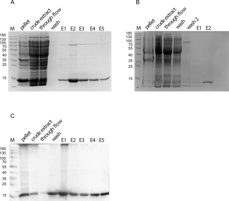
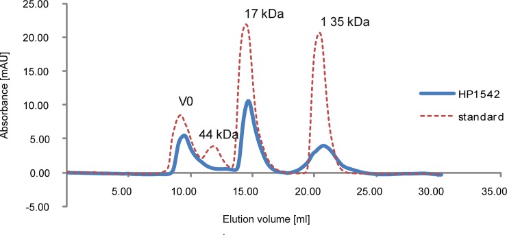
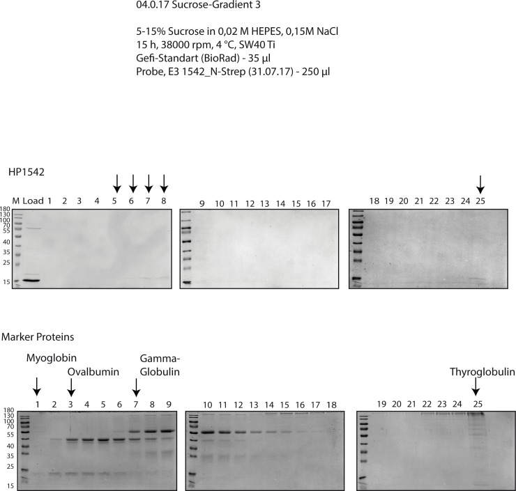
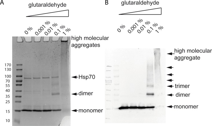
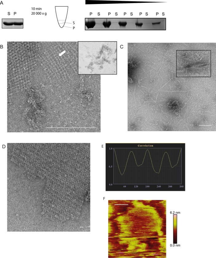
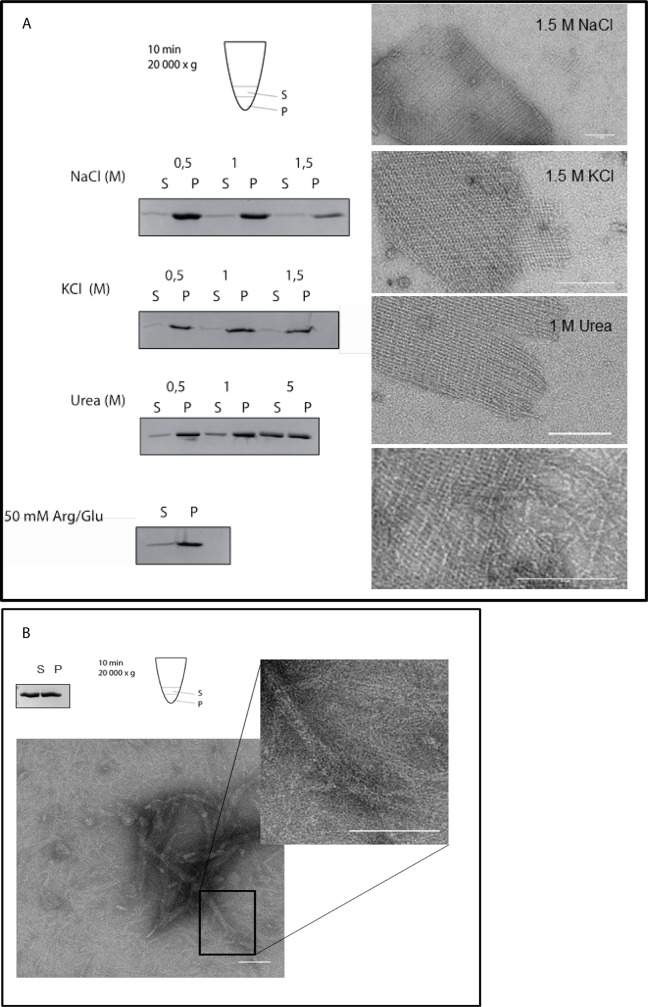
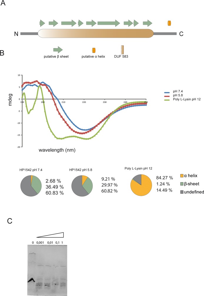
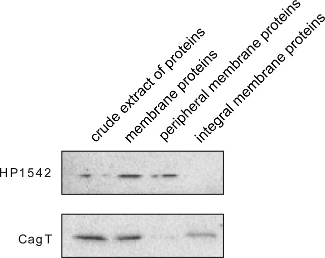
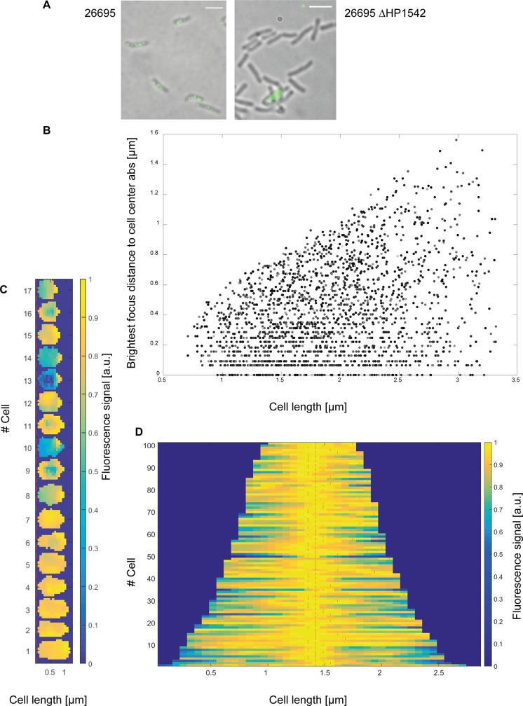
The human pathogen Helicobacter pylori is known for its colonization of the upper digestive system, where it escapes the harsh acidic environment by hiding in the mucus layer. One factor promoting this colonization is the helical cell shape of H. pylori. Among shape determining proteins are cytoskeletal elements like the recently discovered bactofilins. Bactofilins constitute a widespread family of polymer-forming bacterial proteins whose biology is still poorly investigated. Here we describe the first biochemical analysis of the bactofilin HP1542 of H. pylori reference strain 26695. Purified HP1542 forms sheet-like 2D crystalline assemblies, which clearly depend on a natively structured C-terminus. Polymerization properties and protein stability were investigated. Additionally, we also could demarcate HP1542 from amyloid proteins that share similarities with the bactofilin DUF domain. By using zonal centrifugation of total H. pylori cell lysates and immunfluorescence analysis we revealed peripheral membrane association of HP1542 mostly pronounced near mid-cell. Interestingly our results indicate that H. pylori bactofilin does not contribute to cell wall stability. This study might act as a starting point for biophysical studies of the H. pylori bactofilin biology as well as for the investigation of bactofilin cell physiology in this organism. Importantly, this study is the first biochemical analysis of a bactofilin in a human pathogen.


Conflict of interest statement

The authors have declared that no competing interests exist.

Figures

Fig 1
Fig 1. SDS-PAGE analysis of recombinantly produced Strep-HP1542 and HP1542-His from E. coli BL21.
Samples were analyzed on 12% discontinuous polyacrylamide gels. Lane M; low molecular weight marker proteins (Thermo scientific) (A) analysis of recombinant Strep-HP1542 and HP-1542-His (B) through different purification steps. Lanes pellet/ crude extract: cell lysate before purification. Lane through flow: flow through of the cell lysate. Lane wash/ wash 2 after-binding column wash fraction(s). Lane E(n): elution fraction n of elution. (C) Corresponding western blot analysis to (A).
Fig 2
Fig 2. Elution profile on Superdex 75 Increase 10/300 GL/GE of HP1542.
Elution profile on Superdex 75 Increase 10/300 GL/GE (GE Healthcare) of affinity-purified Strep-HP1542 (blue line) and standard proteins (red dashed line, Bio-Rad) as follows: thyroglobulin (670 kDa, 19 S), bovine gamma-globulin (158 kDa, 7.4 S), chicken ovalbumin (44 kDa, 3.5 S), equine myoglobin (17 kDa, 2.0 S), vitamin B12(1.35 kDa). Molecular weights are indicated above. mAU, milliabsorbance units at a wavelength of 280 nm.
Fig 3
Fig 3. Zonal gradient centrifugation.
Zonal centrifugation of Strep-HP1542 (upper panel) and gel filtration standard mix (Bio Rad, lower panel) through 5–15% sucrose gradient. Equal amounts of sample fraction were loaded on SDS-PAGE and stained with Coomassie. Strep-HP1542 containing fractions and standard proteins were indicated by arrowheads and names respectively.
Fig 4
Fig 4. Chemical crosslinking assay.
SDS-PAGE (A) and western blot (B) analysis of cross-linked recombinant Strep-HP1542. Respective concentrations of glutaraldehyde are indicated above. Arrowheads depict detected signals of Strep-HP1542 monomers, dimers as well as higher molecular aggregates. The Hsp70 contamination which is seen in the Coomassie stained SDS PAGE is marked.
Fig 5
Fig 5. Ultrastructure of HP1542.
(A) Spin-down assay of HP1542 after purification from E. coli. Left: coomassie-stained SDS-PAGE of the spin-down assay of freshly purified Strep-HP1542. Middle: pictogram demonstrating the procedure. Samples were split in two equal fractions: the upper one was defined as soluble fraction (S) and the remaining one was defined as pellet fraction (P). Right: coomassie-stained SDS-PAGE of the spin-down assays with decreasing protein concentrations of aggregated Strep-HP1542; (B-D) Transmission electron microscope (TEM) images of purified Strep-HP1542 in high (B, large image) and low magnification (B, small image), of purified HP1542-His (C) and of purified HP1542 without tag (D). (E) Example of symmetry determination of a unit cells via correlation averaging using the ANIMETRA software. (F) Atomic Force microscopy (AFM) topographic image of HP1542 assembled on mica. A height scale bar is shown to the right. Scale bars of all microscopic images are 100 nm.
Fig 6
Fig 6. Evaluation of 2-D crystalline protein assembly of HP1542.
(A) Evaluation of aggregation and 2-D crystalline protein assembly of recombinant Strep-HP1542 in low spin-down assays (left panels) and with electron microscopy (right panels) after various treatments. Conditions were indicated. (B) Upper panel: Spin-down assay of HP15421-339 after purification from E. coli. Pictogram is demonstrating the procedure. Samples were split in two equal fractions: the upper one was defined as soluble fraction (S) and the remaining one was defined as gel-like-pellet fraction (P). Lower panel: transmission electron microscope (TEM) images of purified HP15421-339 in low (and higher magnification (inlet)) Scale bars 100 nm.
Fig 7
Fig 7. Secondary structure analysis.
(A) In silico prediction of the secondary structure of HP1542, as created by the PSIPRED server. The Bactofillin domain (DUF583) is illustrated as orange passage, β-sheets as green arrows and α-helices as orange tube. (B) CD spectroscopy of Strep-HP1542. CD-units are plotted in mDeg against the wavelength in 190–250 nm for HP1542 under mild (blue) and acidic (red) conditions as well as for Poly-L-Lysin in pH 12 (green). The calculated secondary structure contributions are illustrated in pie charts. (C) SDS-PAGE analysis of the digestion of Strep-HP1542 with proteinase K (Sigma-Aldrich) at the indicated concentrations (mg ml−1).
Fig 8
Fig 8. Membrane association of HP1542.
Whole-cell lysate of wild-type strain 26695 was fractionated by ultracentrifugation. Samples from the lysate, the soluble fraction, and the insoluble membrane fraction were analyzed by immunoblotting using anti-HP1542 and anti-CagT antiserum. The integral membrane protein CagT serves as control for fractionation efficiency.
Fig 9
Fig 9. Subcellular localization of HP1542.
(A) 26695 wt and 26695 HP1542 deletion mutant were analyzed by immunofluorescence microscopy (IFM) using an anti-HP1542 primary antibody and an Alexa-Fluor 488-conjugated secondary antibody (scale bar 2 μm). (B- D) Quantitative analysis of the brightest spot of all fluorescence signals using BacStalk software. (B) Scatterplot plot depicting the absolute values of the distance of the brightest spot of all fluorescence signals to mid-cell compared to the cell length of the cells. (C) Demograph of small cells below 1 μm. The fluorescence profiles of individual cells were sorted according to cell length with the shortest cell shown at the top and the longest cell shown at the bottom. (D) Demograph of cells with the brightest focus at a defined mid- cell localization (+/- 0.0 64; n = 101) illustrating additional maxima as indicated with red lines.

References

    1. Cho H. The role of cytoskeletal elements in shaping bacterial cells. Journal of microbiology and biotechnology. 2015;25(3):307–16. Epub 2014/09/30. . - PubMed
    1. Kuhn J, Briegel A, Morschel E, Kahnt J, Leser K, Wick S, et al. Bactofilins, a ubiquitous class of cytoskeletal proteins mediating polar localization of a cell wall synthase in Caulobacter crescentus. The EMBO journal. 2010;29(2):327–39. Epub 2009/12/05. 10.1038/emboj.2009.358 - DOI - PMC - PubMed
    1. Marchler-Bauer A, Anderson JB, Chitsaz F, Derbyshire MK, DeWeese-Scott C, Fong JH, et al. CDD: specific functional annotation with the Conserved Domain Database. Nucleic acids research. 2009;37(Database issue):D205–10. Epub 2008/11/06. 10.1093/nar/gkn845 - DOI - PMC - PubMed
    1. Lin L, Thanbichler M. Nucleotide-independent cytoskeletal scaffolds in bacteria. Cytoskeleton (Hoboken, NJ). 2013;70(8):409–23. Epub 2013/07/16. 10.1002/cm.21126 . - DOI - PubMed
    1. Vasa S, Lin L, Shi C, Habenstein B, Riedel D, Kuhn J, et al. beta-Helical architecture of cytoskeletal bactofilin filaments revealed by solid-state NMR. Proceedings of the National Academy of Sciences of the United States of America. 2015;112(2):E127–36. Epub 2015/01/01. 10.1073/pnas.1418450112 - DOI - PMC - PubMed

Publication types

LinkOut - more resources