. 2019 Jun 24;14(6):e0218854.
doi: 10.1371/journal.pone.0218854. eCollection 2019.

Reciprocal signaling and direct physical interactions between fibroblasts and breast cancer cells in a 3D environment

Affiliations

Reciprocal signaling and direct physical interactions between fibroblasts and breast cancer cells in a 3D environment

Deborah J Wessels et al. PLoS One. .

Abstract

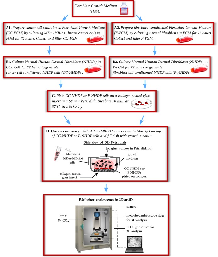
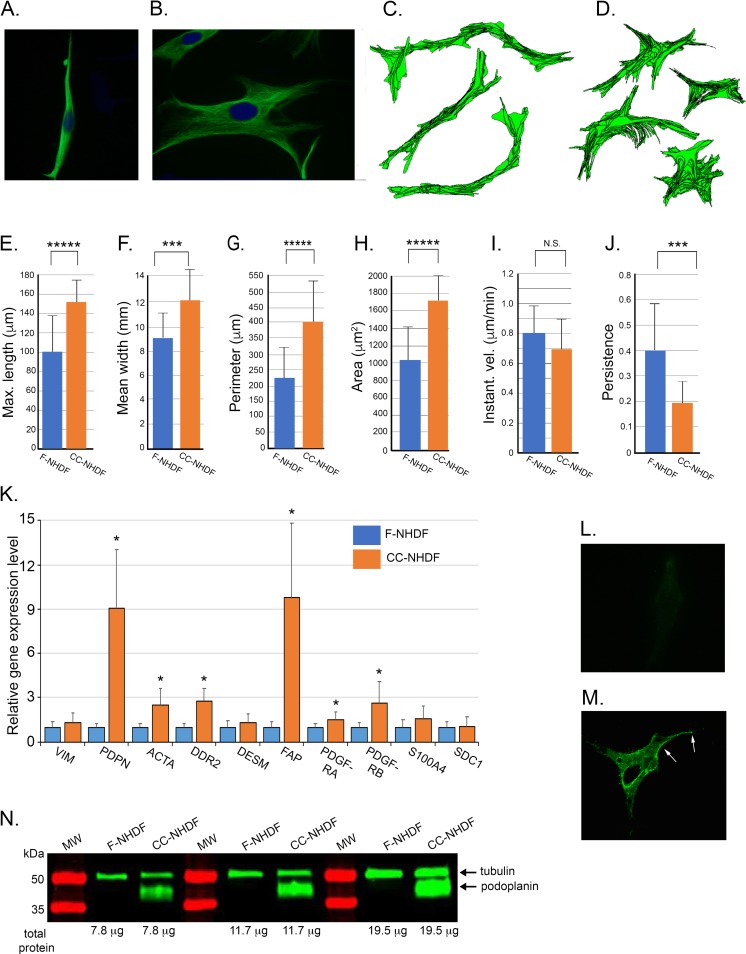
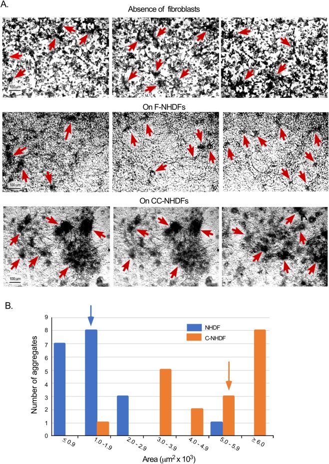
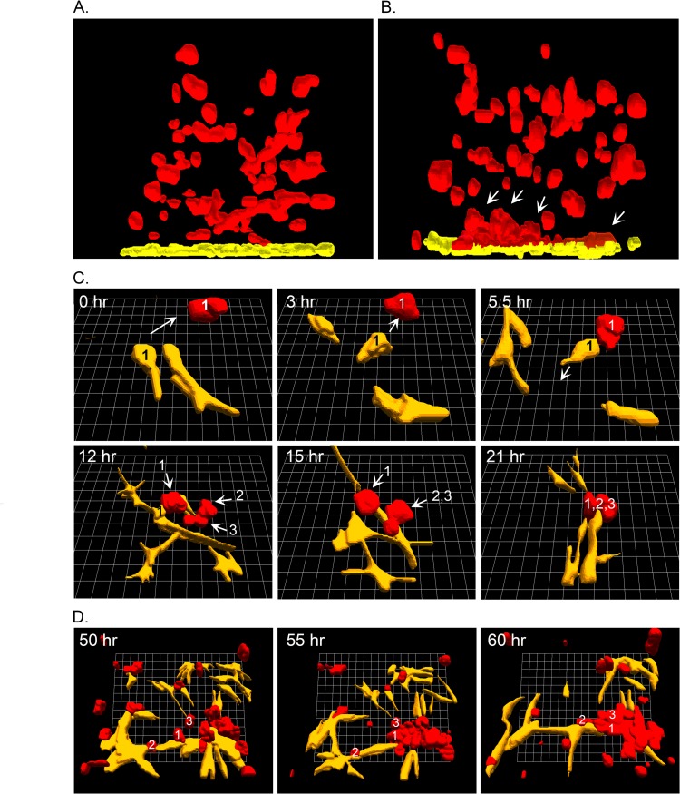
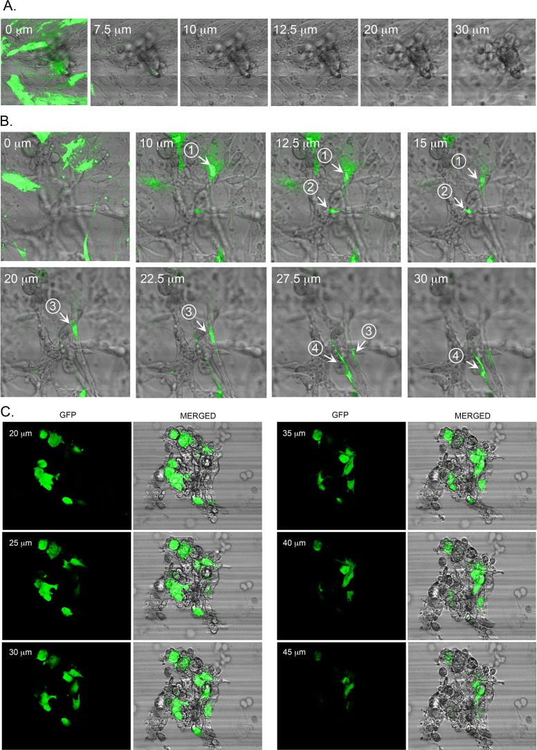
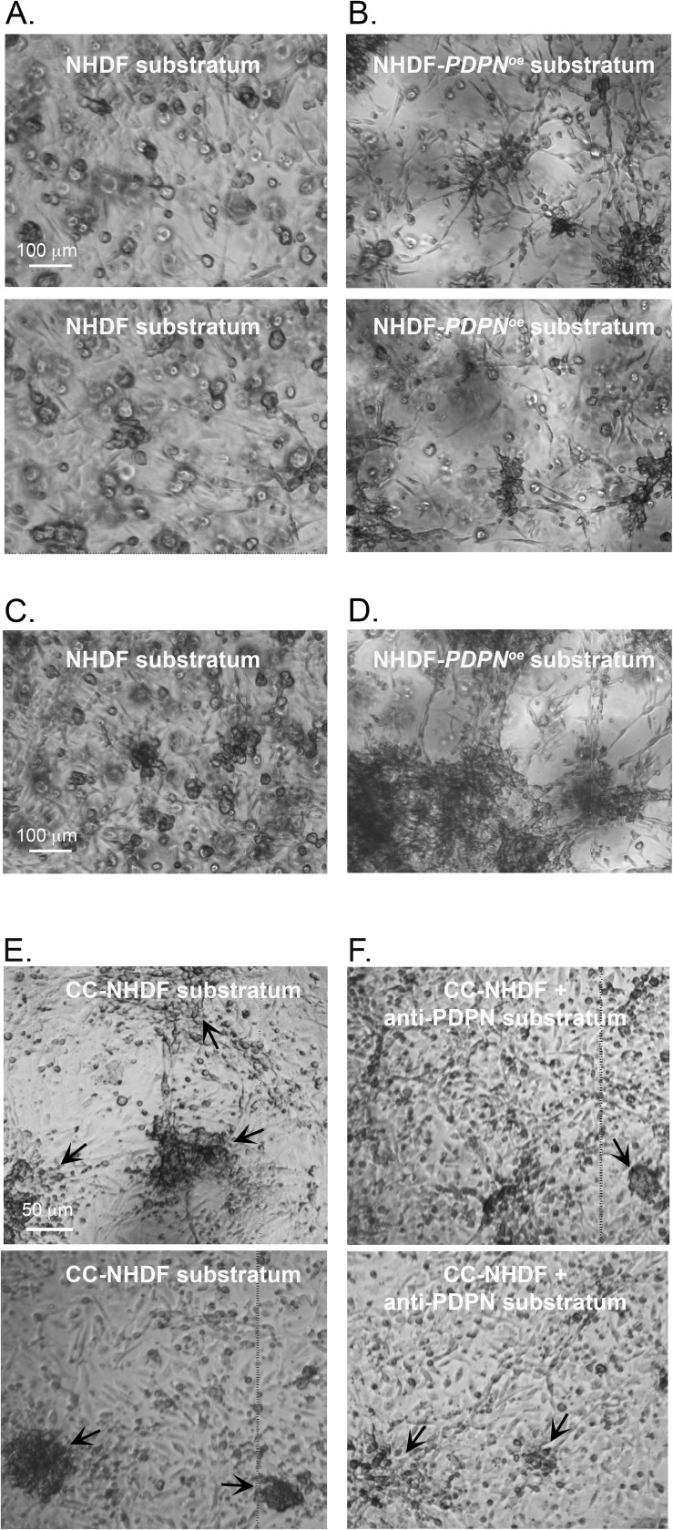
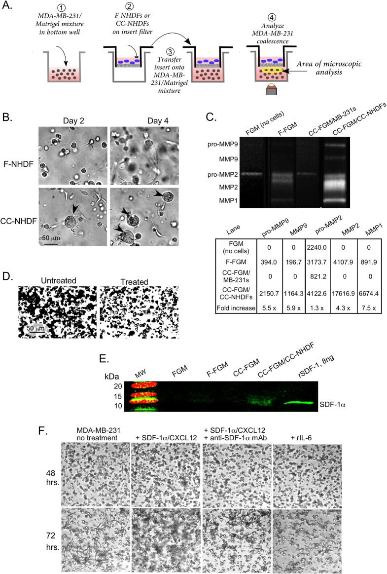
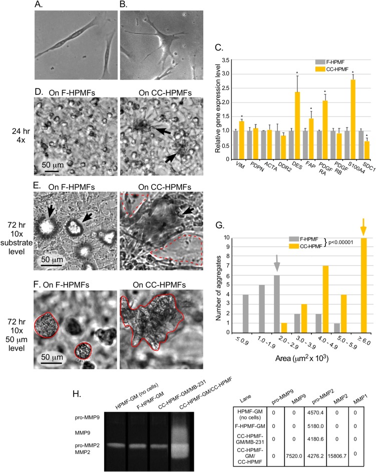
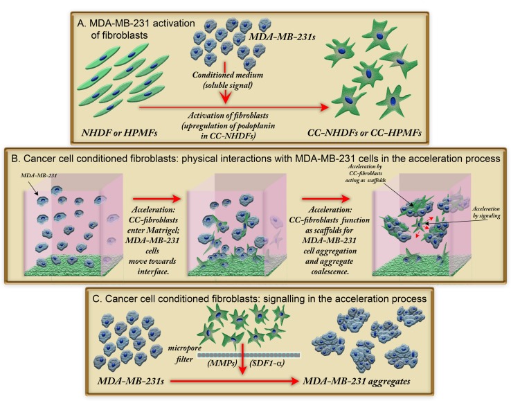
Tumorigenic cells undergo cell aggregation and aggregate coalescence in a 3D Matrigel environment. Here, we expanded this 3D platform to assess the interactions of normal human dermal fibroblasts (NHDFs) and human primary mammary fibroblasts (HPMFs) with breast cancer-derived, tumorigenic cells (MDA-MB-231). Medium conditioned by MDA-MB-231 cells activates both types of fibroblasts, imbuing them with the capacity to accelerate the rate of aggregation and coalescence of MDA-MB-231 cells more than four fold. Acceleration is achieved 1) by direct physical interactions with MDA-MB-231 cells, in which activated fibroblasts penetrate the MDA-MB-231/Matrigel 3D environment and function as supporting scaffolds for MDA-MB-231 aggregation and coalescence, and 2) through the release of soluble accelerating factors, including matrix metalloproteinase (MMPs) and, in the case of activated NHDFs, SDF-1α/CXCL12. Fibroblast activation includes changes in morphology, motility, and gene expression. Podoplanin (PDPN) and fibroblast activation protein (FAP) are upregulated by more than nine-fold in activated NHDFs while activated HPMFs upregulate FAP, vimentin, desmin, platelet derived growth factor receptor A and S100A4. Overexpression of PDPN, but not FAP, in NHDF cells in the absence of MDA-MB-231-conditioned medium, activates NHDFs. These results reveal that complex reciprocal signaling between fibroblasts and cancer cells, coupled with their physical interactions, occurs in a highly coordinated fashion that orchestrates aggregation and coalescence, behaviors specific to cancer cells in a 3D environment. These in vitro interactions may reflect events involved in early tumorigenesis, particularly in cases of field cancerization, and may represent a new mechanism whereby cancer-associated fibroblasts (CAFs) promote tumor growth.


Conflict of interest statement

The authors have declared that no competing interests exist.

Figures

Fig 1
Fig 1. The preparation used to analyze fibroblast cell motility and to compare the effects of F-NHDFs, F-HPMFs, CC-NHDFs and CC-HPMFs on the aggregation and aggregate coalescence of MDA-MB-231 cells.
Fig 2
Fig 2. Medium conditioned by the breast tumor-derived cell line MDA-MB-231 activates NHDFs affecting polarity, cellular translocation, morphology and gene expression.
A, B. F-NHDFs and CC-NHDFs, respectively, stained for vimentin (green) to visualize cell shape, and with DAPI (blue) to visualize nuclei. C-D. Motility tracks at 7.5 minute intervals of F-NHDFs and CC-NHDFs, respectively, generated by J3D-DIAS4.2 software that automatically detects cell perimeters and computes tracks. E- J. J3D-DIAS4.2 computations of length, width, perimeter, area, instantaneous velocity and the persistence of translocation. The means and error bar (standard deviations) are presented in all panels for an N = 50. ***** indicates significance of p<0.00005, *** indicates significance p<0.0005, N.S indicates not significant. K. qRT-PCR analyses of the transcript levels of ten genes associated with fibroblast activation. Means and error bars (standard deviations) are presented for N = 3. Asterisk (*) indicates significance of p <0.05. L,M. F-NHDFs and CC-NHDFs, respectively, stained with anti-PDPN mAb NZ-1. Arrows denote punctate plasma membrane staining. N. Western blot of F-NHDF and CC-NHDF lysates probed with anti-PDPN mAb NZ-1 and the anti-tubulin mAb E7.
Fig 3
Fig 3. CC-NHDFs but not F-NHDFs accelerate MDA-MB-231 cell aggregation and aggregate coalescence in a 3D environment.
A. Representative fields of aggregating MDA-MB-231 cells in which the MDA-MB-231/Matrigel phase is cast over the substrate in the absence of fibroblasts, over F-NHDF cells on collagen and over CC-NHDF cells on collagen. Red arrows indicate MDA-MB-231 aggregates. B. Measurements of aggregate areas at 72 hours in cultures in which the MDA-MB-231/Matrigel phase was cast over F-NHDF cells (blue) or CC-NHDF cells (orange). The median area of each population is noted as a blue or orange arrow, respectively. The difference between MDA-MB-231 aggregate areas was significantly greater (p< 0.00001) in the presence of CC-NHDF when compared to F-NHDFs.
Fig 4
Fig 4. CC-NHDF cells attract MDA-MB-231 cells and function as scaffolds to accelerate aggregation and coalescence of cancer cells.
A, B. Side views of 3D reconstructions of MDA-MB-231 cells (red) in Matrigel cast over NHDF cells (yellow) on collagen or CC-NHDF cells (yellow) on collagen, respectively, after 24 hours of incubation. Arrows point to MDA-MB-231 aggregates forming at or near the collagen/Matrigel interface and in physical contact with CC-NHDFs. C. 3D of CC-NHDFs (yellow) and MDA-MB-231 cells (red) over a 21 hour period, with both cell types tracked by analyzing the optical section stacks at 10 minute intervals. D. 3D reconstructions of CC-NHDFs (yellow) and MDA-MB-231 cells (red) between 50 and 60 hours of incubation. Reconstructions were performed with J3D-DIAS4.2 software [13, 14, 23, 30].
Fig 5
Fig 5. CC-NHDFs act as scaffolds and penetrate MDA-MB-231 aggregates to support and facilitate aggregation.
A. F-NHDF-GFP cells in the basal layer do not penetrate the MDA-MB-231/Matrigel overlay after 72 hours of incubation. B. CC-NHDF-GFP cells in the basal layer penetrate through at least 30 μm of the MB-231/Matrigel upper phase after 72 hours of incubation. Fluorescent images were combined with phase contrast images at ascending depths through the upper phase. Cells or their projections are numbered. C. Optical sections of an aggregate at 96 hours of culture formed in the Matrigel phase of a preparation in which CC-NHDF-GFP cells were plated on the collagen substratum. The fluorescent images of the CC-NHDFs and the phase DIC images of all the cells are merged for each optical section.
Fig 6
Fig 6. Overexpression of podoplanin imbues NHDFs not activated with MDA-MB-231 conditioned medium with the capacity to accelerate aggregation and coalescence, and pretreatment of CC-NHDFs with anti-podoplanin mAb blocks its capacity to accelerate.
A. Aggregation and aggregate coalescence of MDA-MB-231 preparations after 24 hours in which the substratum was NHDF cells on collagen. B. Aggregation and aggregate coalescence of MDA-MB-231 preparations after 24 hours in which the substratum was NHDF-PDPNœ cells on collagen. C, D. Aggregation and coalescence of MDA-MB-231 at 72 hours, in which the substratum was either NHDF (C) or NHDF-PDPNœ cells (D) on collagen. E, F. Aggregation and aggregate coalescence of MDA-MB-231 after 72 hours in which the substratum was either untreated CC-NHDFs (E) or CC-NHDFs pretreated with the anti-podoplanin mAb NZ-1 (F).
Fig 7
Fig 7. CC-NHDFs release soluble signals that accelerate aggregation and coalescence.
A. The transmembrane preparation employed to assess the presence of soluble factors. B. Representative images of aggregation of MDA-MB-231 cells after 2 and 4 days in a preparation with F-NHDFs or CC-NHDFs in the upper insert. C. Gel zymogram and quantitation of pixel intensities of MMPs in FGM directly from the bottle of sterile, unused media (no cells); F-FGM, media from 72 hour cultures of F-NHDFs; CC-FGM/MB-231, media from 72 hour MDA-MB-231 cultures; and CC-FGM/CC-NHDF, media from 72 hour CC-NHDF. D. Aggregation after 48 hours of MDA-MB-231 preparations over a CC-NHDF substratum in the absence and presence of the MMP inhibitor GM6001. E. Western blot of media probed with anti-SDF-1α/CXCL12 mAb. Recombinant SDF-1α/CXCL12 was used as a positive control. F. Coalescence after 48 and 72 hours of untreated MDA-MB-231, MDA-MB-231 cells treated with SDF-1α/CXCL12, MDA-MB-231 cells treated with SDF-1α/CXCL12 plus anti-SDF-1α/CXCL12 mAb, and MDA-MB-231 cells treated with rIL-6.
Fig 8
Fig 8. CC-HPMFs exhibit altered morphology, altered gene expression, accelerate MDA-MB-231 coalescence and release elevated MMPs.
A, B. Representative phase images of F-HPMF and CC-HPMFs. C. qRT-PCR analyses of the transcript levels of ten genes associated with fibroblast activation. Means and error bars (standard deviations) are presented for N = 3. Asterisk (*) indicates significance of p <0.05. D. Representative fields after 24 hrs of aggregating MDA-MB-231 cells in which the MDA-MB-231/Matrigel phase is cast over F-HPMF cells on collagen and over CC-HPMF cells on collagen reveals larger aggregates in the latter. E. Substrate level, 10x magnification of representative fields of aggregating MDA-MB-231 cells over F-HPMF cells on collagen and over CC-HPMF cells on collagen reveals advanced coalescence and considerably more destruction of the CC-HPMF monolayer (dashed red line) in comparison to F-HPMF monolayer. F. Representative images of aggregating MDA-MB-231 cells cast over F-HPMF cells on collagen and over CC-HPMF cells on collagen at 72 hours at the 50 μm level demonstrate significantly larger aggregates over the CC-HPMF layer (solid red line) in comparison to the F-HPMF layer. G. Measurements of aggregate areas at 72 hours in cultures in which the MDA-MB-231/Matrigel phase was cast over F-HPMF cells (gray), and CC-HPMF cells (yellow). The median area of each population is noted as a gray or yellow arrow, respectively. The difference between MDA-MB-231 aggregate areas was significantly greater (p< 0.00001) in the presence of CC-HPMFs when compared to F-HPMFs. H. Gel zymogram and quantitation of pixel intensities of MMPs in HPMF-GM directly from the bottle of sterile, unused media (no cells); F-HPMF-GM, media from 72 hour cultures of F-HPMFs; CC-HPMF-GM/MB-231, media from 72 hour MDA-MB-231 cultures; and CC-HPMF-GM/CC-HPMF, media from 72 hour CC-HPMF cultures.
Fig 9
Fig 9. A model showing how activation of fibroblasts leads to accelerated coalescence of MDA-MB-231 breast cancer cells in a 3D Matrigel environment.
A. Activation of NHDFs and HPMFs to CC-NHDFs and CC-HPMFs, respectively, after exposure to a medium conditioned by MDA-MB-231 cells. B. Physical interactions between MDA-MB-231 cancer cells and CC-fibroblasts occur initially at the collagen 1/Matrigel interface but later throughout the 3D environment with CC-fibroblasts serving as scaffolds for MDA-MB-231 cell coalescence. C. Release of soluble signals by CC-NHDF and CC-HPMF cells accelerates coalescence of MDA-MB-231 cells.

Similar articles

Cited by

References

    1. Spaw M, Anant S, Thomas SM. Stromal contributions to the carcinogenic process. Molecular carcinogenesis. 2017;56(4):1199–213. 10.1002/mc.22583 - DOI - PMC - PubMed
    1. Kalluri R. The biology and function of fibroblasts in cancer. Nat Rev Cancer. 2016;16(9):582–98. 10.1038/nrc.2016.73 - DOI - PubMed
    1. Liakou E, Mavrogonatou E, Pratsinis H, Rizou S, Evangelou K, Panagiotou PN, et al. Ionizing radiation-mediated premature senescence and paracrine interactions with cancer cells enhance the expression of syndecan 1 in human breast stromal fibroblasts: the role of TGF-beta. Aging. 2016;8(8):1650–69. Epub 2016/07/20. 10.18632/aging.100989 - DOI - PMC - PubMed
    1. Yang N, Mosher R, Seo S, Beebe D, Friedl A. Syndecan-1 in breast cancer stroma fibroblasts regulates extracellular matrix fiber organization and carcinoma cell motility. The American journal of pathology. 2011;178(1):325–35. Epub 2011/01/13. 10.1016/j.ajpath.2010.11.039 - DOI - PMC - PubMed
    1. Levental KR, Yu H, Kass L, Lakins JN, Egeblad M, Erler JT, et al. Matrix crosslinking forces tumor progression by enhancing integrin signaling. Cell. 2009;139(5):891–906. Epub 2009/11/26. 10.1016/j.cell.2009.10.027 - DOI - PMC - PubMed

Publication types

MeSH terms