Review
. 2019 Jun;34(6):996-1013.
doi: 10.1002/jbmr.3734. Epub 2019 Jun 24.

Gaucher Disease in Bone: From Pathophysiology to Practice

Affiliations
Review

Gaucher Disease in Bone: From Pathophysiology to Practice

Derralynn Hughes et al. J Bone Miner Res. 2019 Jun.

Abstract

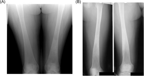
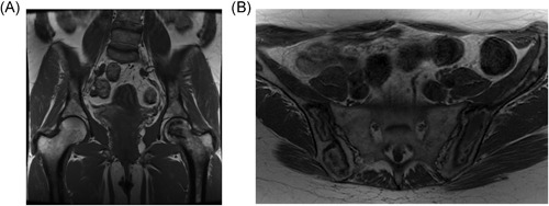
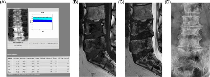
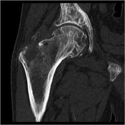
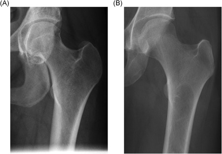
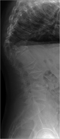
Gaucher disease (GD) is a rare, genetic lysosomal disorder leading to lipid accumulation and dysfunction in multiple organs. Involvement of the skeleton is one of the most prevalent aspects of GD and a major cause of pain, disability, and reduced quality of life. Uniform recommendations for contemporary evaluation and management are needed. To develop practical clinical recommendations, an international group of experienced physicians conducted a comprehensive review of 20 years' of the literature, defining terms according to pathophysiological understanding and pointing out best practice and unmet needs related to the skeletal features of this disorder. Abnormalities of bone modeling, reduced bone density, bone infarction, and plasma cell dyscrasias accompany the displacement of healthy adipocytes in adult marrow. Exposure to excess bioactive glycosphingolipids appears to affect hematopoiesis and the balance of osteoblast and osteoclast numbers and activity. Imbalance between bone formation and breakdown induces disordered trabecular and cortical bone modeling, cortical bone thinning, fragility fractures, and osteolytic lesions. Regular assessment of bone mineral density, marrow infiltration, the axial skeleton and searching for potential malignancy are recommended. MRI is valuable for monitoring skeletal involvement: It provides semiquantitative assessment of marrow infiltration and the degree of bone infarction. When MRI is not available, monitoring of painful acute bone crises and osteonecrosis by plain X-ray has limited value. In adult patients, we recommend DXA of the lumbar spine and left and right hips, with careful protocols designed to exclude focal disease; serial follow-up should be done using the same standardized instrument. Skeletal health may be improved by common measures, including adequate calcium and vitamin D and management of pain and orthopedic complications. Prompt initiation of specific therapy for GD is crucial to optimizing outcomes and preventing irreversible skeletal complications. Investing in safe, clinically useful, and better predictive methods for determining bone integrity and fracture risk remains a need. © 2019 The Authors. Journal of Bone and Mineral Research Published by Wiley Periodicals Inc.

Keywords: BIOMARKERS; BONE DISEASE; GAUCHER DISEASE; OSTEONECROSIS; OSTEOPOROSIS; RADIOLOGY; THERAPEUTICS.


Figures

Figure 1
Figure 1
Erlenmeyer flask deformity. (A) Typical appearance. Radiograph of the lower femora, showing the triangular outline of the metaphysis. Note the indistinct boundary between the cortex and medulla, typical of the Erlenmeyer flask deformity in Gaucher disease (GD). Incidentally, there is an area of serpiginous sclerosis in the left femoral metaphysis, suggestive of osteonecrosis. (B) Atypical appearance. Radiograph of the lower femora of a woman with GD who began enzyme replacement therapy at the age of 12 years. The proximal metaphysis has features similar to those in A, but the distal metaphysis has the more normal, trumpet‐shaped outline of the distal femur, with a clear border between the cortex and medulla. We speculate that the normal modeling process of endochondral ossification took place from the time of initiation of therapy
Figure 2
Figure 2
Osteonecrosis. (A) Typical. T1‐weighted MR image of the pelvis, showing a geographic area of low signal in the head of the left femur. Note that the joint surface remains intact in this case; therefore, there is no deformity or degenerative change in the hip joint. (B) Atypical. T1‐weighted MR image of the pelvis in a different patient showing diffuse low geographic area of low and high signal through the pelvic bones bilaterally. The radiologic changes in (B) occurred gradually over years, on enzyme therapy, and without the typical symptoms of bone crisis
Figure 3
Figure 3
The lumbar spine of a patient with GD and recurrent bone crises despite therapy. All images taken several years after last acute bone crisis. (A) DXA image. Note the increased BMD in L3 and L4. These vertebrae were not excluded from the report, despite the variation in BMD among vertebrae, contrary to best practice. The total lumbar vertebral BMD reported is therefore artifactually elevated. (B) T1‐weighted MR image showing widespread, low‐signal geographic changes in L4, L5, and the sacrum, features indicative of osteonecrosis. Central endplate depression is also seen. (C) T2‐weighted image showing double‐line sign indicative of osteonecrosis best seen in S1 and S2 segments and areas of low signal in the bodies of L4 and L4. Low signal in both T1‐ and T2‐weighted images indicates osteosclerosis. (D) Plain anteroposterior lumbar spine radiograph in the same patient show osteosclerosis of the body of L4. The increased density at L4 and L5 is therefore a result of osteosclerosis, consequent upon osteonecrosis, although a contribution from endplate depression cannot be excluded
Figure 4
Figure 4
CT image of the right hip, showing a lytic and expanding lesion (Gaucheroma) of the greater trochanter, superiorly displacing fragments of bone
Figure 5
Figure 5
Plain radiographs of the proximal left femora of two patients with Gaucher disease. (A) Normal cortical thickness in the femoral shaft of the left proximal femur. (B) Reduced cortical thickness in the femoral shaft of the left proximal femur. Note the thinner cortex in comparison with A
Figure 6
Figure 6
Lateral radiograph of the spine of a patient with type 3 GD, showing an abruptly angulated thoracic kyphosis

References

    1. Hruska KS, LaMarca ME, Sidransky E Gaucher disease: molecular biology and genotype–phenotype correlations In: Futerman AH, & Zimran A, editors, Gaucher disease. Volume 1. Boca Raton, FL: CRC Press, 2007. Chapter 2, p.13.
    1. Altarescu G, Hill S, Wiggs E, et al. The efficacy of enzyme replacement therapy in patients with chronic neuronopathic Gaucher's disease. J Pediatr. 2001;138(4):539–47. - PubMed
    1. Davies EH, Erikson A, Collin‐Histed T, et al. Outcome of type III Gaucher disease on enzyme replacement therapy: Review of 55 cases. J Inherit Metab Dis. 2007;30(6):935–42. - PubMed
    1. Bultron G, Kacena K, Pearson D, et al. The risk of Parkinson's disease in type 1 Gaucher disease. J Inherit Metab Dis. 2010;33(2):167–73. - PMC - PubMed
    1. AHV Schapira. Glucocerebrosidase and Parkinson disease: Recent advances. Mol Cell Neurosci. 2015;66:37–42. - PMC - PubMed

Publication types

MeSH terms