. 2019 Sep:131:255-261.
doi: 10.1016/j.urology.2019.06.016. Epub 2019 Jun 22.

"Pin the Tumor on the Kidney:" An Evaluation of How Surgeons Translate CT and MRI Data to 3D Models

Affiliations

"Pin the Tumor on the Kidney:" An Evaluation of How Surgeons Translate CT and MRI Data to 3D Models

Nicole Wake et al. Urology. 2019 Sep.

Abstract

Objective: To quantify how surgeons translate 2-dimensional (2D) computed tomography (CT) or magnetic resonance imaging (MRI) data to a 3-dimensional (3D) model and evaluate if 3D printed models improve tumor localization.

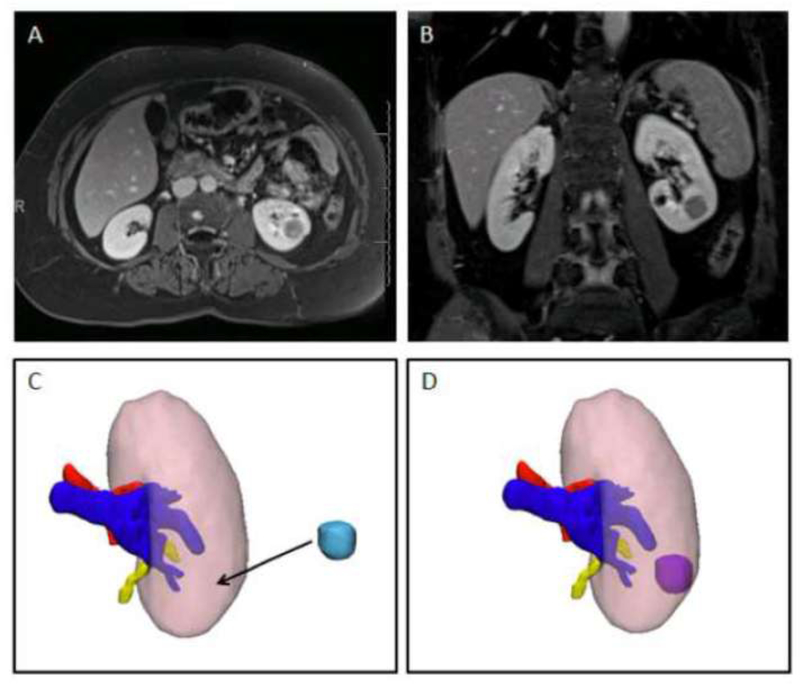
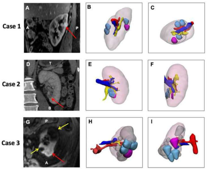
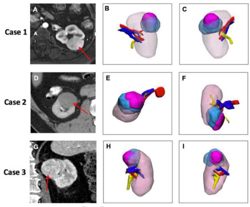
Materials and methods: Twenty patients with renal masses were randomly selected from our institutional review board approved prospective 3D modeling study. Three surgeons reviewed the clinically available CT or MRI data; and using computer-aided design software, translated the renal tumor to the position on the kidney that corresponded with the image interpretation. The renal tumor location determined by each surgeon was compared to the true renal mass location determined by the segmented imaging data and the Dice Similarity Coefficient (DSC) was calculated to evaluate the spatial overlap accuracy. The exercise was repeated for a subset of patients with a 3D printed model.

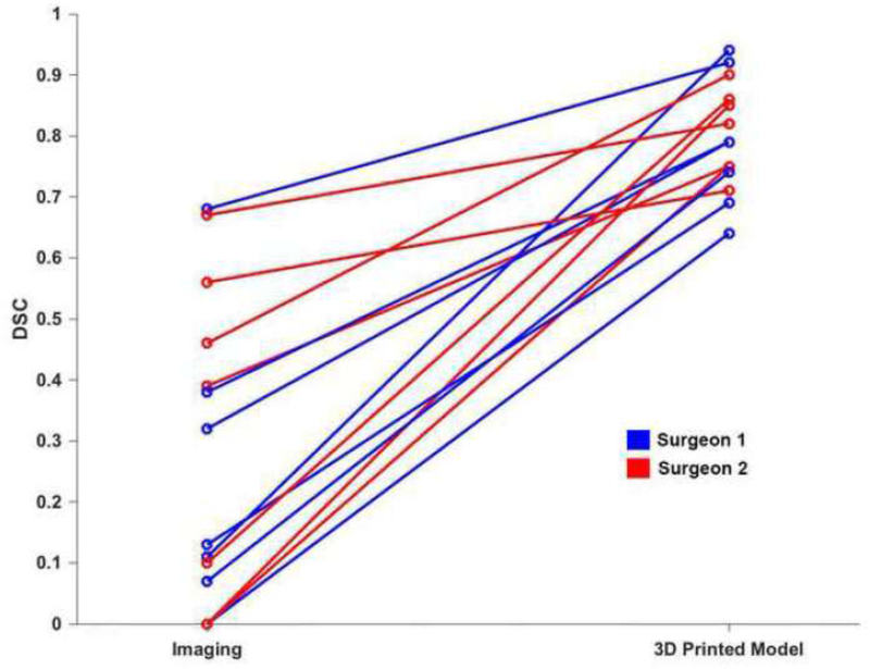
Results: The mean DSC was 0.243 ± 0.236 for the entire cohort (n = 60). There was no overlap between the actual renal tumor and renal tumor identified by the surgeons in 16 of 60 cases (26.67%). Seven cases were reviewed again by 2 surgeons in a different setting with a 3D printed renal cancer model. For these cases, the DSC improved from 0.277 ± 0.248 using imaging only to 0.796 ± 0.090 with the 3D printed model (P < .01).

Conclusion: In this study, cognitive renal tumor localization based on CT and MRI data was poor. This study demonstrates that experienced surgeons cannot always translate 2D imaging studies into 3D. Furthermore, 3D printed models can improve tumor localization and potentially assist with appropriate surgical approach.


Figures

Figure 1
Figure 1
Single case example showing A) axial and B) coronal images of a left endophytic renal mass. C) 3D kidney model with the kidney – pink, artery-red, vein- blue, and collecting system – yellow. The tumor (light blue) has been removed so the surgeon can translate the location where he believes it is after reviewing the images. D) 3D kidney tumor model with tumor in its correct location.
Figure 2:
Figure 2:
Three cases with no overlap (zero correlation) between actual lesion location and cognitive localization by surgeon after reviewing available images. All 3D models have the following color scheme: kidney – light pink, actual segmented lesion – magenta, surgeon placed lesions -light blue, artery- red, vein- blue, collecting system – yellow. Case 1 is shown in the top row: (A), Sagittal Post VIBE MRI with lesion purple. (B) 3D model demonstrating the kidney in the same orientation as imaging slice, and (C) 3D model in axial orientation demonstrating that there is no overlap. Case 2 is in the middle row: (D) Coronal CT with the lesion –red arrow, (E) 3D model demonstrating the kidney in the same orientation as imaging slice, (F) 3D model in sagittal orientation demonstrating that there is no overlap. Case 3 is in the bottom row. (G) Axial Post VIBE MRI with lesion-red arrow and cysts – yellow arrows, (H) 3D model demonstrating the kidney in the same orientation as imaging slice, and (I) Posterior coronal view of 3D model demonstrating that there is no overlap.
Figure 3:
Figure 3:
Three cases with the maximum overlap (highest correlation) between actual lesion location and cognitive localization by surgeon after reviewing available images. The same color scheme as Figure 2 is used here. Case 1 is shown in the top row: (A) Axial post VIBE MRI with the lesion –red arrow, (B) 3D model demonstrating the kidney in the coronal orientation (anterior) and (C) 3D model posterior coronal view. Case 2 is shown in the middle row: (D) Axial CT with the lesion –red arrow, (E) 3D model demonstrating the kidney in the same orientation as imaging slice, (F) 3D model in coronal orientation demonstrating overlap between surgeon placed lesions and true lesion. Case 3 is shown in the bottom row: (G) Axial CT with lesion-red arrow, (H) 3D model coronal orientation, and (I) Sagittal view of 3D model demonstrating the overlap.
Figure 4:
Figure 4:
Line graph demonstrating the improvement in DSC after reviewing the 3D printed model for the seven patients in which the exercise was completed in both scenarios. Results for surgeon 1 are shown in blue and for surgeon 2 in red.

References

    1. Smith SJ, Bosniak MA, Megibow AJ et al. Renal cell carcinoma: earlier discovery and increased detection. Radiology 1989; 170: 699–703. - PubMed
    1. Bosniak MA. The small (less than or equal to 3.0 cm) renal parenchymal tumor: detection, diagnosis, and controversies. Radiology 1991; 179: 307–17. - PubMed
    1. Campbell S, Uzzo RG, Allaf ME et al. Renal Mass and Localized Renal Cancer: AUA Guideline. The Journal of urology 2017; 198: 520–9. - PubMed
    1. Wake N, Rude T, Kang SK et al. 3D printed renal cancer models derived from MRI data: application in pre-surgical planning. Abdom Radiol (NY) 2017; 42: 1501–9. - PMC - PubMed
    1. Hegarty M; Keehner MC C; Montello DR,; Lippa Y The Role of Spatial Cognition in Medicine: Applications for Selecting and Training Professionals In Allen GL (Ed) Applied spatial cognition: From research to cognitive technology 2007; Mahwah, NJ, US: Lawrence Erlbaum Associates Publishers.: 285–315.