Review
. 2019 Aug:59:147-158.
doi: 10.1016/j.ceb.2019.05.003. Epub 2019 Jun 22.

Lysosome-related organelles as functional adaptations of the endolysosomal system

Affiliations
Review

Lysosome-related organelles as functional adaptations of the endolysosomal system

Cédric Delevoye et al. Curr Opin Cell Biol. 2019 Aug.

Erratum in

Abstract

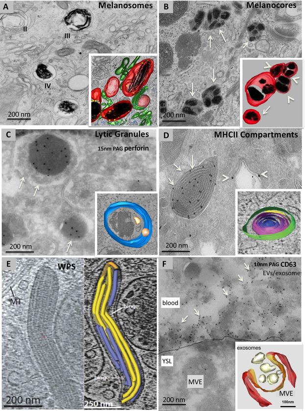
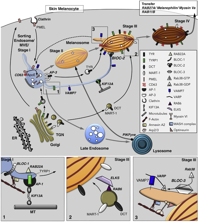
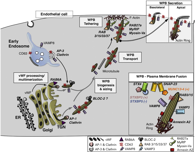
Unique functions of specialised cells such as those of the immune and haemostasis systems, skin, blood vessels, lung, and bone require specialised compartments, collectively referred to as lysosome-related organelles (LROs), that share features of endosomes and lysosomes. LROs harbour unique morphological features and cell type-specific contents, and most if not all undergo regulated secretion for diverse functions. Ongoing research, largely driven by analyses of inherited diseases and their model systems, is unravelling the mechanisms involved in LRO generation, maturation, transport and secretion. A molecular understanding of these features will provide targets and markers that can be exploited for diagnosis and therapy of a myriad of diseases.


Figures

Figure 1.
Figure 1.. Examples of ELRO ultrastructure.
Conventional electron microscopy (A, B), immunogold labelling on ultrathin cryosections (C, D, F), cryomicroscopy (E) and three-dimensional (3D) reconstructions of electron tomograms of model ELROs and their derivatives (A, B, C, D, E, F). A, stage II immature and stage III and IV mature melanosomes in an MNT-1 human melanoma cell fixed by high pressure freezing and embedded in plastic by freeze substitution. Note the striated appearance in stages II and III. Inset: 3D reconstruction showing dark melanin on fibrillar structures (black) in stage III/ IV melanosomes. Unpigmented fibrillar structures present in stage II melanosomes are shown in white. Melanosomes are surrounded by tubular membranes (green), corresponding to endosomal transport carriers.Ribosomes are in blue B, melanocore containing organelles (arrows) in a keratinocyte in human skin biopsies. Inset: 3D reconstruction. Note several melanocores (black) enclosed by a single membrane (red). These organelles appear isolated (arrow) or in a network of clusters (arrowheads). C, lytic granules (arrows) of a human cytotoxic T cell depicting the characteristic dense core containing perforin, immunolabelled with 15 nm protein A gold particles (PAG), surrounded by small membrane vesicles. Inset: 3D reconstruction showing the limiting membrane (blue) and ILVs (yellow). The dense core is not pseudocoloured. D, an ultrathin cryosection of a mouse dendritic cell showing MHC class II (MHCII) compartments (arrows) immunolabeled for MHCII molecules with 10 nm PAG and endosomes (arrowheads) immunolabeled for the endosomal protein EEA1 with 15 nm PAG. Inset: 3D reconstruction showing the multiple concentric membrane layers of an MHCII compartment. E, Cigar-shaped WPBs in a thick section of a human umbilical vein endothelial cell visualized by cryomicroscopy (left panel). The right panel shows a 3D reconstruction of the vWF tubules (blue and yellow) contained within the WPB. F, ultrathin cryosection of a zebrafish embryo expressing CD63-pHluorin in the yolk syncytial layer and labelled for GFP with 10 nm PAG. The plasma membrane of the yolk sac layer is indicated by the dashed line. Note the MVE in the yolk cell and numerous membrane vesicles labelled for CD63 (arrows) in the blood. Inset: 3D reconstruction of an MVE in a HeLa cell in the process of fusing with the plasma membrane. Magnifications are indicated in the panels.
Figure 2.
Figure 2.
Model of membrane dynamics during melanosome biogenesis. Shown are pathways of membrane transport from the Golgi and early endosomes to generate different stages of melanosomes. Melanosomes mature from stage I (equivalent to vacuolar domain of a sorting early endosome) to stage IV by progressive acquisition of protein cargoes, mirrored by morphological changes. Following clathrin-dependent endocytosis from the plasma membrane, PMEL is targeted to stage I melanosomes within which it forms fibrils, with the assistance of CD63 and ApoE, on intraluminal vesicles (white circles). The fibrils then mature to fully assembled sheets in stage II in a process requiring collisions with lysosomes mediated by PIKFyve (thick arrow). Melanin synthesis begins upon acquisition of Tyrosinase (TYR), additional enzymes (e.g. TYRP1 and DCT), and transporters that neutralize the luminal pH. The newly generated melanin accumulates on the sheets in stage III and throughout the organelle in stage IV. Cargo transport from endosomes occurs by an AP-3-dependent vesicular pathway taken by TYR and a tubular pathway taken by TYRP1 and other cargoes that requires BLOC-1, RAB22A, AP-1 and KIF13A for membrane tubule formation along microtubules (Box 1), BLOC-2 for targeting to maturing stage III melanosomes, and the vSNARE VAMP7 for fusion with melanosomes. An additional pathway to target DCT and MART-1 to melanosomes from the Golgi requires RAB6 for vesicle formation and its effector ELKS for docking to melanosomes (Box 2). VAMP7 is recycled from melanosomes in tubules that require RAB38, its GEF BLOC-3, and the scaffold protein VARP for formation and/ or VAMP7 incorporation, and that require Myosin VI, optineurin, the WASH complex, Arp2/3 and actin for tubule constriction, scission and release (Box 3); the target organelle for these tubules remains speculative (dashed arrow). Mature stage IV melanosomes in skin melanocytes are captured in the periphery by the RAB27A/ Melanophilin/ Myosin Va complex, and may fuse with the plasma membrane in a RAB11B-dependent manner to release melanocores for uptake by neighbouring keratinocytes.
Figure 3.
Figure 3.
Model of membrane dynamics during Weibel-Palade body (WPB) biogenesis. WPB biogenesis begins with the assembly of tubular multimers of von Willebrand factor (vWF) in the Golgi. The quantal length of the multimers is limited by the length of the Golgi cisterna within an individual stack, the size of which is controlled by RAB6A; vWF multimer quanta are then assembled into longer tubules depending on the integrity of the Golgi ribbon. Immature WPBs encasing vWF tubules bud from the Golgi in a process requiring AP-1 and clathrin, and then mature by fusion with vesicles bearing CD63 and the vSNARE, VAMP8, originating from early endosomes in an AP-3-dependent process. BLOC-2 also contributes to WPB maturation in as yet unknown ways. Small WPBs are secreted basolaterally. By contrast, maturing large WPBs are transported to the apical plasma membrane of endothelial cells along microtubules and tethered to the plasma membrane through the action of the RAB27A/ MyRIP/ MyosinVa complex. Mature WPBs are decorated by several RABs (e.g. RAB3, 15, 33 and 37), some of which (e.g. RAB3 and RAB15) might positively regulate the release step. Apical secretion of large WPBs (Box) requires actin polymerization at the basal tip mediated by Annexin A2, calcium-dependent tethering mediated by MUNC13–4, and fusion mediated by the Syntaxin 4/ SNAP-23 tSNARE and either VAMP3 or VAMP8 as the vSNARE. The latter step is facilitated by STXBP3 (Munc18c) and antagonized by STXBP5.

Similar articles

Cited by

References

    1. Klumperman J, Raposo G: The complex ultrastructure of the endolysosomal system. Cold Spring Harb. Perspect. Biol 2014, 6:a016857. - PMC - PubMed
    1. Dell’Angelica EC, Mullins C, Caplan S, Bonifacino JS: Lysosome-related organelles. FASEB J. 2000, 14:1265–1278. - PubMed
    1. Marks MS, Heijnen HFG, Raposo G: Lysosome-related organelles: unusual compartments become mainstream. Curr. Opin. Cell Biol 2013, 25:495–505. - PMC - PubMed
    1. van Niel G, D’Angelo G, Raposo G: Shedding light on the cell biology of extracellular vesicles. Nat. Rev. Mol. Cell Biol 2018, 19:213–228. - PubMed
    1. Raposo G, Marks MS, Cutler DF: Lysosome-related organelles: driving post-golgi compartments into specialisation. Curr. Opin. Cell Biol 2007, 19:394–401. - PMC - PubMed

Publication types