. 2019 Jun 23;20(12):3070.
doi: 10.3390/ijms20123070.

Analysis of Expression and Functional Activity of Aromatic L-Amino Acid Decarboxylase (DDC) and Serotonin Transporter (SERT) as Potential Sources of Serotonin in Mouse Ovary

Affiliations

Analysis of Expression and Functional Activity of Aromatic L-Amino Acid Decarboxylase (DDC) and Serotonin Transporter (SERT) as Potential Sources of Serotonin in Mouse Ovary

Denis A Nikishin et al. Int J Mol Sci. .

Abstract

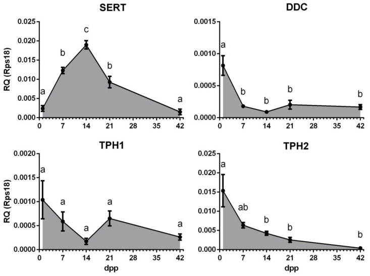
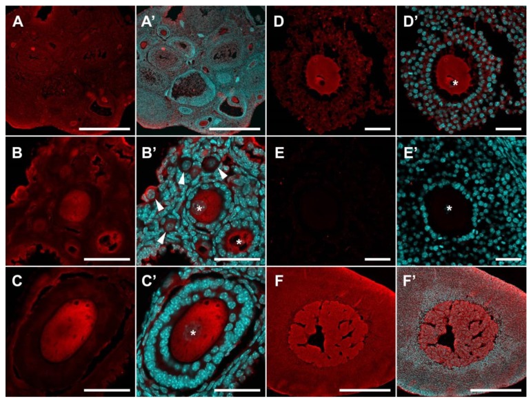
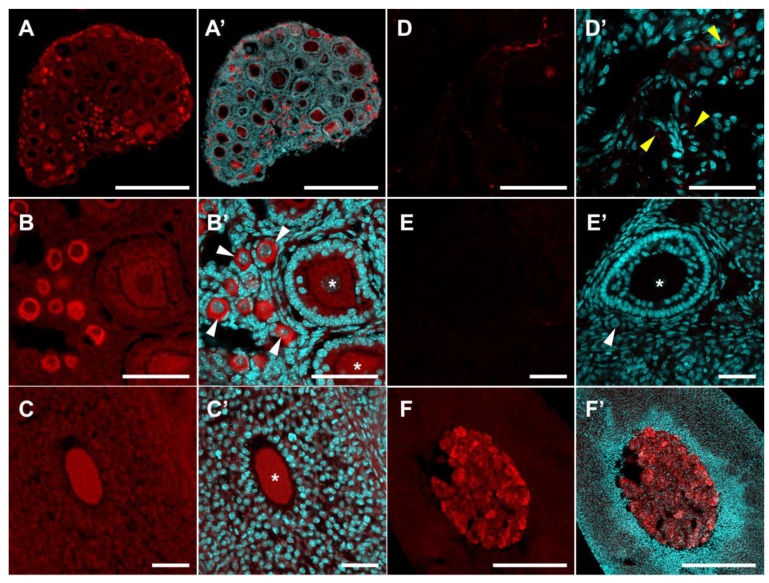
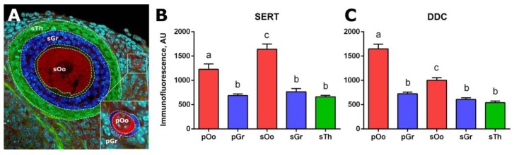
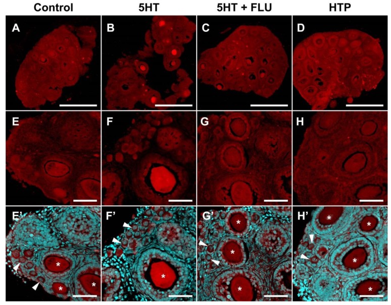
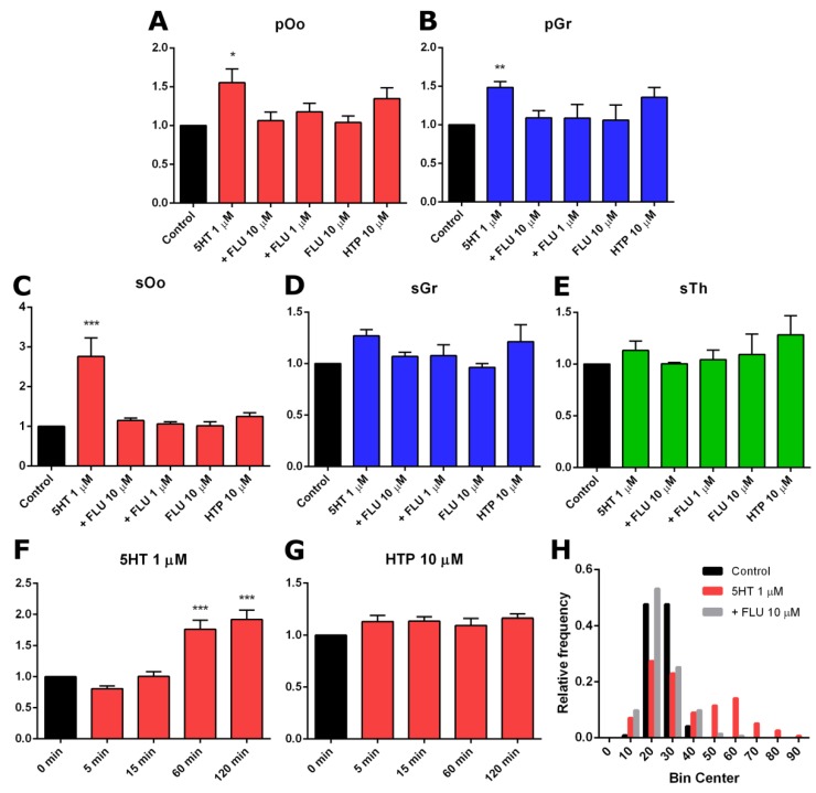
The origin of serotonin in the ovary is the key question for understanding mechanisms of serotonergic regulation of reproductive function. We performed a study of the expression and functional activity of the serotonin transporter (SERT) and the enzyme for the synthesis of serotonin, aromatic l-amino acid decarboxylase (DDC) in mouse ovary. A pronounced peak of SERT mRNA expression occurs at the age of 14 days, but serotonin synthesis enzymes are expressed at the maximum level in the ovaries of newborn mice. SERT is detected immunohistochemically in all cellular compartments of the ovary with a maximum level of immunostaining in the oocytes of growing ovarian follicles. DDC immunolocalization, in contrast, is detected to a greater extent in primordial follicle oocytes, and decreases at the later stages of folliculogenesis. Serotonin synthesis in all cellular compartments occurs at very low levels, whereas specific serotonin uptake is clearly present, leading to a significant increase in serotonin content in the oocytes of growing primary and secondary follicles. These data indicate that the main mechanism of serotonin accumulation in mouse ovary is its uptake by the specific SERT membrane transporter, which is active in the oocytes of the growing ovarian follicles.

Keywords: DDC; SERT; fluoxetine; mouse; ovary; serotonin.


Conflict of interest statement

The authors declare no conflict of interest.

Figures

Figure 1
Figure 1
Gene expression profiles of the serotonin transporter SERT and enzymes of serotonin synthesis DDC, TPH1 and TPH2, during postnatal development of the mouse ovary. The relative quantity (RQ) was calculated using 2−ΔCt method relative to the reference gene RPS18 (M ± SEM). Different letters denote statistical significance between groups at p < 0.05, according to ordinary one-way ANOVA with Holm-Sidak’s multiple comparisons test.
Figure 2
Figure 2
Immunohistochemical detection of SERT in the mouse ovary. (AD) SERT immunoreactivity in the ovary of prepubertal (B,C) and adult mice (A,D). (E) Negative control: Ovary immunostaining without primary antibodies. (F) Positive control: Adrenal gland with SERT immunoreactivity in the medullar region. (A’F’) Images merged with nuclear staining. Twenty-μm cryosections were stained for SERT (red false colour) and nuclei were dyed with DAPI counterstain (cyan false colour). Arrowheads point to primordial follicles. Asterisks mark oocytes in growing follicles. Scale bars: (A,F) 500 µm; (BE) 50 µm.
Figure 3
Figure 3
Immunohistochemical detection of DDC in the mouse ovary. (AD) DDC immunoreactivity in the ovary of prepubertal (A,B,D) and adult mice (C). (E) Negative control: Ovary immunostaining without primary antibodies. (F) Positive control: Adrenal gland with DDC immunoreactivity in the medullar region. (A’F’) Images merged with nuclear staining. Twenty-μm cryosections were stained for DDC (red false colour) and nuclei were dyed with DAPI counterstain (cyan false colour). White arrowheads point to primordial follicles. Asterisks mark oocytes in growing follicles. Yellow arrowheads point to nerve fibres in ovarian stromal tissue. Scale bars: (A,F) 500 µm; (BE) 50 µm.
Figure 4
Figure 4
Quantitative analysis of anti-SERT and anti-DDC immunoreactivity in individual cellular compartments of mouse ovaries. (A) Cell compartments of the ovary. Dashed lines mark the areas to be analysed: sOo–oocyte, sGr–granulosa cells, sTh–theca cells of the secondary (preantral) follicle; pOo–oocyte, pGr–granulosa cells of the primordial follicle. (B) Quantitative analysis of SERT immunoreactivity. (C) Quantitative analysis of DDC immunoreactivity. AU–arbitrary units of immunofluorescence. Different letters denote statistical significance between groups at p < 0.05, according to ordinary one-way ANOVA with Holm-Sidak’s multiple comparisons test.
Figure 5
Figure 5
Functional activity of uptake and synthesis of serotonin in the prepubertal (14 dpp) mouse ovary. (A,E) Control. (B,F) Two-h incubation with serotonin (5HT) (1 μM). (C,G) Two-h incubation with 5HT (1 μM) with previous addition of fluoxetine (FLU) (10 μM). (D,H) Two-h incubation with 5-hydroxytryptophan HTP (10 μM). (E’H’) Images merged with nuclear staining. Twenty-μm cryosections were stained for serotonin (red false colour) and nuclei were dyed with DAPI counterstain (cyan false colour). White arrowheads point to primordial follicles. Asterisks mark oocytes in growing follicles. Scale bars: (AD) 500 µm; (EH) 50 µm.
Figure 6
Figure 6
Quantitative analysis of serotonin accumulation in the study of the activity of the systems of its synthesis and uptake in mouse ovaries. (AE) Quantitative analysis of anti-serotonin immunoreactivity in individual cellular compartments of mouse ovaries after incubation with serotonin (5HT), with 5HT and previous addition of fluoxetine (+FLU), with fluoxetine only (FLU) and with 5-hydroxytryptophan (HTP): sOo–oocyte, sGr–granulosa cells and sTh–theca cells of the secondary (preantral) follicle; pOo–oocyte and pGr–granulosa cells of primordial follicle (M ± SEM). The level of immunofluorescence in the control sample is taken as 1. * Denotes statistical significance at p < 0.05, ** at p < 0.01 and *** at p < 0.005, between control and treated groups, using the Friedman test. (F,G) The temporal dynamics of serotonin accumulation in oocytes of growing follicles (sOo) during incubation with 5HT (1 μM) and HTP (10 μM) (M ± SEM). *** Denotes statistical significance at p < 0.005, between control and treated groups, using the Friedman test. (H) Frequency distribution histogram of serotonin accumulation in oocytes of growing follicles (sOo). A bimodal distribution is observed in the 5HT group.

Similar articles

Cited by

References

    1. Clausell D.E., Soliman K.F. Ovarian serotonin content in relation to ovulation. Experientia. 1978;34:410–411. doi: 10.1007/BF01923065. - DOI - PubMed
    1. Bòdis J., Bognàr Z., Hartmann G., Török A., Csaba I.F. Measurement of noradrenaline, dopamine and serotonin contents in follicular fluid of human graafian follicles after superovulation treatment. Gynecol. Obstet. Investig. 1992;33:165–167. doi: 10.1159/000294873. - DOI - PubMed
    1. Amireault P., Dubé F. Serotonin and its antidepressant-sensitive transport in mouse cumulus-oocyte complexes and early embryos. Biol. Reprod. 2005;73:358–365. doi: 10.1095/biolreprod.104.039313. - DOI - PubMed
    1. Buznikov G.A., Nikitina L.A., Galanov A.Y., Malchenko L.A., Trubnikova O.B. The control of oocyte maturation in the starfish and amphibians by serotonin and its antagonists. Int. J. Dev. Biol. 1993;37:363–364. - PubMed
    1. Terranova P.F., Uilenbroek J.T., Saville L., Horst D., Nakamura Y. Serotonin enhances oestradiol production by hamster preovulatory follicles in vitro: effects of experimentally induced atresia. J. Endocrinol. 1990;125:433–438. doi: 10.1677/joe.0.1250433. - DOI - PubMed

MeSH terms

LinkOut - more resources