. 2019 Jun 24;19(1):140.
doi: 10.1186/s12866-019-1506-7.

Stationary phase persister formation in Escherichia coli can be suppressed by piperacillin and PBP3 inhibition

Affiliations

Stationary phase persister formation in Escherichia coli can be suppressed by piperacillin and PBP3 inhibition

Sandra J Aedo et al. BMC Microbiol. .

Abstract

Background: Persisters are rare phenotypic variants within a bacterial population that are capable of tolerating lethal antibiotic concentrations. Passage through stationary phase is associated with the formation of persisters (type I), and a major physiological response of Escherichia coli during stationary phase is cell wall restructuring. Given the concurrence of these processes, we sought to assess whether perturbation to cell wall synthesis during stationary phase impacts type I persister formation.

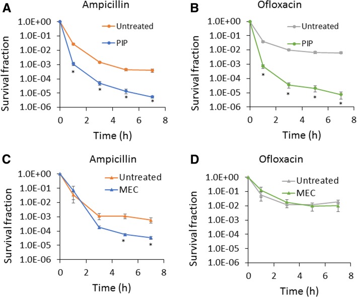
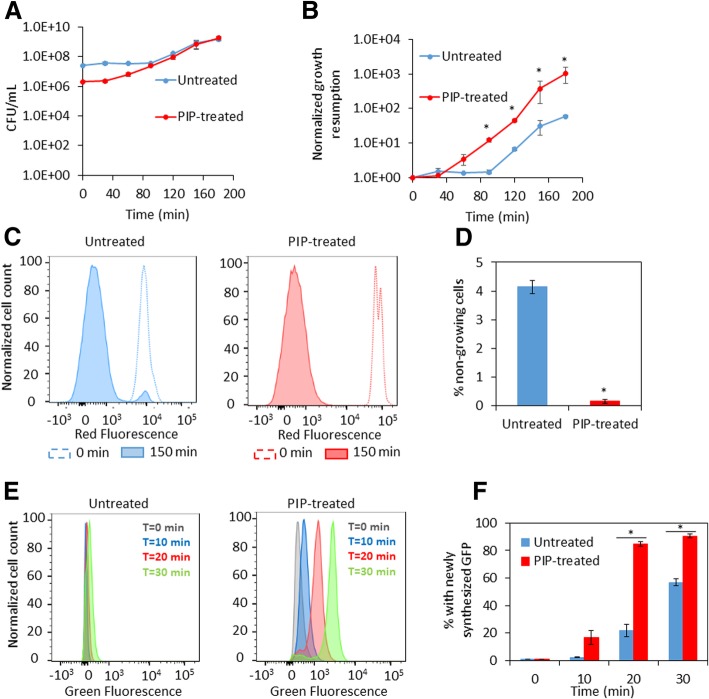
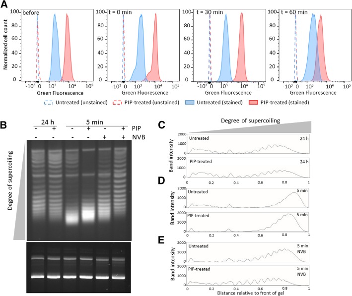
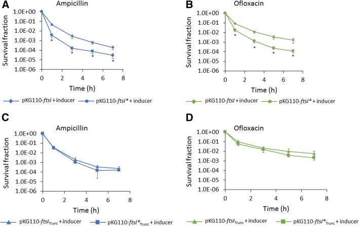
Results: We tested a panel of cell wall inhibitors and found that piperacillin, which primarily targets penicillin binding protein 3 (PBP3 encoded by ftsI), resulted in a significant reduction in both β-lactam (ampicillin, carbenicillin) and fluoroquinolone (ofloxacin, ciprofloxacin) persister levels. Further analyses showed that piperacillin exposure through stationary phase resulted in cells with more ATP, DNA, RNA, and protein (including PBPs) than untreated controls; and that their physiology led to more rapid resumption of DNA gyrase supercoiling activity, translation, and cell division upon introduction into fresh media. Previously, PBP3 inhibition had been linked to antibiotic efficacy through the DpiBA two component system; however, piperacillin suppressed persister formation in ΔdpiA to the same extent as it did in wild-type, suggesting that DpiBA is not required for the phenomenon reported here. To test the generality of PBP3 inhibition on persister formation, we expressed FtsI Ser307Ala to genetically inhibit PBP3, and suppression of persister formation was also observed, although not to the same magnitude as that seen for piperacillin treatment.

Conclusions: From these data we conclude that stationary phase PBP3 activity is important to type I persister formation in E. coli.

Keywords: E. coli; Fluoroquinolone; PBP3; Persisters; Piperacillin; β-Lactam.


Conflict of interest statement

None to declare.

Figures

Fig. 1
Fig. 1
Treatment of stationary-phase cultures with specific β-lactams impairs persister formation. Cell cultures were treated with 200 μg/mL piperacillin (PIP) (a and b) or mecillinam (MEC) (c and d) at t = 4 h. Cells in control culture were treated with an equal volume of solvent, which was water (untreated). At 24 h, cultures were washed to remove chemicals and diluted in fresh LB containing 200 μg/mL ampicillin or 5 μg/mL ofloxacin. Survival fractions were monitored at the indicated time points. CFU/mL at the indicated time points are provided in Additional file 10: Figure S19. * p < 0.05 (t- test). Data represent three or more biological replicates. Each data point was denoted as mean ± s.e
Fig. 2
Fig. 2
Biochemical characterization of PIP-treated stationary phase cells. Cell cultures were treated with piperacillin (PIP-treated) or water (untreated) at t = 4 h. At t = 24 h, measurements of DNA (a), ATP, protein, and RNA content (b) were carried out. a Cell cultures were fixed and stained with PicoGreen for DNA content and chromosome number assessment using flow cytometry. Numbers above the brackets indicate chromosome copy number. The chromosome number scale was determined with stationary-phase cell cultures of known DNA content (Additional file 7: Figure S7). b Cells were pelleted for RNA extraction, sonicated for protein concentration determination by the Bradford assay, or diluted to an OD600 of ~ 0.1 for ATP measurements using the BacTiter-Glo assay. Abundances were calculated on a per cell basis, with cell number quantified by flow cytometry, and are presented relative to untreated. * p < 0.05 (t-test). Data represent three or more biological replicates. Each data point was denoted as mean ± s.e
Fig. 3
Fig. 3
Growth resumption of PIP-treated stationary phase cells. Cell cultures were treated with piperacillin (PIP-treated) or water (untreated) at t = 4 h. At t = 24 h, measurements of growth resumption, cell division, and protein synthesis were initiated. a Cells were washed to remove piperacillin and diluted in fresh media. CFU/mL were monitored at the indicated time points. b Normalized growth resumption from A were plotted at the indicated time points. c Cell cultures carrying mCherry expression system were grown in LB with inducer (1 mM IPTG) until t = 24 h. At 24 h, cultures were washed and diluted in fresh LB without inducer. mCherry levels in cells at t = 0 min and t = 150 min were detected with flow cytometry (a representative replicate is shown). d The percentages of non-growing cells were calculated from fractions of mCherry positive cells in entire cell populations at t = 150 min. e Cell cultures carrying pQE-80 L-gfp (without IPTG) were diluted at t = 24 h in fresh LB with inducer (1 mM IPTG) for GFP expression. GFP was monitored at indicated time points with flow cytometry (a representative replicate is shown). f Percentage of cells with newly-synthesized GFP were plotted with respect to time. * p < 0.05 (t-test). Data represent three or more biological replicates. Each data point was denoted as mean ± s.e
Fig. 4
Fig. 4
PIP-treated stationary phase cells contain more PBPs and exhibit more uniform DNA gyrase supercoiling activity upon dilution into fresh media. a Cell cultures were treated with piperacillin (PIP-treated) or water (untreated) at t = 4 h. At t = 24 h, an aliquot was removed for staining with Bocillin-FL (before sample). Further, at t = 24 h, cell cultures were washed to remove piperacillin and diluted in fresh LB. Aliquots were taken at the indicated time points for staining with Bocillin-FL (t = 0, 30, and 60 min). Bocillin-FL stained samples were analyzed by flow cytometry (solid lines and filled histogram) and unstained samples were used to control for autofluorescence (dashed lines and hollow histogram). Data shown correspond to one replicate of at least three biological replicates. b Cultures of MG1655 with pQE-80 L-kan were treated with piperacillin or water at t = 4.5 h (OD600 ~ 1). Piperacillin was removed at t = 24 h by washes in fresh LB, in the presence or absence of novobiocin, before dilution and incubation. Plasmid DNA was extracted at t = 24 h and after 5 min incubation in fresh media with or without novobiocin. Plasmid DNA concentration was determined and equal amounts of plasmid DNA were loaded onto an agarose gel containing chloroquine (top gel) and an agarose gel without intercalator as a loading control (bottom gel). Top and bottom gels were run for 21 h and 1 h, respectively. c-e Densitometry scans of untreated and PIP-treated samples that were either processed at t = 24 h (c), washed and incubated in fresh LB for 5 min (d), or washed and incubated for 5 min in fresh LB in the presence of novobiocin (NVB) prior to plasmid extraction (e). Two more replicates are presented in Additional file 8: Figure S8
Fig. 5
Fig. 5
Expression of an inactivated PBP3 during stationary phase inhibits persister formation. Cultures of MG1655 carrying pKG110-ftsI (wild-type ftsI) or pKG110-ftsI* (catalytically inactive mutant) were treated with 100 μM sodium salicylate (inducer) at t = 3 h (a and b). At 24 h, cell cultures were washed to remove the inducer and diluted in fresh LB containing 200 μg/mL ampicillin (a) or 5 μg/mL ofloxacin (b). In (c) and (d) cultures of MG1655 carrying pKG110-ftsITrunc or pKG110-ftsI*Trunc (each devoid of their transmembrane and cytoplasmic domains) were treated as in a and b, and ampicillin (c) and ofloxacin (d) persister assays were carried out. Survival fractions were monitored at the indicated time points. CFU/mL at the indicated time points are provided in Additional file 14: Figure S12E and S12H. * p < 0.05 (t-test). Data represent three or more biological replicates. Each data point was denoted as mean ± s.e

References

    1. Balaban NQ, Merrin J, Chait R, Kowalik L, Leibler S. Bacterial persistence as a phenotypic switch. Science. 2004;305(5690):1622–1625. doi: 10.1126/science.1099390. - DOI - PubMed
    1. Keren I, Kaldalu N, Spoering A, Wang Y, Lewis K. Persister cells and tolerance to antimicrobials. FEMS Microbiol Lett. 2004;230(1):13–18. doi: 10.1016/S0378-1097(03)00856-5. - DOI - PubMed
    1. Wallis RS, Patil S, Cheon SH, Edmonds K, Phillips M, Perkins MD, et al. Drug tolerance in Mycobacterium tuberculosis. Antimicrob Agents Chemother. 1999;43(11):2600–2606. doi: 10.1128/AAC.43.11.2600. - DOI - PMC - PubMed
    1. Meylan S, Andrews IW, Collins JJ. Targeting antibiotic tolerance, pathogen by pathogen. Cell. 2018;172(6):1228–1238. doi: 10.1016/j.cell.2018.01.037. - DOI - PubMed
    1. Mulcahy LR, Burns JL, Lory S, Lewis K. Emergence of Pseudomonas aeruginosa strains producing high levels of persister cells in patients with cystic fibrosis. J Bacteriol. 2010;192(23):6191–6199. doi: 10.1128/JB.01651-09. - DOI - PMC - PubMed

Publication types

MeSH terms