. 2019 Jun 24;19(1):618.
doi: 10.1186/s12885-019-5824-9.

Inhibition of PI3K/Akt/mTOR signaling pathway alleviates ovarian cancer chemoresistance through reversing epithelial-mesenchymal transition and decreasing cancer stem cell marker expression

Affiliations

Inhibition of PI3K/Akt/mTOR signaling pathway alleviates ovarian cancer chemoresistance through reversing epithelial-mesenchymal transition and decreasing cancer stem cell marker expression

Junli Deng et al. BMC Cancer. .

Abstract

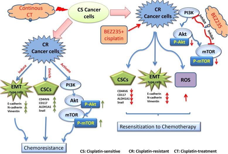
Background: Ovarian cancer is the most common malignant tumor of the female reproductive tract. Chemoresistance is a major challenge for current ovarian cancer therapy. However, the mechanism underlying epithelial ovarian cancer (EOC) chemoresistance is not completely uncovered. The phosphatidylinositol-3-kinase (PI3K)/Akt/mammalian target of rapamycin (mTOR) signaling is an important intracellular pathway in regulating cell cycle, quiescence, and proliferation. The aim of this study is to investigate the role of PI3K/Akt/mTOR signaling pathway and its association with epithelial-mesenchymal transition (EMT) and cancer stem cell (CSC) marker expression in EOC chemoresistance.

Methods: The expressions of EMT and CSC markers were detected by immunofluorescence, western blot, and quantitative real-time PCR. BEZ235, a dual PI3K/mTOR inhibitor, was employed to investigate the role of PI3K/Akt/ mTOR signaling in regulating EMT and CSC marker expression. Students' t test and one-way ANOVA with Tukey's post-hoc test were used to compare the data from different groups.

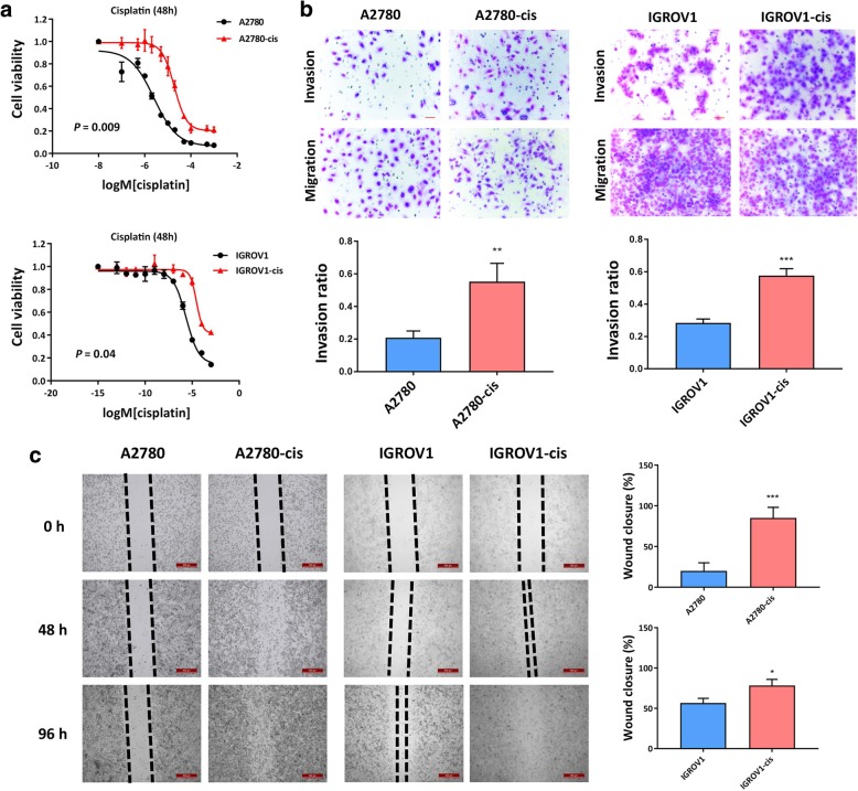
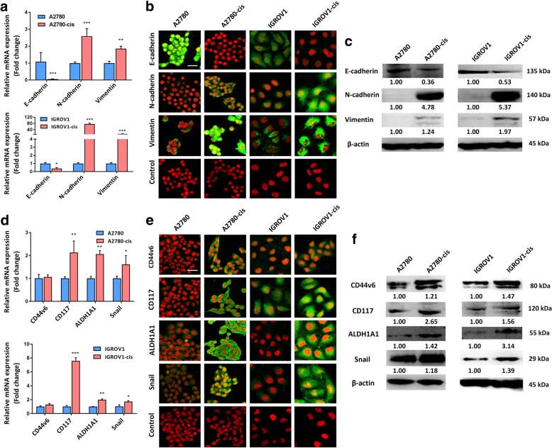
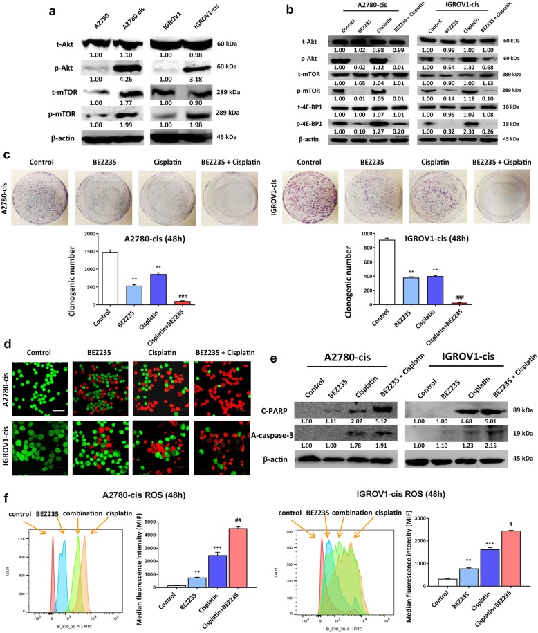
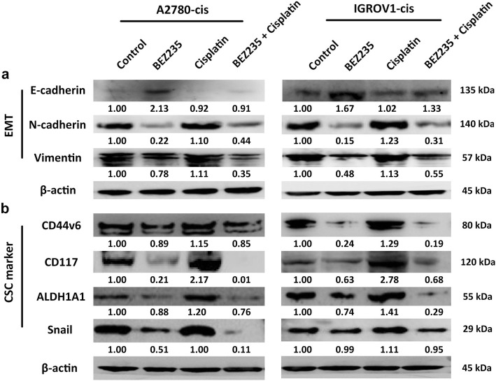
Results: We found that EMT and CSC marker expression were significantly enhanced in chemoresistant EOC cells, which was accompanied by the activation of PI3K/Akt/mTOR signaling. Compared with single cisplatin treatment, combined treatment with BEZ235 and cisplatin significantly disrupted the colony formation ability, induced higher ROS level and more apoptosis in chemoresistant EOC cells. Furthermore, the combination approach effectively inhibited PI3K/Akt/mTOR signaling pathway, reversed EMT, and decreased CSC marker expression in chemoresistant EOC cells compared with cisplatin mono-treatment.

Conclusions: Our results first demonstrate that EMT and enhanced CSC marker expression triggered by activated PI3K/Akt/mTOR signaling are involved in the chemoresistance of EOC, and BEZ235 in combination with cisplatin might be a promising treatment option to reverse EOC chemoresistance.

Keywords: CSC; Chemoresistance; EMT; Ovarian cancer; PI3K/Akt/mTOR signaling.


Conflict of interest statement

The authors declare that they have no competing interests.

Figures

Fig. 1
Fig. 1
EOC-cis cell lines showed enhanced resistance to cisplatin and enhanced metastatic potential. a Cells were treated with different concentrations of cisplatin for 48 h and cell viability was tested using cell proliferation assay. EOC-cis cells showed significant resistance to cisplatin. b The invasion ability of cells within 48 h was detected using Matrigel invasion assay. Representative images for EOC cell invasion and migration were photogrphed at 200 x magnification. The invasive potential of EOC-cis cell lines was significantly increased as compared to parental cell lines. c The migration ability of cells within 96 h was detected by wound-healing assay. Typical images were obtained at 40x amplification at 0 h, 48 h, and 96 h and the percentage of wound closure area in 96 h was compared. A2780-cis and IGROV1-cis had a higher wound closure rate than the corresponding parental cells. All data were expressed as mean ± SD. **P < 0.01 and ***P < 0.001 versus parental cells (n = 3)
Fig. 2
Fig. 2
EOC-cis cells showed higher expressions of EMT and CSC markers. a mRNA expressions of EMT markers were detected by qRT-PCR. GAPDH was used as a control. b Typical images of immnuflourescence for the expressions of EMT markers in EOC-cis and parental cells were obtained at 200x magnification (Green). Red indicates nuclei stain. c Protein expressions of EMT markers were detected by western blot. β-actin was used as a loading control. d mRNA expressions of CSC markers in EOC-cis cell lines were detected by qRT-PCR. GAPDH was used as a control. e Typical images of immnuflourescence for the expressions of CSC markers in EOC-cis and parental cells were obtained at 200x magnification (Green). Red indicates nuclei stain. f Protein expressions of CSC markers were detected by western blot. β-actin was used as a loading control. All data were expressed as mean ± SD. *P < 0.05, **P < 0.01, and ***P < 0.001 versus parental cells (n = 3)
Fig. 3
Fig. 3
PI3K/Akt/mTOR signaling was activated in EOC-cis cells and BEZ235 can sensitize EOC-cis cells to cisplatin. a The protein expressions of Akt and mTOR were detected by western blot. β-actin was used as a loading control. b Combination treatment of BEZ235 and cisplatin significantly reduced the expressions of PI3K/Akt/mTOR signaling pathway proteins (p-AKT, pmTOR and p-4E-BP1) as compared to cisplatin mono-treatment group. c Colony formation ability of EOC-cis cell lines was detected after treatment with BEZ235 (½ IC50), cisplatin (½ IC50), or in combination for 48 h. Typical images of colony for different treatment groups are shown. d The apoptosis of EOC-cis cells after different treatments was tested with AO/EB assay. Red indicates cells undergoing apoptosis, while green indicates normal cells. Photos were obtained at 200x magnification. e The expressions of pre-apoptotic proteins in EOC-cis cells after different treatments were detected by western blot. β-actin was used as a loading control. f Quantitation analysis of ROS was performed using flow cytometry with median fluorescence intensity (MFI) detection. Fluorescence emission spectra of ROS in control and treatment groups of EOC-cis cells was shown. ROS level was significantly increased in the combination treatment group compared with single cisplatin treatment in both EOC-cis cell lines. All data were expressed as mean ± SD. **P < 0.01 and ***P < 0.01 versus control group, while #P < 0.05, ##P < 0.01, and ###P < 0.001 versus cisplatin treatment group (n = 3)
Fig. 4
Fig. 4
Combination treatment of BEZ235 and cisplatin inhibited EMT and decreased CSC marker expression. a The protein expressions of EMT markers in EOC-cis cells after treatment with BEZ235 (½ IC50), cisplatin (½ IC50), or in combination for 48 h were detected by western blot. β-actin was used as a loading control. Combination treatment of BEZ235 and cisplatin obviously reversed EMT in EOC-cis cells. b The protein expressions of CSC markers in EOC-cis cells after treatment with BEZ235 (½ IC50), cisplatin (½ IC50), or in combination for 48 h were detected by western blot. β-actin was used as a loading control. Combination treatment of BEZ235 and cisplatin markedly inhibited CSC marker expression in EOC-cis cells
Fig. 5
Fig. 5
The potential mechanisms underlying EOC chemoresistance and the sensitizing effect of PI3K inhibition

Similar articles

Cited by

References

    1. Torre LA, Bray F, Siegel RL, Ferlay J, Lortet-Tieulent J, Jemal A. Global cancer statistics, 2012. CA Cancer J Clin. 2015;65(2):87–108. doi: 10.3322/caac.21262. - DOI - PubMed
    1. Jelovac D, Armstrong DK. Recent progress in the diagnosis and treatment of ovarian cancer. CA Cancer J Clin. 2011;61(3):183–203. doi: 10.3322/caac.20113. - DOI - PMC - PubMed
    1. Deng J, Wang L, Chen H, Hao J, Ni J, Chang L, Duan W, Graham P, Li Y. Targeting epithelial-mesenchymal transition and cancer stem cells for chemoresistant ovarian cancer. Oncotarget. 2016;7(34):55771–55788. doi: 10.18632/oncotarget.9908. - DOI - PMC - PubMed
    1. Thiery JP, Acloque H, Huang RY, Nieto MA. Epithelial-mesenchymal transitions in development and disease. Cell. 2009;139(5):871–890. doi: 10.1016/j.cell.2009.11.007. - DOI - PubMed
    1. Taube JH, Herschkowitz JI, Komurov K, Zhou AY, Gupta S, Yang J, Hartwell K, Onder TT, Gupta PB, Evans KW, et al. Core epithelial-to-mesenchymal transition interactome gene-expression signature is associated with claudin-low and metaplastic breast cancer subtypes. Proc Natl Acad Sci U S A. 2010;107(35):15449–15454. doi: 10.1073/pnas.1004900107. - DOI - PMC - PubMed

MeSH terms