

A novel function of artesunate on inhibiting migration and invasion of fibroblast-like synoviocytes from rheumatoid arthritis patients
- PMID: 31234900
- PMCID: PMC6591920
- DOI: 10.1186/s13075-019-1935-6
A novel function of artesunate on inhibiting migration and invasion of fibroblast-like synoviocytes from rheumatoid arthritis patients
Abstract
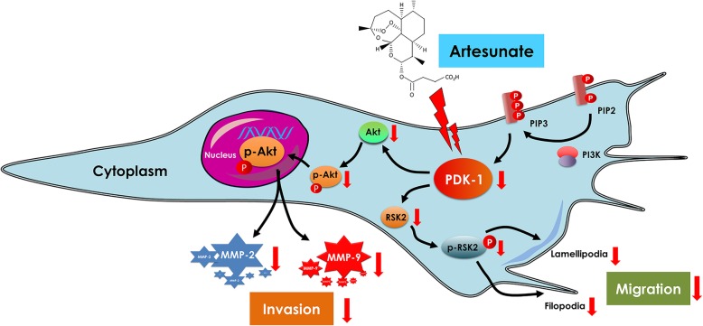
Introduction: Anti-malarial drug artesunate can suppress inflammation and prevent cartilage and bone destruction in collagen-induced arthritis model in rats-suggesting it may be a potent drug for rheumatoid arthritis (RA) therapy. We aimed to investigate its effect on the invasive property of fibroblast-like synoviocytes (FLS) from patients with RA.
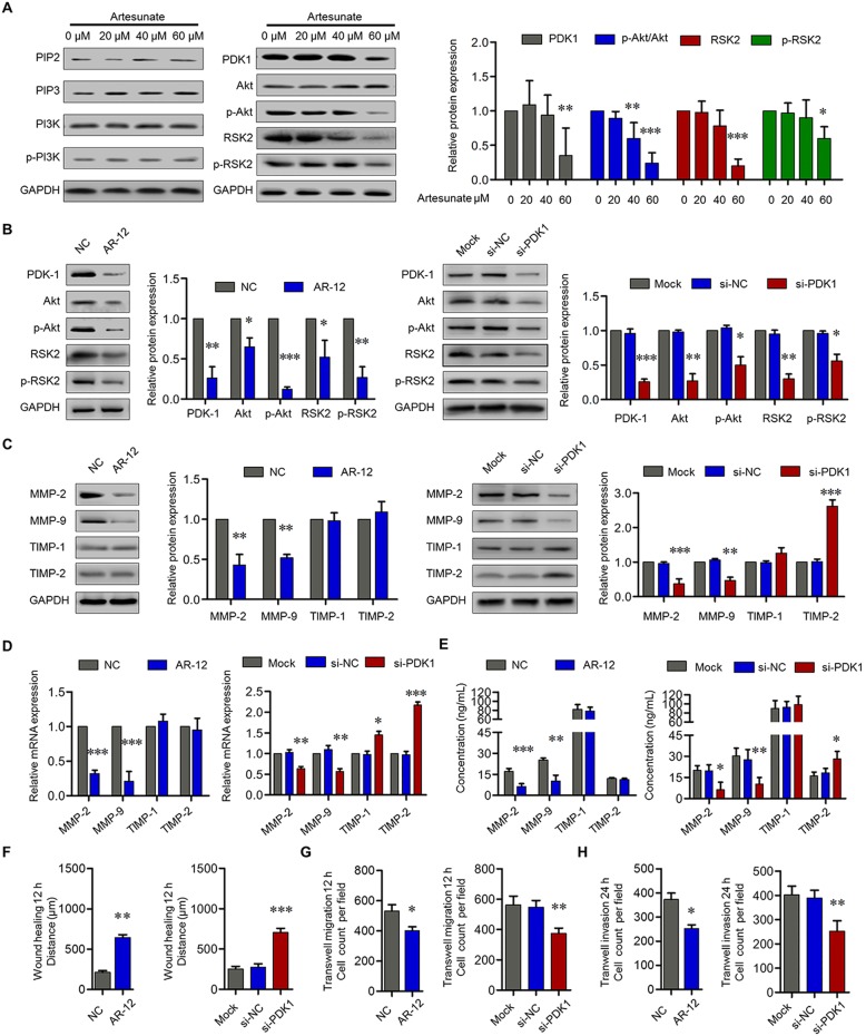
Methods: Synovial tissues were obtained by closed needle biopsy from active RA patients, and FLS were isolated and cultured in vitro. RA-FLS were treated with artesunate at various concentrations, while methotrexate or hydroxychloroquine was employed as comparator drugs. Cell viability, proliferation, cell cycle, apoptosis, migration, invasion, and pseudopodium formation of RA-FLS were assessed by CCK-8 assays, EdU staining, Annexin V-FITC/PI staining, transwell assays, or F-actin staining, respectively. Further, relative changes of expressed proteases were analyzed by Proteome profiler human protease array and verified by quantitative real-time PCR (qPCR), Western blot, and ELISA. The expression of signaling molecules of MAPK, NF-κB, AP-1, and PI3K/Akt pathways were measured by qPCR and Western blot. PDK-1 knockdown by specific inhibitor AR-12 or siRNA transfection was used to verify the pharmacological mechanism of artesunate on RA-FLS.
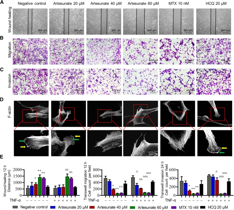
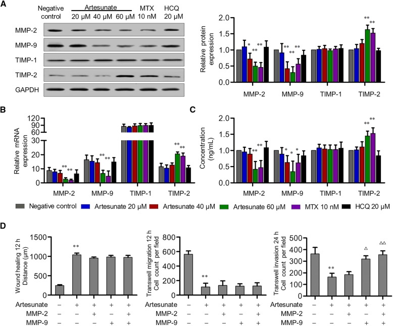
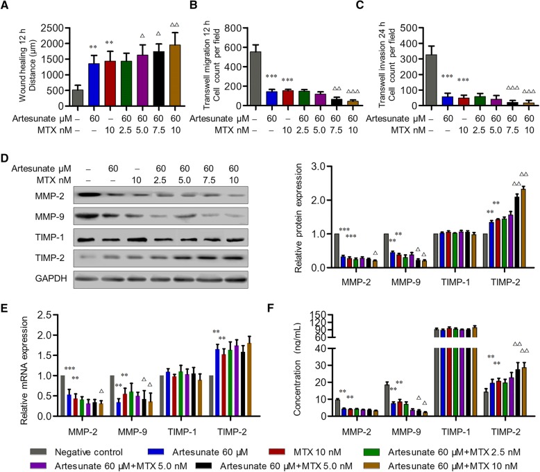
Results: Artesunate significantly inhibited the migration and invasion of RA-FLS in a dose-dependent manner with or without TNF-α stimulation. The effect was mediated through artesunate inhibition of MMP-2 and MMP-9 production, and pre-treatment with exogenous MMP-9 reversed the inhibitory effect of artesunate on RA-FLS invasion. Artesunate had a stronger inhibitory effect on migration and invasion of RA-FLS as well as greater anti-inflammatory effect than those of hydroxychloroquine. Similar inhibitory effect was detected between artesunate and methotrexate, and synergy was observed when combined. Mechanistically, artesunate significantly inhibited PDK-1 expression as well as Akt and RSK2 phosphorylation-in a similar manner to PDK-1-specific inhibitor AR-12 or PDK-1 knockdown by siRNA transfection. This inhibition results in suppression of RA-FLS migration and invasion as well as decreased MMP-2 and MMP-9 expression.
Conclusions: Our study demonstrates artesunate is capable of inhibiting migration and invasion of RA-FLS through suppression of PDK1-induced activation of Akt and RSK2 phosphorylation-suggesting that artesunate may be a potential disease-modifying anti-rheumatic drug for RA.
Keywords: Artesunate; Fibroblast-like synoviocytes; Invasion; Migration; Rheumatoid arthritis.
Conflict of interest statement
The authors declare that they have no competing interests.
Figures

Similar articles
-
Piperlongumine inhibits the proliferation, migration and invasion of fibroblast-like synoviocytes from patients with rheumatoid arthritis.Inflamm Res. 2018 Mar;67(3):233-243. doi: 10.1007/s00011-017-1112-9. Epub 2017 Nov 8. Inflamm Res. 2018. PMID: 29119225
-
PPARγ agonist rosiglitazone inhibits migration and invasion by downregulating Cyr61 in rheumatoid arthritis fibroblast-like synoviocytes.Int J Rheum Dis. 2017 Oct;20(10):1499-1509. doi: 10.1111/1756-185X.12913. Epub 2016 Jul 26. Int J Rheum Dis. 2017. PMID: 27456070
-
Isorhapontigenin suppresses inflammation, proliferation and aggressiveness of rheumatoid arthritis fibroblast-like synoviocytes by targeting farnesyl diphosphate synthase.Int Immunopharmacol. 2025 Jun 26;159:114894. doi: 10.1016/j.intimp.2025.114894. Epub 2025 May 23. Int Immunopharmacol. 2025. PMID: 40412131
-
Unveiling the Therapeutic Potential: Targeting Fibroblast-like Synoviocytes in Rheumatoid Arthritis.Expert Rev Mol Med. 2025 Jun 5;27:e18. doi: 10.1017/erm.2025.11. Expert Rev Mol Med. 2025. PMID: 40468839 Free PMC article. Review.
-
E3 ubiquitin ligase gene BIRC3 modulates TNF-induced cell death pathways and promotes aberrant proliferation in rheumatoid arthritis fibroblast-like synoviocytes.Front Immunol. 2024 Sep 5;15:1433898. doi: 10.3389/fimmu.2024.1433898. eCollection 2024. Front Immunol. 2024. PMID: 39301019 Free PMC article. Review.
Cited by
-
Artemisinin and its derivatives as promising therapies for autoimmune diseases.Heliyon. 2024 Mar 11;10(7):e27972. doi: 10.1016/j.heliyon.2024.e27972. eCollection 2024 Apr 15. Heliyon. 2024. PMID: 38596057 Free PMC article. Review.
-
Artesunate inhibits melanoma progression in vitro via suppressing STAT3 signaling pathway.Pharmacol Rep. 2021 Apr;73(2):650-663. doi: 10.1007/s43440-021-00230-6. Epub 2021 Feb 20. Pharmacol Rep. 2021. PMID: 33609273
-
MAPK/ERK signaling pathway in rheumatoid arthritis: mechanisms and therapeutic potential.PeerJ. 2025 Jul 14;13:e19708. doi: 10.7717/peerj.19708. eCollection 2025. PeerJ. 2025. PMID: 40677749 Free PMC article. Review.
-
Rspo2 exacerbates rheumatoid arthritis by targeting aggressive phenotype of fibroblast-like synoviocytes and disrupting chondrocyte homeostasis via Wnt/β-catenin pathway.Arthritis Res Ther. 2023 Nov 9;25(1):217. doi: 10.1186/s13075-023-03198-1. Arthritis Res Ther. 2023. PMID: 37946278 Free PMC article.
-
Artemisinin derivative SM934 in the treatment of autoimmune and inflammatory diseases: therapeutic effects and molecular mechanisms.Acta Pharmacol Sin. 2022 Dec;43(12):3055-3061. doi: 10.1038/s41401-022-00978-4. Epub 2022 Sep 1. Acta Pharmacol Sin. 2022. PMID: 36050518 Free PMC article. Review.
References
-
- Ma JD, Wei XN, Zheng DH, Mo YQ, Chen LF, Zhang X, et al. Continuously elevated serum matrix metalloproteinase-3 for 3 ~ 6 months predict one-year radiographic progression in rheumatoid arthritis: a prospective cohort study. Arthritis Res Ther. 2015;17:289. doi: 10.1186/s13075-015-0803-2. - DOI - PMC - PubMed
Publication types
MeSH terms
Substances
Grants and funding
- 81671612/National Natural Science Foundation of China/International
- 81801606/National Natural Science Foundation of China/International
- 2017A030313576/Natural Science Foundation of Guangdong Province/International
- 2017A030310236 and 2018A030313541/Natural Science Foundation of Guangdong Province/International
- A2017109/Guangdong Medical Research Foundation (CN)/International
LinkOut - more resources
Full Text Sources
Medical
Research Materials
Miscellaneous
