. 2019 Aug 8;201(17):e00154-19.
doi: 10.1128/JB.00154-19. Print 2019 Sep 1.

Guanine Quadruplex DNA Regulates Gamma Radiation Response of Genome Functions in the Radioresistant Bacterium Deinococcus radiodurans

Affiliations

Guanine Quadruplex DNA Regulates Gamma Radiation Response of Genome Functions in the Radioresistant Bacterium Deinococcus radiodurans

Shruti Mishra et al. J Bacteriol. .

Abstract

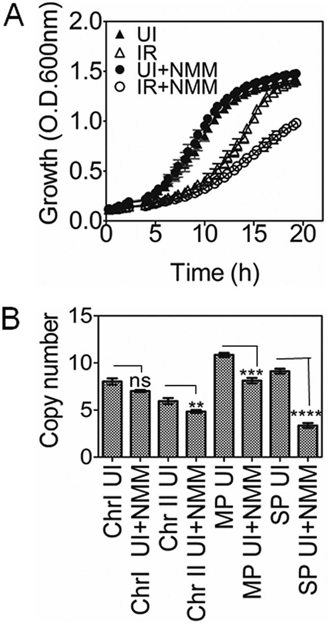
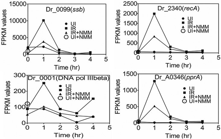
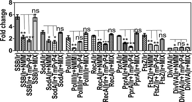
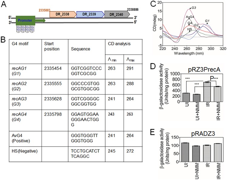
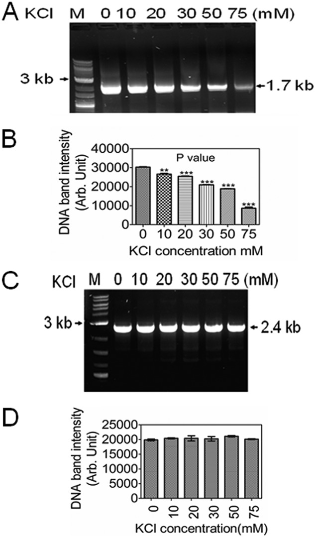
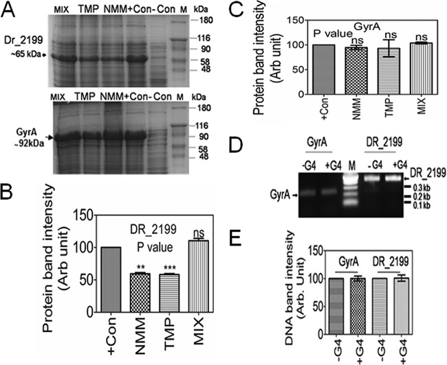
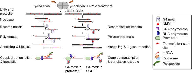
Guanine quadruplex (G4) DNA/RNA are secondary structures that regulate the various cellular processes in both eukaryotes and bacteria. Deinococcus radiodurans, a Gram-positive bacterium known for its extraordinary radioresistance, shows a genomewide occurrence of putative G4 DNA-forming motifs in its GC-rich genome. N-Methyl mesoporphyrin (NMM), a G4 DNA structure-stabilizing drug, did not affect bacterial growth under normal conditions but inhibited the postirradiation recovery of gamma-irradiated cells. Transcriptome sequencing analysis of cells treated with both radiation and NMM showed repression of gamma radiation-responsive gene expression, which was observed in the absence of NMM. Notably, this effect of NMM on the expression of housekeeping genes involved in other cellular processes was not observed. Stabilization of G4 DNA structures mapped at the upstream of recA and in the encoding region of DR_2199 had negatively affected promoter activity in vivo, DNA synthesis in vitro and protein translation in Escherichia coli host. These results suggested that G4 DNA plays an important role in DNA damage response and in the regulation of expression of the DNA repair proteins required for radioresistance in D. radioduransIMPORTANCEDeinococcus radiodurans can recover from extensive DNA damage caused by many genotoxic agents. It lacks LexA/RecA-mediated canonical SOS response. Therefore, the molecular mechanisms underlying the regulation of DNA damage response would be worth investigating in this bacterium. D. radiodurans genome is GC-rich and contains numerous islands of putative guanine quadruplex (G4) DNA structure-forming motifs. Here, we showed that in vivo stabilization of G4 DNA structures can impair DNA damage response processes in D. radiodurans Essential cellular processes such as transcription, DNA synthesis, and protein translation, which are also an integral part of the double-strand DNA break repair pathway, are affected by the arrest of G4 DNA structure dynamics. Thus, the role of DNA secondary structures in DNA damage response and radioresistance is demonstrated.

Keywords: DSB repair; Deinococcus; guanine quadruplex DNA; radioresistance; transcriptomics.


Figures

FIG 1
FIG 1
Effect of G4 DNA-stabilizing ligand (NMM) on growth kinetics and genome copy number in D. radiodurans. (A) The growth of D. radiodurans was monitored under normal conditions (solid symbols) and after exposure to gamma radiation (open symbols) in TYG (triangles) and TYG supplemented with NMM (circles) as described in Materials and Methods. (B) Similarly, the effects of G4 stabilization on the genome copy numbers of chromosome I (ChrI), chromosome II (ChrII), megaplasmid (MP), and small plasmid (SP) were determined in cells grown under normal conditions in TYG (UI) or in TYG supplemented with NMM (UI+NMM) as described in Materials and Methods. The data shown in panel A are means ± the standard deviations (SD; n = 9), whereas the data in panel B are means ± standard errors of the mean (SEM). A t test was performed to determine the statistical significance of the copy numbers of untreated (UI) and NMM-treated (UI+NMM) samples. ns (not significant), P > 0.05; **, P ≤ 0.01; ***, P ≤ 0.001; ****, P ≤ 0.0001.
FIG 2
FIG 2
Effect of NMM on gamma radiation-responsive genes expression in D. radiodurans. D. radiodurans cells were grown under unirradiated (UI), irradiated (IR), irradiated followed by NMM, and unirradiated followed by NMM conditions, as described in Materials and Methods. Total RNAs were sequenced from these cells were collected at different time intervals after treatment. The FPKM values for these cells were calculated and plotted to check the fold change in expression of DNA repair genes such as ssb (DR_0099), recA (DR_2340), pprA (DR_A0346), and the DNA PolIII beta subunit gene (DR_0001) in response to gamma radiation as a function of the G4-stabilizing ligand.
FIG 3
FIG 3
Real-time PCR validation of transcriptomics data. D. radiodurans cells were grown in the absence or presence of G4-stabilizing ligands (NMM and TmPyP4) and a non-G4-stabilizing agent identical MIX, as described for Fig. 2. Total RNAs from these cells were obtained and converted to cDNA by reverse transcriptase, as described in Materials and Methods. Real-time PCR was carried out for the transcripts of some of the candidate proteins such as single-strand DNA-binding protein (SSB), Mn superoxide dismutase (SodA), polymerase III subunit (PolIII), RecA (RecA), PprA (PprA), and cell division proteins FtsZ (FtsZ) and DivIVA (DivIVA). PCR products were quantified, and the fold changes with respect to the corresponding controls (e.g., gamma radiation against unirradiated control, NMM-treated gamma radiation against NMM-treated unirradiated controls, TmPyP4-treated gamma radiation against TmPyP4-treated unirradiated controls) and MIX-treated gamma radiation against MIX-treated unirradiated controls) were calculated and plotted. The data show means ± the SD (n = 6). A t test was performed to analyze the statistical significance in the fold change between (i) gamma-irradiated (Ir) and NMM-treated/gamma-irradiated (Ir+NMM), (ii) gamma-irradiated (Ir) and TmPyP4-treated/gamma-irradiated (Ir+TmPyP4), and (iii) gamma-irradiated (Ir) and MIX-treated/gamma-irradiated (Ir+MIX) samples for each gene. ns (not significant), P > 0.05; **, P ≤ 0.01; ***, P ≤ 0.001; ****, P ≤ 0.0001.
FIG 4
FIG 4
Effect of NMM on G4 DNA motif-containing recA promoter activity in response to gamma radiation in D. radiodurans. (A) The 2G runs forming the G4 DNA structure are found in the upstream region of the recA encoding sequence at various positions. (B) Their sequences and λmax and λmin values obtained by CD analysis for putative recA G4 motifs, along with control DNA sequences, as mentioned in Materials and Methods, are also shown. (C) Putative G4 DNA motifs present in the upstream region of recA were characterized for the formation of G4 structures by CD, along with a known G4-forming sequence (AvG4) and a non-G4-forming sequence (HS). The recA promoter was transcriptionally fused with lacZ in pRZ3PrecA, and the expression of lacZ was monitored in the presence or absence of NMM. (D and E) The expression of β-galactosidase activity under the control of upstream sequence of recA in pRZ3PrecA (D) and under the groESL promoter in pRADZ3 (E) was monitored in unirradiated (UI), unirradiated plus NMM (UI+NMM), and irradiated (IR) followed by treatment with NMM (IR+NMM) D. radiodurans, as described in Materials and Methods. The β-galactosidase activity is represented as the mean units/mg of protein ± the SD (n = 9). The significance of the difference was analyzed using a t test; P values were obtained at 95% confidence intervals. ***, P < 0.0001.
FIG 5
FIG 5
Effect of NMM on in vitro DNA synthesis. PCR amplification of the DR_2199 template carrying the G4 motif (A) and the DR_1913 (GyrA) template as the control (C) was carried out in the presence of increasing concentrations of KCl (0 to 75 mM), a G4 structure-stabilizing ion, in vitro. PCR products were analyzed on agarose, and the DNA band intensities of the PCR-amplified products were quantitated by ImageJ software and plotted in GraphPad Prism 5.0 for DR_2199 (B) and DR_1913 (GyrA) (D). The experiment was repeated three independent times, and a t test was performed to determine the statistical significance between the agarose band intensity at 0 mM and at 10, 20, 30, 50, and 75 mM KCl concentrations. **, P ≤ 0.01; ***, P ≤ 0.001.
FIG 6
FIG 6
Effect of G4 DNA-binding ligands on gene expression in heterologous host. The role of G4 DNA dynamics on translatability of G4 motif containing transcript of DR_2199 was evaluated in E. coli host and compared to deinococcal GyrA (without the G4 motif) expression as a control as described in Materials and Methods. DR_2199 expressing plasmids pET2199 and pETGyrA were transformed in the E. coli strain BL21(DE3)/pLysS strain, and the expression of protein was checked in the presence of the G4 DNA-binding ligands NMM, TmPyP4, and MIX. Samples without any ligand treatment were taken as a positive control (+Con); those without IPTG induction were used as a negative control (–Con). (A) Total proteins from these samples were analyzed using 10% PAGE. The recombinant protein band intensity under all of these conditions was quantitated using ImageJ software and is plotted as the means ± the SD (n = 3) in GraphPad Prism 5.0. We used DR_2199 protein as a positive control (+Con) as 100 (B) and deinococcal GyrA (C). A Student t test (paired t test) was performed comparing +Con and treated samples (NMM, TmPyP4, and MIX) to determine the statistical significance. Similarly, the effect of the G4 DNA structure stabilization on in vitro transcription was assayed using a T7 expression cassette of 1.79-kbp DR_2199 coding sequences containing G4 motif and the 2.4-kbp DR_1913 coding sequence as a control. These cassettes present on plasmids were subjected to conditions that would favor G4 DNA structure folding (+) and unfolding (–). (D) The levels of the transcripts were estimated by RT-PCR using internal primers, and PCR products were analyzed on a 1.2% agarose gel. (E) DNA band intensity was quantitated using ImageJ software and plotted in GraphPad Prism 5.0 as the means ± the SD (n = 3), taking the DNA intensity of the unfolded substrate (–) as 100. ns (not significant), P > 0.05; **, P ≤ 0.01; ***, P ≤ 0.001; ****, P ≤ 0.0001.
FIG 7
FIG 7
Proposed model explaining probable G4 DNA roles in radiation resistance phenotype in D. radiodurans. The extended synthesis-dependent strand annealing (ESDSA) mechanism during PIR, followed by coupled transcription and translation, provide the most plausible explanation for the extraordinary radioresistance in D. radiodurans . ESDSA requires several crucial enzymes of DNA metabolism and involves the formation of long stretches of single-strand DNA precursors, which would be prone to G4 DNA structure formation but might have not affected the wild-type bacterium because of its regulated dynamicity by indigenous mechanisms. Since the arrest of G4 DNA structural dynamics affects DNA synthesis, recombination repair, and gamma radiation-responsive gene expression, the potential steps in the ESDSA mechanism that may be affected by the synthetic arrest of G4 DNA dynamics are highlighted and represented. This might explain the selective effect of G4 DNA dynamics arrest during gamma radiation-stressed growth in D. radiodurans.

Similar articles

Cited by

References

    1. Huppert JL. 2010. Structure, location, and interactions of G-quadruplexes. FEBS J 277:3452–3458. doi:10.1111/j.1742-4658.2010.07758.x. - DOI - PubMed
    1. Bochman ML, Paeschke K, Zakian VA. 2012. DNA secondary structures: stability and function of G-quadruplex structures. Nat Rev Genet 13:770–780. doi:10.1038/nrg3296. - DOI - PMC - PubMed
    1. Maizels N, Gray LT. 2013. The G4 genome. PLoS Genet 9:e1003468. doi:10.1371/journal.pgen.1003468. - DOI - PMC - PubMed
    1. Tomas S, Petr P, Mikael K. 1998. DNA tetraplex formation in the control region of c-myc. Nucleic Acids Res 26:1167–1172. - PMC - PubMed
    1. Cogoi S, Xodo LE. 2006. G-quadruplex formation within the promoter of the KRAS proto-oncogene and its effect on transcription. Nucleic Acids Res 34:2536–2549. doi:10.1093/nar/gkl286. - DOI - PMC - PubMed

MeSH terms

LinkOut - more resources