. 2019 Jul 9;116(28):14119-14128.
doi: 10.1073/pnas.1900147116. Epub 2019 Jun 24.

Chemical depletion of phagocytic immune cells in Anopheles gambiae reveals dual roles of mosquito hemocytes in anti- Plasmodium immunity

Affiliations

Chemical depletion of phagocytic immune cells in Anopheles gambiae reveals dual roles of mosquito hemocytes in anti- Plasmodium immunity

Hyeogsun Kwon et al. Proc Natl Acad Sci U S A. .

Abstract

Mosquito immunity is composed of both cellular and humoral factors that provide protection from invading pathogens. Immune cells known as hemocytes, have been intricately associated with phagocytosis and innate immune signaling. However, the lack of genetic tools has limited hemocyte study despite their importance in mosquito anti-Plasmodium immunity. To address these limitations, we employ the use of a chemical-based treatment to deplete phagocytic immune cells in Anopheles gambiae, demonstrating the role of phagocytes in complement recognition and prophenoloxidase production that limit the ookinete and oocyst stages of malaria parasite development, respectively. Through these experiments, we also define specific subtypes of phagocytic immune cells in An. gambiae, providing insights beyond the morphological characteristics that traditionally define mosquito hemocyte populations. Together, this study represents a significant advancement in our understanding of the roles of mosquito phagocytes in mosquito vector competence and demonstrates the utility of clodronate liposomes as an important tool in the study of invertebrate immunity.

Keywords: Anopheles gambiae; hemocytes; innate immunity; malaria; phagocyte depletion.


Conflict of interest statement

The authors declare no conflict of interest.

Figures
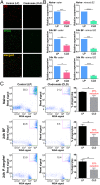
Fig. 1.
Fig. 1.
Mosquito phagocytes are significantly depleted following clodronate liposome (CLD) treatment. Following the injection of control (LP) or clodronate liposomes (CLD), mosquitoes were challenged with P. berghei and hemocytes were perfused 24 h postinfection. Hemocytes were stained with the hemocyte-specific markers, DiI (red) and WGA (green), to visualize the effects of CLD treatment on hemocyte populations (A). Additional experiments with molecular markers of phagocytic cells, eater and nimrod B2, were used to further evaluate phagocyte depletion. Relative transcript levels of eater and nimrod B2 expression were significantly reduced in CLD-treated mosquitoes (B). Further validation was performed using flow cytometry to confirm the depletion of phagocytic hemocyte populations under naive, 24-h blood-fed (24-h BF), and 24-h P. berghei-infected (24-h P.b) conditions (C). Representative flow cytometry experiments display the depletion of phagocytic hemocytes (as determined by the uptake of fluorescent beads and WGA staining), with bar graphs depicting a significant decrease in phagocytes following CLD treatment from 3 independent experiments (C). Blood-feeding, independent of pathogen challenge, caused phagocytes to be more susceptible to CLD treatment (C). Bars represent mean ± SEM of 3 independent replicates. Data were analyzed by unpaired t test using GraphPad Prism 6.0. Asterisks denote significance (*P < 0.05, **P < 0.01, ***P < 0.001, ****P < 0.0001). (Scale bar: 20 μm.)
Fig. 2.
Fig. 2.
Depletion of phagocytic cells influences mosquito survival after bacterial challenge. Mosquitoes were treated with either control or CLDs, and then subjected to injury (sterile PBS injection) or bacterial challenge. Survivorship was monitored in mosquitoes every day for 10 d to evaluate the effects of injury (A), S. marcescens (B), or S. aureus (C) challenge. Phagocyte depletion (CLD) in mosquitoes results in a high susceptibility to bacterial infections. Error bars represent the mean ± SEM of 3 independent replicates. In each replicate, 30 female mosquitoes were used for each experimental treatment. Data were analyzed by a log-rank (Mantel-Cox) test using GraphPad Prism 6.0.
Fig. 3.
Fig. 3.
Effects of phagocyte depletion on P. berghei development. One day before challenge with P. berghei (pretreatment), mosquitoes were treated with control (LP) or clodronate liposomes (CLD). Ten days postinfection, Plasmodium oocyst numbers were evaluated to determine the effects of phagocyte depletion on malaria parasite numbers (A). To determine the temporal components that influence this increase in parasite survival, day 2 early oocyst numbers were examined in LP- and CLD-treated mosquitoes (B). Although phagocyte depletion increased early oocyst numbers, levels of TEP1 protein did not differ between LP and CLD treatments in either naive or 24-h P. berghei-infected hemolymph samples where serpin 3 (SRPN3) was used as a protein loading control (C). Nonspecific bands were denoted by an asterisk. Evaluation of TEP1 binding to invading ookinetes (∼22–24 h post-P. berghei infection) by immunofluorescence after phagocyte depletion demonstrates that TEP1 binding (green; indicated by arrows) to ookinetes (α-Pbs 21; red) is significantly impaired (D). To examine oocyst survival, oocyst numbers were examined at 2 and 10 d post-P. berghei infection in mosquitoes treated with control liposomes (E) or CLDs (F). Oocyst numbers were measured by fluorescence using the same cohort of mosquitoes for both time points. Clodronate treatment after the establishment of infection (24 h post-P. berghei infection) had no effect on malaria parasite survival (G). Three or more independent experiments were performed for all infection experiments, and data were analyzed using Mann–Whitney test with GraphPad Prism 6.0. Median oocyst numbers are indicated by the horizontal red line, and asterisks denote significance (*P < 0.05, **P < 0.01, ***P < 0.001); ns, not significant; n, number of midguts examined.
Fig. 4.
Fig. 4.
Phagocyte depletion reduces of prophenoloxidase (PPO) expression. RNA-seq analyses following clodronate treatment revealed 50 differentially regulated genes in abdomen tissues 24 h post-P. berghei infection and grouped by gene ontology (A). Comprising the largest category of affected genes, the annotations and logtwofold change of specific immune genes with significant differential regulation are displayed (B). This includes several PPO genes, therefore leading us to examine the expression of all 9 PPO family members by qRT-PCR analyses in the carcass (C) and hemocyte (D) samples in control liposome (LP) and clodronate-treated (CLD) samples. Data were analyzed using an unpaired t test to determine differences in relative gene expression of each respective PPO gene between LP and CLD treatments (C and D). Due to the importance of PPOs in phenoloxidase (PO) activation, PO activity was measured in hemolymph samples derived from LP and CLD samples in naive, blood-fed, and P. berghei-infected conditions (E). Measurements (OD490) were taken for DOPA conversion assays at 5-min intervals from 0 to 30 min, and then again using a final readout at 60 min. Data were analyzed using a two-way repeated-measures ANOVA followed by Sidak’s multiple comparisons using GraphPad Prism 6.0. Bars represent mean ± SEM of 3 independent experiments. Asterisks denote significance (*P < 0.05, **P < 0.01, ***P < 0.001, ****P < 0.0001).
Fig. 5.
Fig. 5.
The silencing of PPOs influences Plasmodium oocyst survival. The influence of PPO2, PPO3, and PPO9 silencing on Plasmodium oocysts numbers in An. gambiae was evaluated 8 d postinfection compared with dsGFP controls (A). To determine whether gene silencing influences the success of ookinete invasion, similar experiments were performed in which early oocyst numbers were used as a readout of ookinete survival 2 d postinfection (B). For all experiments, each dot represents the number of parasites on an individual midgut, with the median value denoted by a horizontal red line. Data were pooled from 3 or more independent experiments with statistical analysis determined by a Mann–Whitney test using GraphPad Prism 6.0. Asterisks denote significance (*P < 0.05, ***P < 0.001).
Fig. 6.
Fig. 6.
Clodronate treatment differentially impacts mosquito phagocyte subpopulations. PPO6 staining was evaluated by immunofluorescence in perfused hemocytes after the injection of fluorescent beads from control liposome (LP) or clodronate-treated (CLD) mosquitoes ∼24 h postinfection with P. berghei. After adherence and fixation, hemocytes were stained with a PPO6 antibody (A). PPO6+ phagocytes (green) that have phagocytosed red fluorescent beads were compared between LP and CLD treatments (A), as well as the proportion of nonphagocytic PPO6+ cells (B). Closer examination of PPO6+ phagocytic cells revealed distinct populations of immune cells distinguished by PPO6 signal intensity (high or low) and morphological features (elongated/spread, small rounded) (C). Two independent experiments of immunofluorescent assays were performed. Data were analyzed by Mann–Whitney test using GraphPad Prism 6.0. Median is indicated by the horizontal red line (A). Bars represent mean ± SEM (B and C). Asterisks denote significance (**P < 0.01, ****P < 0.0001). (Scale bar: 10 μm.)
Fig. 7.
Fig. 7.
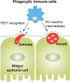
Multimodal contributions of phagocytes on anti-Plasmodium immunity. Experiments with CLDs establish integral roles of phagocytic immune cells in malaria parasite killing, which include the role of phagocytes in the recognition of invading ookinetes and the production of PPOs that limit oocyst survival.

Similar articles

Cited by

References

    1. Salzet M., Vertebrate innate immunity resembles a mosaic of invertebrate immune responses. Trends Immunol. 22, 285–288 (2001). - PubMed
    1. Gold K. S., Brückner K., Macrophages and cellular immunity in Drosophila melanogaster. Semin. Immunol. 27, 357–368 (2015). - PMC - PubMed
    1. Williams M. J., Drosophila hemopoiesis and cellular immunity. J. Immunol. 178, 4711–4716 (2007). - PubMed
    1. Honti V., Csordás G., Kurucz É., Márkus R., Andó I., The cell-mediated immunity of Drosophila melanogaster: Hemocyte lineages, immune compartments, microanatomy and regulation. Dev. Comp. Immunol. 42, 47–56 (2014). - PubMed
    1. Castillo J. C., Robertson A. E., Strand M. R., Characterization of hemocytes from the mosquitoes Anopheles gambiae and Aedes aegypti. Insect Biochem. Mol. Biol. 36, 891–903 (2006). - PMC - PubMed

Publication types

MeSH terms