. 2019 Jul;30(7):1220-1237.
doi: 10.1681/ASN.2018080860. Epub 2019 Jun 24.

Tyrosine Phosphorylation of CD2AP Affects Stability of the Slit Diaphragm Complex

Affiliations

Tyrosine Phosphorylation of CD2AP Affects Stability of the Slit Diaphragm Complex

Irini Tossidou et al. J Am Soc Nephrol. 2019 Jul.

Abstract

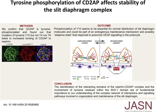
Background: CD2-associated protein (CD2AP), a slit diaphragm-associated scaffolding protein involved in survival and regulation of the cytoskeleton in podocytes, is considered a "stabilizer" of the slit diaphragm complex that connects the slit diaphragm protein nephrin to the cytoskeleton of the cell. Tyrosine phosphorylation of slit diaphragm molecules can influence their surface expression, but it is unknown whether tyrosine phosphorylation events of CD2AP are also physiologically relevant to slit diaphragm stability.

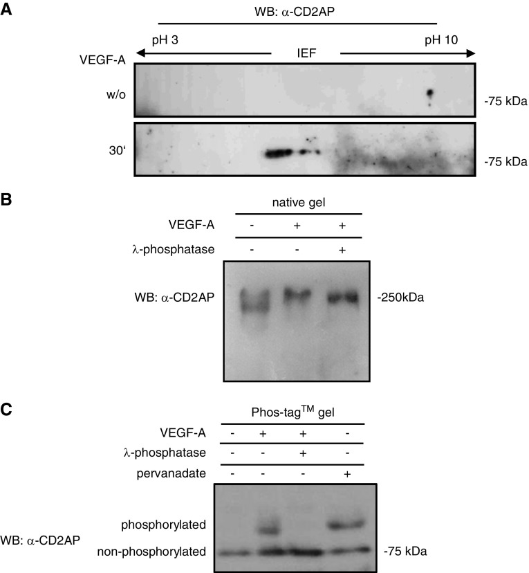
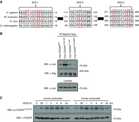
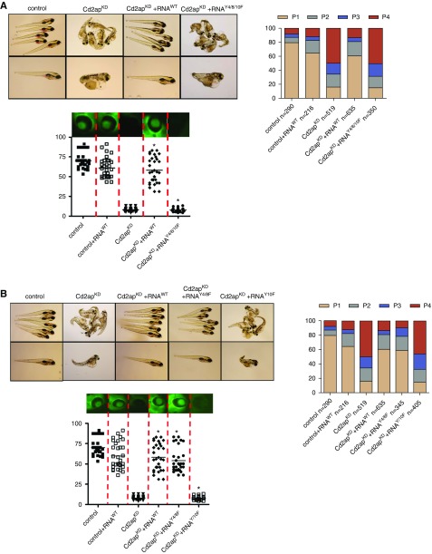
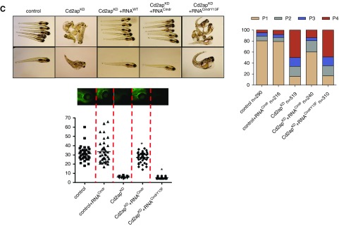
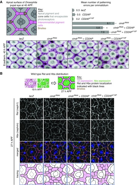
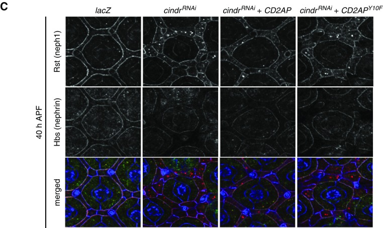
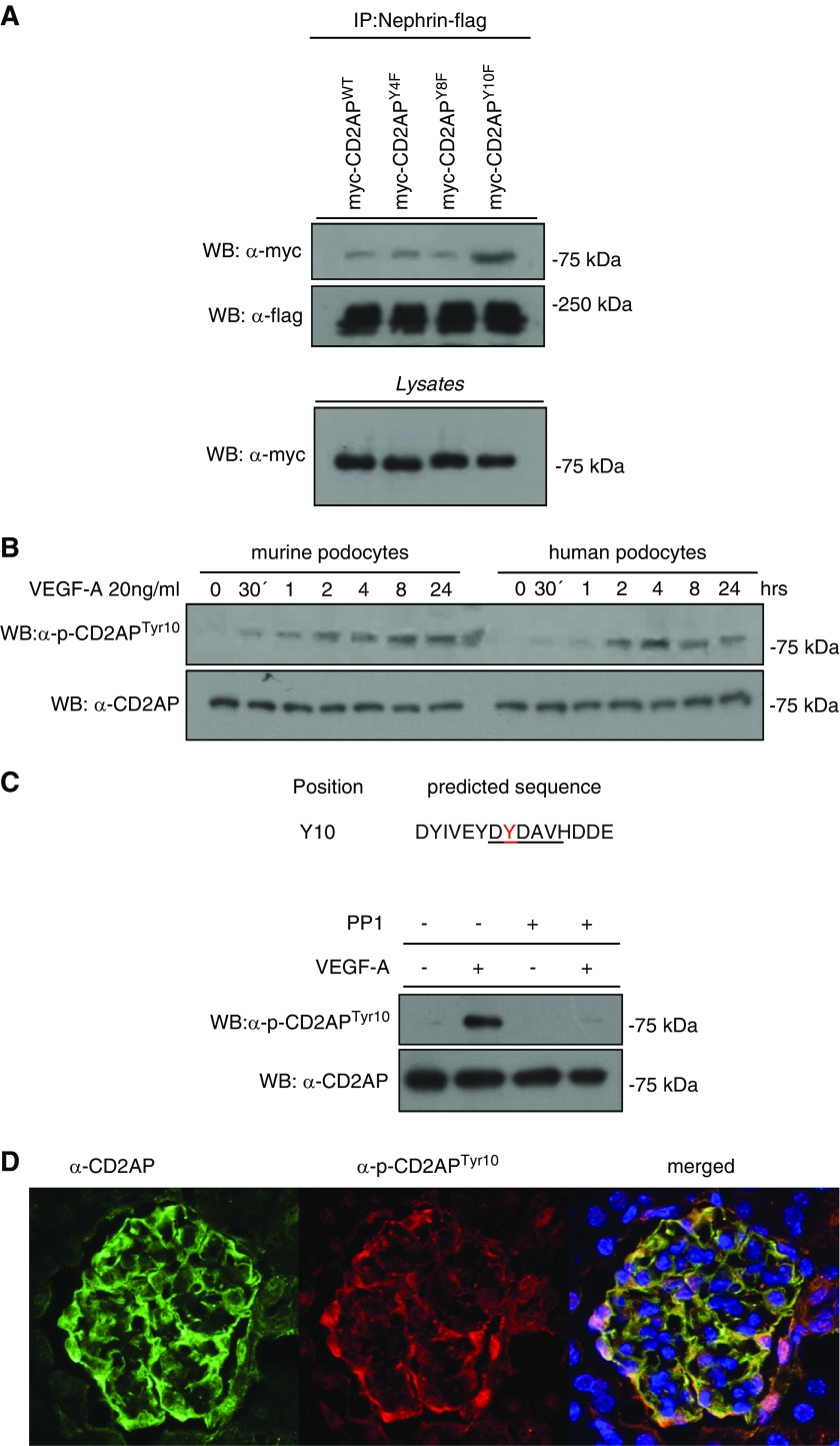
Methods: We used isoelectric focusing, western blot analysis, and immunofluorescence to investigate phosphorylation of CD2AP, and phospho-CD2AP antibodies and site-directed mutagenesis to define the specific phosphorylated tyrosine residues. We used cross-species rescue experiments in Cd2apKD zebrafish and in Drosophila cindrRNAi mutants to define the physiologic relevance of CD2AP phosphorylation of the tyrosine residues.

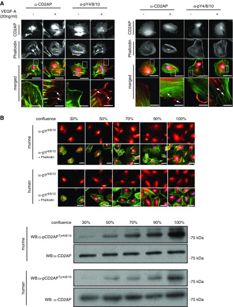
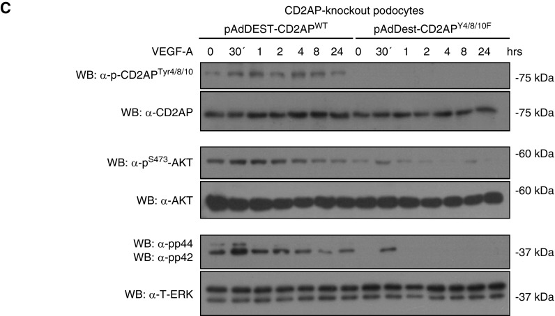
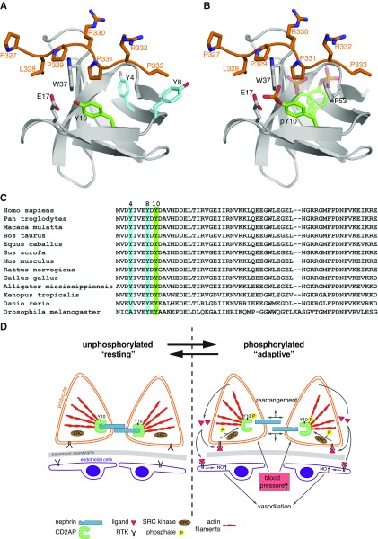
Results: We found that VEGF-A stimulation can induce a tyrosine phosphorylation response in CD2AP in podocytes, and that these phosphorylation events have an important effect on slit diaphragm protein localization and functionality in vivo. We demonstrated that tyrosine in position Y10 of the SH3-1 domain of CD2AP is indispensable for CD2AP function in vivo. We found that the binding affinity of nephrin to CD2AP is significantly enhanced in the absence of Y10; however, unexpectedly, this increased affinity leads not to stabilization but to functional impairment of the glomerular filtration barrier.

Conclusions: Our findings provide insight into CD2AP and its phosphorylation in the context of slit diaphragm functionality, and indicate a fine-tuned affinity balance of CD2AP and nephrin that is influenced by receptor tyrosine kinase stimulation.

Keywords: CD2AP; Phosphorylation; nephrin; podocyte.


Figures

None
Graphical abstract
Figure 1.
Figure 1.
CD2AP is tyrosine phosphorylated after VEGF-stimulation in podocytes. (A) Two-dimensional isoelectric electrophoresis using an anti-CD2AP antibody shows a shift of CD2AP after treatment with VEGF-A after 30 minutes. (B) Native gel electrophoresis using an anti-CD2AP antibody shows a weight shift after VEGF-A stimulation. Treatment with λ-phosphatase partially restores the weight shift. (C) Phos-tag gel shows phosphorylated and nonphosphorylated proteins as different bands. λ-phosphatase dephosphorylates and pervanadate phosphorylates CD2AP. Western blot was analyzed with an anti-CD2AP antibody. IEF, isoelectric focusing; WB, western blotting; w/o, without.
Figure 2.
Figure 2.
Phosphorylation of tyrosines Y4/8/10 has a characteristic activation profile and antagonizes interactions with nephrin. (A) Phylogenetic and sequence analysis of indicated species shows evolutionarily conserved tyrosines in each of the three SH3 domains. (B) Myc-tagged CD2AP DNA was point-mutated on indicated tyrosine sites. HEK-293T cells transfected with myc-CD2AP, myc-CD2AP tyrosine mutants, and flag-Nephrin were immunoprecipitated with a flag-antibody and western blot was performed with a Myc-antibody. (C) Differentiated podocytes were treated with 20 ng/ml VEGF-A for 24 hours. Western blot analysis of lysates was performed using a p-CD2APtyr4/8/10 antibody. CD2AP was used as loading control. D. rerio, Danio rerio; D. melanogaster, Drosophila melanogaster; H. sapiens, Homo sapiens; IP, immunoprecipitation; M. musculus, Mus musculus; WB, western blotting.
Figure 3.
Figure 3.
Phosphorylated CD2AP is found at the leading edges and in focal contacts, and cellular confluency enhances CD2AP phosphorylation. (A) Differentiated murine and human podocytes untreated and treated with VEGF-A were stained with CD2AP or p-CD2APtyr4/8/10 antibodies (red) and costained with phalloidin (green) and DAPI (blue). Scale bars, 20 µm. Boxed regions are enlarged (bottom panel). White arrows depict phosphorylated CD2AP at leading edges. Scale bars, 2 µm. (B) Murine and human podocytes cultured to 30%–100% confluency were stained with anti–p-CD2APtyr4/8/10, phalloidin, and DAPI. Western blot of podocyte lysates at different confluence percentages, probed with p-CD2APtyr4/8/10 and CD2AP antibodies. Scale bars, 10 µm. (C) CD2AP-knockout podocytes were transduced with adenoviral vectors expressing CD2APWT and CD2APY4/8/10F. Podocytes were treated with 20 ng/ml VEGF-A for 24 hours. Western blot analysis of lysates was performed using p-CD2APtyr4/8/10, p-AKTS273, and p-ERKThr202/Tyr204 antibodies. CD2AP, AKT, and ERK were used as loading controls. WB, western blotting.
Figure 3.
Figure 3.
Phosphorylated CD2AP is found at the leading edges and in focal contacts, and cellular confluency enhances CD2AP phosphorylation. (A) Differentiated murine and human podocytes untreated and treated with VEGF-A were stained with CD2AP or p-CD2APtyr4/8/10 antibodies (red) and costained with phalloidin (green) and DAPI (blue). Scale bars, 20 µm. Boxed regions are enlarged (bottom panel). White arrows depict phosphorylated CD2AP at leading edges. Scale bars, 2 µm. (B) Murine and human podocytes cultured to 30%–100% confluency were stained with anti–p-CD2APtyr4/8/10, phalloidin, and DAPI. Western blot of podocyte lysates at different confluence percentages, probed with p-CD2APtyr4/8/10 and CD2AP antibodies. Scale bars, 10 µm. (C) CD2AP-knockout podocytes were transduced with adenoviral vectors expressing CD2APWT and CD2APY4/8/10F. Podocytes were treated with 20 ng/ml VEGF-A for 24 hours. Western blot analysis of lysates was performed using p-CD2APtyr4/8/10, p-AKTS273, and p-ERKThr202/Tyr204 antibodies. CD2AP, AKT, and ERK were used as loading controls. WB, western blotting.
Figure 4.
Figure 4.
Phosphorylation of CD2AP at tyrosine 10 is physiologically relevant for a functional glomerular filtration barrier. Fertilized zebrafish eggs were injected at the 1–4-cell stage with a CD2AP-morpholino and murine CD2AP-capped mRNA. (A) Images (left) and phenotypic analyses (right) of larval zebrafish 120 hours after injection. Phenotypes were categorized into four groups: P1, no edema; P2, mild edema; P3, severe edema; and P4, very severe edema. Fluorescent images of the retinal vessel plexus (bottom panel) and bar graph of fluorescence intensity of eGFP-DBP in fish eyes (at least 30 animals for each treatment were analyzed). Error bars show the mean±SEM; *P≤0.001 by unpaired t test. CD2APtY4/8/10F mRNA could not rescue the phenotypes associated with CD2APKD, whereas coinjection of CD2APWT mRNA restored wild-type phenotypes. (B) Larval zebrafish 120 hours after injection (left) and phenotypic analyses (right). Fluorescent images and eGFP-DBP analyses of the retinal vessel plexus of at least 30 animals (bottom panel). Error bars indicate mean±SEM; *P≤0.001 by unpaired t test. Injection of CD2APY10F into CD2APKD larvae failed to restore wild-type phenotypes, unlike injection of both CD2APWT mRNA and CD2APY4/8F mRNA. (C) Larval zebrafish 120 hours after injection (left) and phenotypic analyses (right). Fluorescent images and eGFP-DBP analyses of the retinal vessel plexus of at least 30 animals (bottom panel). Error bars indicate mean±SEM; *P≤0.001 by unpaired t test. Injection of Cindr RNA into CD2APKD larvae restored wild-type phenotypes, unlike injection of CindrY13F mRNA.
Figure 4.
Figure 4.
Phosphorylation of CD2AP at tyrosine 10 is physiologically relevant for a functional glomerular filtration barrier. Fertilized zebrafish eggs were injected at the 1–4-cell stage with a CD2AP-morpholino and murine CD2AP-capped mRNA. (A) Images (left) and phenotypic analyses (right) of larval zebrafish 120 hours after injection. Phenotypes were categorized into four groups: P1, no edema; P2, mild edema; P3, severe edema; and P4, very severe edema. Fluorescent images of the retinal vessel plexus (bottom panel) and bar graph of fluorescence intensity of eGFP-DBP in fish eyes (at least 30 animals for each treatment were analyzed). Error bars show the mean±SEM; *P≤0.001 by unpaired t test. CD2APtY4/8/10F mRNA could not rescue the phenotypes associated with CD2APKD, whereas coinjection of CD2APWT mRNA restored wild-type phenotypes. (B) Larval zebrafish 120 hours after injection (left) and phenotypic analyses (right). Fluorescent images and eGFP-DBP analyses of the retinal vessel plexus of at least 30 animals (bottom panel). Error bars indicate mean±SEM; *P≤0.001 by unpaired t test. Injection of CD2APY10F into CD2APKD larvae failed to restore wild-type phenotypes, unlike injection of both CD2APWT mRNA and CD2APY4/8F mRNA. (C) Larval zebrafish 120 hours after injection (left) and phenotypic analyses (right). Fluorescent images and eGFP-DBP analyses of the retinal vessel plexus of at least 30 animals (bottom panel). Error bars indicate mean±SEM; *P≤0.001 by unpaired t test. Injection of Cindr RNA into CD2APKD larvae restored wild-type phenotypes, unlike injection of CindrY13F mRNA.
Figure 5.
Figure 5.
CD2AP but not CD2APY10F rescues cindrRNAi eye-mispatterning. (A) Illustration of an ommatidium at 40 hours APF, with cell types labeled. Small regions of eyes expressing UAS-lacZ (abbreviated to GMR>lacZ), cindrRNAi (which severely disrupted patterning), CD2AP and cindrRNAi (which partly rescued patterning), and CD2APY10F and cindrRNAi. Quantification of the mean number of patterning defects per ommatidium: x axis plots error number for each genotype, as shown. Seventy-five ommatidia were analyzed per genotype. Error bars indicate SEM. Asterisk represents statistically significant differences between patterning defects in GMR>cindrRNAi and GMR>cindrRNAi +CD2AP eyes. GMR>cindrRNAi, CD2APY10F did not differ significantly from GMR>cindrRNAi. (B) Illustrations of rst (neph1) and hbs (nephrin) expression and localization at 19 hours APF and 27 hours APF. Immunofluorescence of ECadherin (blue), Rst (red), and Hbs (green) in a control GMR>lacZ eye at 27 hours APF and eyes expressing cindrRNAi (which disrupted Rst and Hbs localization), cindrRNAi and CD2AP (which partly rescued Rst/Hbs localization), and cindrRNAi and CD2APY10F. In tracings of each image (bottom panels), interommatidial cells are pink and density of black cell outlines represents density of Rst/Hbs detected at membranes. (C) Immunofluorescence of ECadherin (blue), Rst (red), and Hbs (green) in a control GMR>lacZ eye at 40 hours APF and eyes expressing cindrRNAi (which disrupted Rst localization; little Hbs is observed at cell membranes), cindrRNAi and CD2AP (which partly rescued Rst localization but not Hbs, which is not robustly detected at cell membranes), and cindrRNAi and CD2APY10F.
Figure 5.
Figure 5.
CD2AP but not CD2APY10F rescues cindrRNAi eye-mispatterning. (A) Illustration of an ommatidium at 40 hours APF, with cell types labeled. Small regions of eyes expressing UAS-lacZ (abbreviated to GMR>lacZ), cindrRNAi (which severely disrupted patterning), CD2AP and cindrRNAi (which partly rescued patterning), and CD2APY10F and cindrRNAi. Quantification of the mean number of patterning defects per ommatidium: x axis plots error number for each genotype, as shown. Seventy-five ommatidia were analyzed per genotype. Error bars indicate SEM. Asterisk represents statistically significant differences between patterning defects in GMR>cindrRNAi and GMR>cindrRNAi +CD2AP eyes. GMR>cindrRNAi, CD2APY10F did not differ significantly from GMR>cindrRNAi. (B) Illustrations of rst (neph1) and hbs (nephrin) expression and localization at 19 hours APF and 27 hours APF. Immunofluorescence of ECadherin (blue), Rst (red), and Hbs (green) in a control GMR>lacZ eye at 27 hours APF and eyes expressing cindrRNAi (which disrupted Rst and Hbs localization), cindrRNAi and CD2AP (which partly rescued Rst/Hbs localization), and cindrRNAi and CD2APY10F. In tracings of each image (bottom panels), interommatidial cells are pink and density of black cell outlines represents density of Rst/Hbs detected at membranes. (C) Immunofluorescence of ECadherin (blue), Rst (red), and Hbs (green) in a control GMR>lacZ eye at 40 hours APF and eyes expressing cindrRNAi (which disrupted Rst localization; little Hbs is observed at cell membranes), cindrRNAi and CD2AP (which partly rescued Rst localization but not Hbs, which is not robustly detected at cell membranes), and cindrRNAi and CD2APY10F.
Figure 6.
Figure 6.
Phosphorylation of CD2AP at Tyr10 is physiologically relevant in podocytes. (A) HEK-293T cells were transfected with myc-tagged CD2AP, mutated CD2APY4F, CD2APY8F, or CD2APY10F and cotransfected with flag-tagged Nephrin. Coimmunoprecipitation was performed with a flag-antibody and western blot with a myc-antibody. (B) Western blot of lysates of murine and human differentiated podocytes treated for 24 hours with VEGF-A, probed with anti-pCD2APtyr10 and anti-CD2AP. (C) The Src-recognition sequence is predicted to contain tyrosine 10 in CD2AP. Differentiated murine podocytes were treated or untreated with VEGF-A (20 ng/ml) and the specific Src inhibitor PP1 (5 µm, preincubated 1 hour before VEGF-A treatment). Western blot was performed with anti-pCD2APtyr10 and anti-CD2AP. (D) Cryosections of murine glomeruli stained with anti-CD2AP (green), anti-pCD2APtyr10 (red), and DAPI (blue). IP, immunoprecipitation; WB, western blotting.
Figure 7.
Figure 7.
Phosphorylation of Tyr10 induces conformational changes that alter ligand binding. (A) Model of the CD2AP-SH3A (CMS-SH3–1) domain in complex with a CD2 peptide (residues 327–333). Crystal structure (Protein Data Bank entry code 2J6O) of SH3-A is shown in gray and the CD2 peptide in orange. The side chains of Y4 and Y8 are shown in blue, and the side chain of Y10 in green. (B) Addition of a phosphate group to the aromatic hydroxyl group of Y10 induces electrostatic repulsion between Y10 and E17 of SH3-A. Repositioning of the side chain of Y10 (indicated in transparent green) would lead to severe steric clashes with P331 of the peptide and F53 of SH3-A, respectively. Hence, phosphorylation of Y10 would modify interactions with proline-rich peptides and ligands including nephrin. (C) Alignment of conserved protein sequence of CD2AP/CMS from different species. Nonphosphorylated tyrosines 4 and 8 are highlighted in blue, and Y10 is highlighted in green. Y4 is not strictly conserved between all sequences shown. (D) Schematic overview of proposed CD2AP phosphorylation. RTK activation (e.g., by VEGF) leads to phosphorylation of CD2AP on tyrosine 10 via Src-kinase. This leads to steric changes and rearrangement between the connection of CD2AP to nephrin and actin filaments. Thus, autocrine VEGF can lead to an adaptive state in podocytes compensating for vasodilatation- and BP-induced dynamics. RTK, receptor tyrosine kinase.

Similar articles

Cited by

References

    1. Kestilä M, Lenkkeri U, Männikkö M, Lamerdin J, McCready P, Putaala H, et al. .: Positionally cloned gene for a novel glomerular protein--nephrin--is mutated in congenital nephrotic syndrome. Mol Cell 1: 575–582, 1998 - PubMed
    1. Jones N, Blasutig IM, Eremina V, Ruston JM, Bladt F, Li H, et al. .: Nck adaptor proteins link nephrin to the actin cytoskeleton of kidney podocytes. Nature 440: 818–823, 2006 - PubMed
    1. Martin CE, Jones N: Nephrin signaling in the podocyte: An updated view of signal regulation at the slit diaphragm and beyond. Front Endocrinol (Lausanne) 9: 302, 2018 - PMC - PubMed
    1. Tryggvason K, Pikkarainen T, Patrakka J: Nck links nephrin to actin in kidney podocytes. Cell 125: 221–224, 2006 - PubMed
    1. Palmén T, Lehtonen S, Ora A, Kerjaschki D, Antignac C, Lehtonen E, et al. .: Interaction of endogenous nephrin and CD2-associated protein in mouse epithelial M-1 cell line. J Am Soc Nephrol 13: 1766–1772, 2002 - PubMed

Publication types

LinkOut - more resources