. 2019 Jul 15;146(14):dev177659.
doi: 10.1242/dev.177659.

Sall4 regulates neuromesodermal progenitors and their descendants during body elongation in mouse embryos

Affiliations

Sall4 regulates neuromesodermal progenitors and their descendants during body elongation in mouse embryos

Naoyuki Tahara et al. Development. .

Abstract

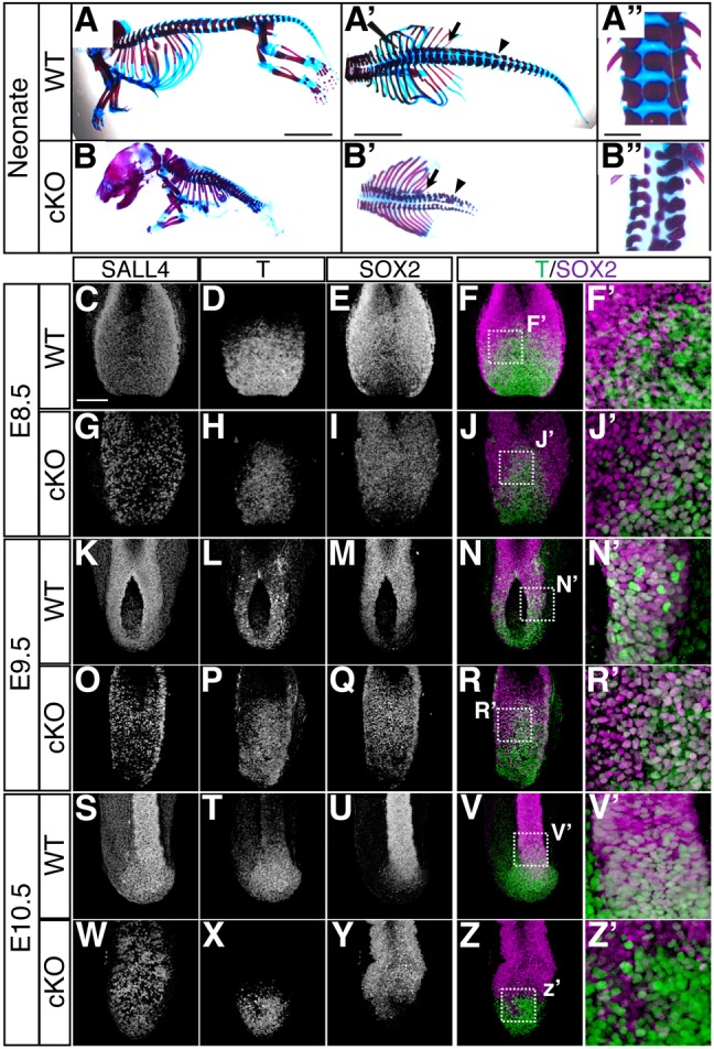
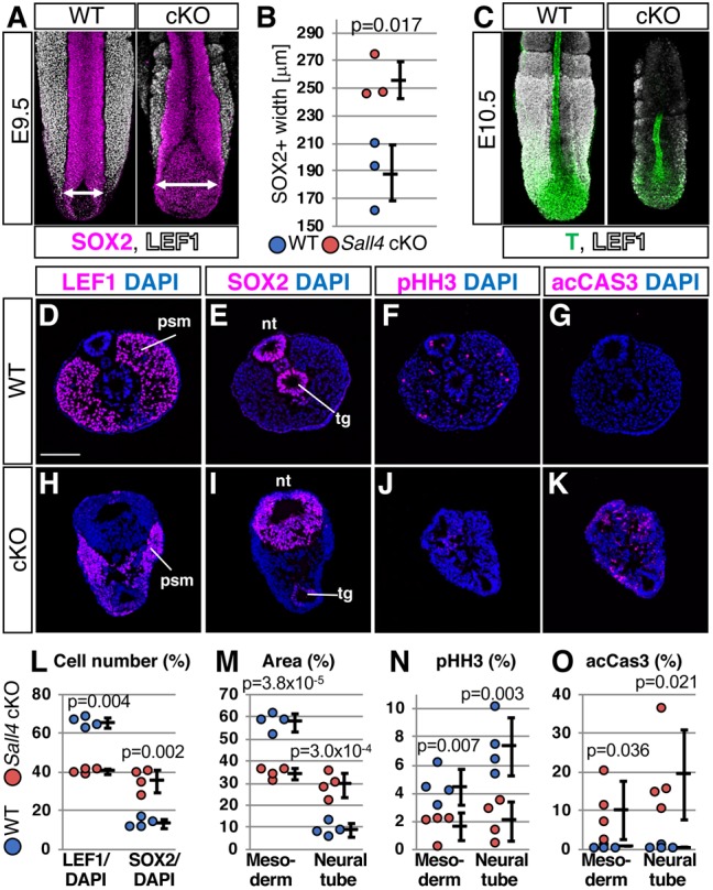
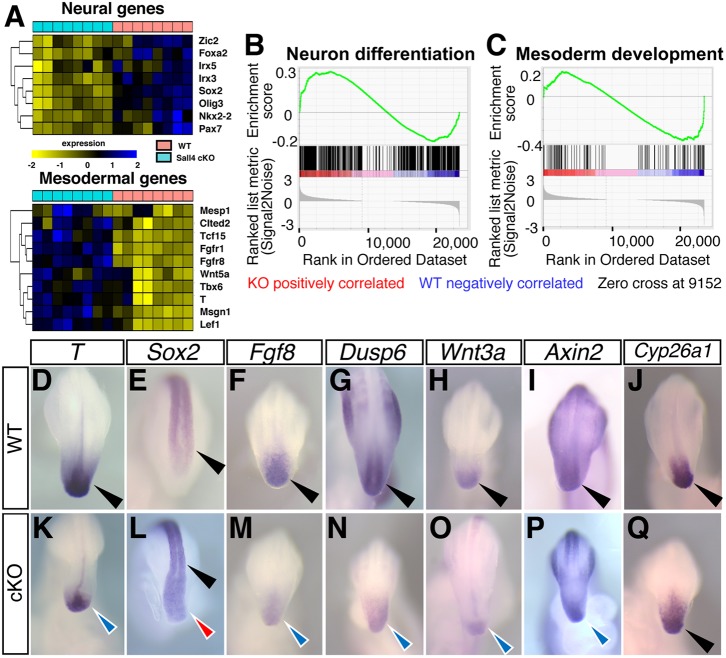
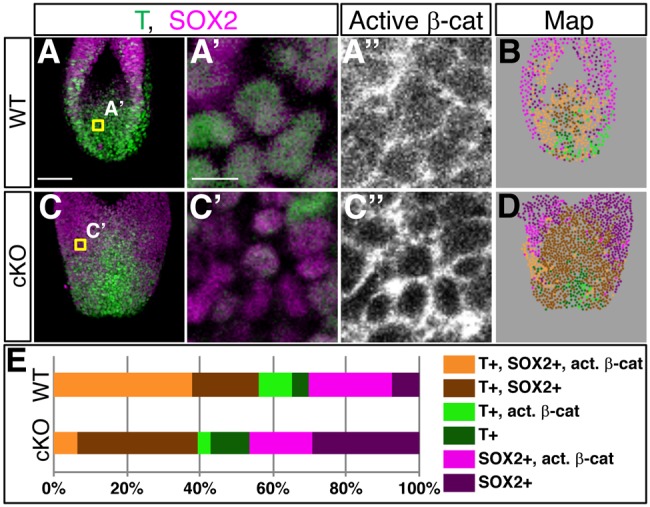
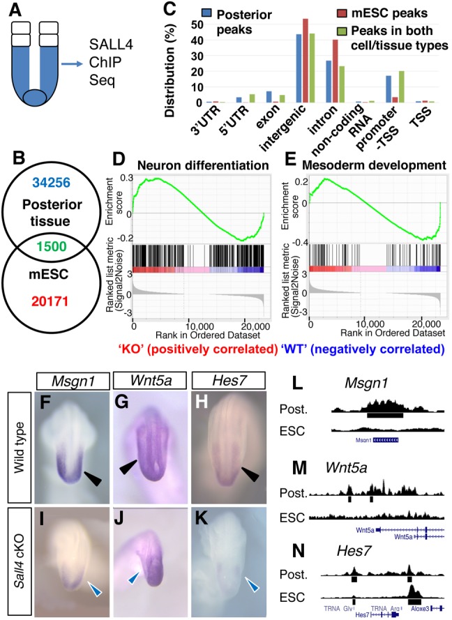
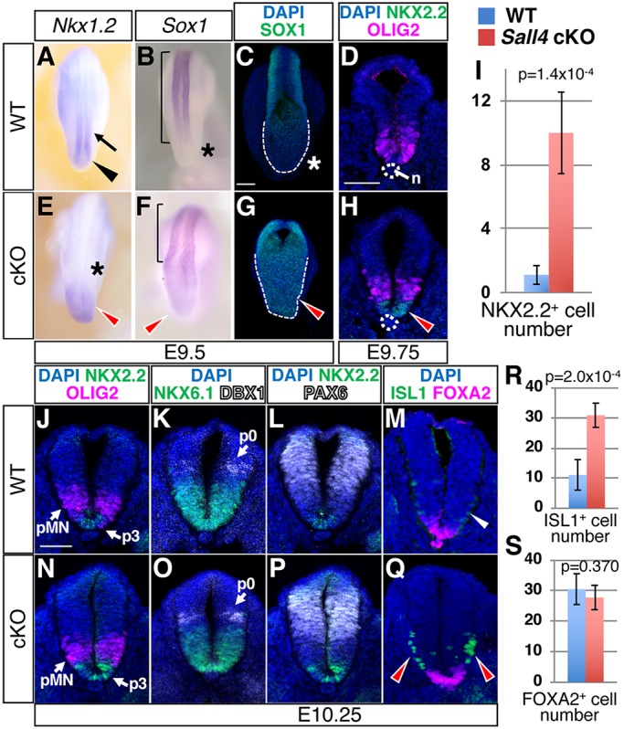
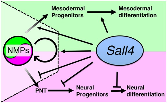
Bi-potential neuromesodermal progenitors (NMPs) produce both neural and paraxial mesodermal progenitors in the trunk and tail during vertebrate body elongation. We show that Sall4, a pluripotency-related transcription factor gene, has multiple roles in regulating NMPs and their descendants in post-gastrulation mouse embryos. Sall4 deletion using TCre caused body/tail truncation, reminiscent of early depletion of NMPs, suggesting a role of Sall4 in NMP maintenance. This phenotype became significant at the time of the trunk-to-tail transition, suggesting that Sall4 maintenance of NMPs enables tail formation. Sall4 mutants exhibit expanded neural and reduced mesodermal tissues, indicating a role of Sall4 in NMP differentiation balance. Mechanistically, we show that Sall4 promotion of WNT/β-catenin signaling contributes to NMP maintenance and differentiation balance. RNA-Seq and SALL4 ChIP-Seq analyses support the notion that Sall4 regulates both mesodermal and neural development. Furthermore, in the mesodermal compartment, genes regulating presomitic mesoderm differentiation are downregulated in Sall4 mutants. In the neural compartment, we show that differentiation of NMPs towards post-mitotic neuron is accelerated in Sall4 mutants. Our results collectively provide evidence supporting the role of Sall4 in regulating NMPs and their descendants.

Keywords: Body/tail elongation; Mesodermal progenitors and neural progenitors; Neuromesodermal progenitors; Sall4; WNT/β-catenin signaling.


Conflict of interest statement

Competing interestsThe authors declare no competing or financial interests.

Figures

Fig. 1.
Fig. 1.
Sall4 deletion leads to early NMP depletion and body truncation. (A-B″) Lateral (A,B) and dorsal (A′,B′) views of WT and Sall4 cKO neonatal mice stained with Alcian Blue and Alizarin Red. Arrows and arrowheads in A′ and B′ point to the most posterior thoracic and lumber vertebrae, respectively. In the upper-left corner of A′ the edge of the forceps can be seen. A″ and B″ show dorsal views of vertebrae at the thoracic to lumber level. (C-Z′) Whole-mount immunofluorescence images of the caudal part of the body of WT and Sall4 cKO embryos at E8.5 (C-J′), E9.5 (K-R′) and E10.5 (S-Z′). Immunoreactivities for the indicated antibodies are shown. F′,J′,N′,R′,V′,Z′ are magnifications of the boxed areas in F,J,N,R,V,Z, which are shown as overlays of T (green) and SOX2 (magenta) signals. Scale bars: 5 mm (A-B′); 1 mm (A″,B″);100 µm (C-Z).
Fig. 2.
Fig. 2.
Sall4 deletion causes increased neural tissue and decreased mesodermal tissue. (A) Whole-mount WT and Sall4 cKO embryos at E9.5 stained with antibodies for SOX2 (magenta) and LEF1 (white). (B) Graph of the width of the widest region of the posterior neural plate/tube at E9.5 (shown by double-headed arrows in A, n=3). Shown are mean±s.d. The P-value by unpaired t-test is shown. (C) Whole-mount WT and Sall4 cKO embryos at E10.5 stained with antibodies for T (green) and LEF1 (white). (D-K) Immunofluorescence of LEF1, SOX2, phosphohistone H3 (pHH3) and activated caspase 3 (acCAS3) in the PSM levels of WT (D-G) and Sall4 cKO (H-K) embryos at E10.5. (L) Quantification of LEF1+ nuclei and SOX2+ nuclei in the neural tube without tail gut per total nuclei of the section. (M) Quantification of LEF1+ mesoderm area and SOX2+ neural tube area per all DAPI+ area of the section. (N) Quantification of pHH3+ cells in the mesoderm area and in the neural tube area per total nuclei of the section. (O) Quantification of acCAS3+ cells in the mesoderm area and in the neural tube area per total nuclei of the section. Shown are mean±s.d. P-values are shown in each panel (unpaired t-test). n=4 for both WT and Sall4 cKO. nt, neural tube; psm, presomitic mesoderm; tg, tail gut. Scale bar: 100 µm.
Fig. 3.
Fig. 3.
Sall4 deletion causes changes of expression of genes related to NMP maintenance and neural and mesodermal genes. (A) Heat map of genes with GO term as neural genes (upper) and mesoderm genes (lower) in WT versus Sall4 cKO. (B,C) GSEA analysis of genes with GO terms of neural differentiation (B) and mesoderm development (C) among genes that are differentially expressed in WT versus Sall4 cKO posterior tissue. (D-Q) Whole-mount in situ hybridization of the indicated genes in WT (D-J) and Sall4 cKO (K-Q) at E9.5. Black arrowheads point to normal expression in the posterior tip of the body in WT embryos and Sox2 and Cyp26a1 expression in the Sall4 cKO embryo. Blue arrowheads point to reduced expression in Sall4 cKO embryos (K,M-P). Red arrowhead in L points to upregulated Sox2 expression in the Sall4 cKO embryo.
Fig. 4.
Fig. 4.
Downregulation of WNT/β-catenin signaling in Sall4 cKO embryos. (A,C) Stacked confocal images of whole-mount-stained WT and Sall4 cKO embryos at E9.5. (A′,A″,C′,C″) Single-layer images of embryos stained for T (green), SOX2 (magenta) and active β-catenin. Shown are high magnifications of single-layer images in the areas indicated in A or C. (B,D) Stacked maps of cells with different combinations of T, SOX2 and active β-catenin with the color code shown in E. (E) Quantification of cells with different combinations of T, SOX2 and active β-catenin (as shown in B and D). n=4 for WT, n=3 for Sall4 cKO. Scale bars: 100 µm (A,C); 10 µm (A′,A″,C′,C″).
Fig. 5.
Fig. 5.
SALL4 ChIP-Seq analysis of the posterior tissue and downregulation of mesoderm differentiation genes in Sall4 cKO embryos. (A) Schematic of dissected posterior tissue (blue) for SALL4 ChIP-Seq analysis. (B) Venn diagram for SALL4-enriched sequence numbers in the posterior tissue and mESCs. (C) Distributions of SALL4-enriched sequences in the posterior tissue, mESCs and both cell/tissue types. (D,E) GSEA analysis of genes with GO terms of neural differentiation (D) and mesoderm development (E) among genes that are enriched by SALL4 in the posterior tissue. (F-K) Whole-mount in situ hybridization of the indicated mesodermal genes in WT and Sall4 cKO embryos at E9.5. Black arrowheads point to normal expression in WT PSM (F-H). Blue arrowheads point to reduced expression in the PSM in Sall4 cKO embryos (I-K). (L-N) Visual representation of SALL4 ChIP-Seq results of the indicated gene loci. Post., the posterior tissue.
Fig. 6.
Fig. 6.
Accelerated neural patterning and differentiation in the posterior region of Sall4 cKO embryos. (A,B,E,F) In situ hybridization of Nkx1.2 and Sox1 at E9.5 in WT (A,B) and Sall4 cKO (E,F) embryos. Black arrowhead and arrow in A point to expression in the posterior neural plate and the posterior neural tube, respectively. Red arrowheads point to increased (E) or ectopic (F) expression. Asterisks mark lack of expression. Bracket indicates Sox1 expression in the neural tube. (C,G) Confocal images of SOX1 expression in the posterior of whole-mount embryos. Dashed lines indicate posterior end of the neural plate. Red arrowhead points to ectopic expression of SOX1 (G). Asterisk indicates a lack of SOX1 signals at the posterior end of the neural plate (C). (D,H) Immunofluorescence of OLIG2 (magenta) and NKX2.2 (green) in WT (D) and Sall4 cKO (H) embryos at E9.75 (27/28 somite stage). Red arrowhead points to NKX2.2+ cells in Sall4 cKO embryos. Dotted circle indicates the notochord (n). (I) Quantification of NKX2.2+ cells at the posterior hindlimb level at E9.75. Graphs shows numbers of cells (mean±s.d.) per section. P-values by unpaired t-test are shown. n=5 for WT, n=6 for Sall4 cKO. (J-Q) Immunofluorescence of the indicated markers in WT (J-M) and Sall4 cKO (N-Q) embryos at E10.25 (33/34 somite stage). White and red arrowheads point to ISL1+ cells at the posterior hindlimb level in WT (M) and Sall4 cKO (Q) embryos, respectively. In K and O, DBX1 signals are shown in white. (R,S) Quantification of ISL1+ cells (R) and FOXA2+ cells (S) at the posterior hindlimb level at E10.25. Graphs show numbers of cells (mean±s.d.) per section. P-values by unpaired t-test are shown within each panel. n=5 for both WT and Sall4 cKO. Scale bars: 100 µm (C,G); 50 µm (D,H,J-Q).
Fig. 7.
Fig. 7.
A model of Sall4 function in NMPs and their descendants in the mouse embryo. Proposed functions of Sall4 for NMP maintenance and regulation of differentiation between mesodermal versus neural fate, promotion of mesodermal differentiation and restriction of neural differentiation of NMP descendants. For more detail see Discussion.

Similar articles

Cited by

References

    1. Abu-Abed S., Dolle P., Metzger D., Beckett B., Chambon P. and Petkovich M. (2001). The retinoic acid-metabolizing enzyme, CYP26A1, is essential for normal hindbrain patterning, vertebral identity, and development of posterior structures. Genes Dev. 15, 226-240. 10.1101/gad.855001 - DOI - PMC - PubMed
    1. Aires R., Jurberg A. D., Leal F., Nóvoa A., Cohn M. J. and Mallo M. (2016). Oct4 is a key regulator of vertebrate trunk length diversity. Dev. Cell 38, 262-274. 10.1016/j.devcel.2016.06.021 - DOI - PubMed
    1. Aires R., Dias A. and Mallo M. (2018). Deconstructing the molecular mechanisms shaping the vertebrate body plan. Curr. Opin. Cell Biol. 55, 81-86. 10.1016/j.ceb.2018.05.009 - DOI - PubMed
    1. Aires R., de Lemos L., Nóvoa A., Jurberg A. D., Mascrez B., Duboule D. and Mallo M. (2019). Tail bud progenitor activity relies on a network comprising Gdf11, Lin28, and Hox13 genes. Dev. Cell 48, 383-395.e388. 10.1016/j.devcel.2018.12.004 - DOI - PubMed
    1. Akiyama R., Kawakami H., Wong J., Oishi I., Nishinakamura R. and Kawakami Y. (2015). Sall4-Gli3 system in early limb progenitors is essential for the development of limb skeletal elements. Proc. Natl. Acad. Sci. USA 112, 5075-5080. 10.1073/pnas.1421949112 - DOI - PMC - PubMed

Publication types