. 2019 Aug 14;39(33):6449-6459.
doi: 10.1523/JNEUROSCI.3193-18.2019. Epub 2019 Jun 24.

GABAergic Inputs to POMC Neurons Originating from the Dorsomedial Hypothalamus Are Regulated by Energy State

Affiliations

GABAergic Inputs to POMC Neurons Originating from the Dorsomedial Hypothalamus Are Regulated by Energy State

Andrew R Rau et al. J Neurosci. .

Abstract

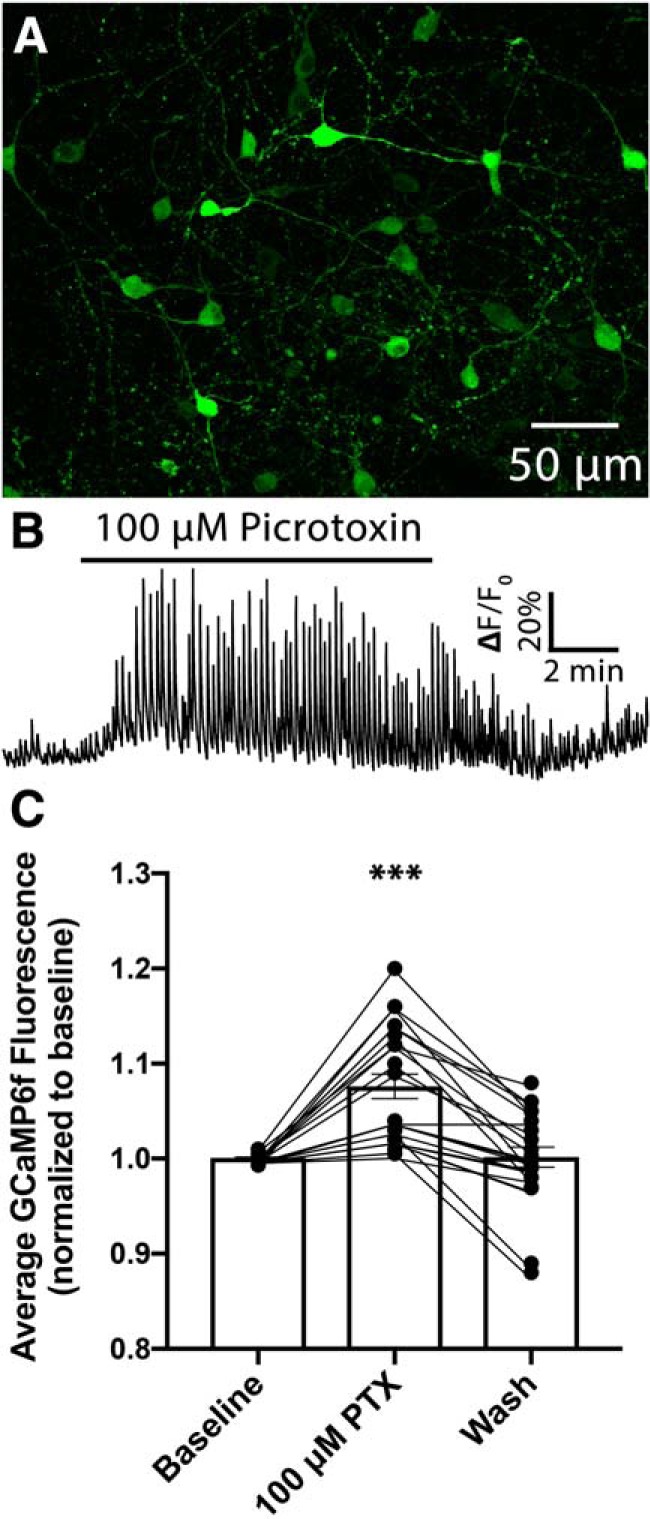
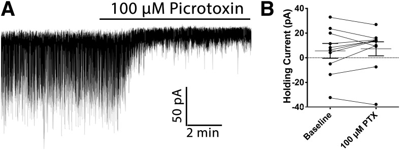
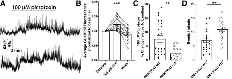
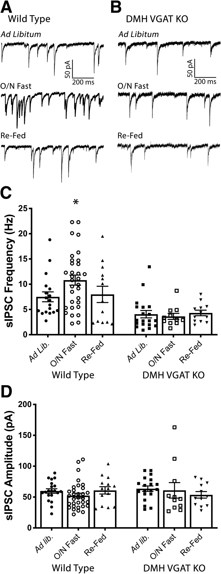
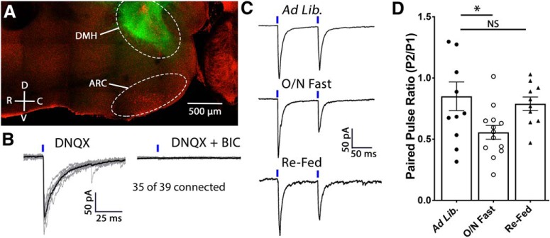
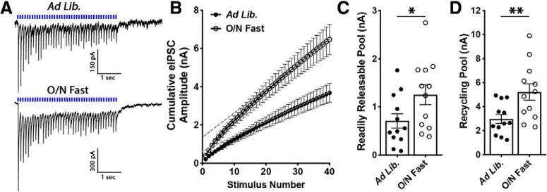
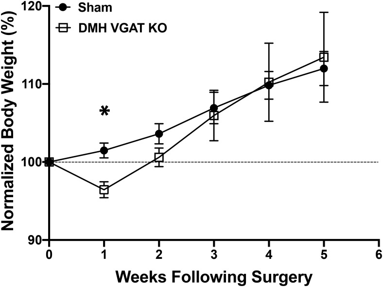
Neuronal circuits regulating hunger and satiety synthesize information encoding the energy state of the animal and translate those signals into motivated behaviors to meet homeostatic needs. Proopiomelanocortin (POMC) neurons in the arcuate nucleus of the hypothalamus are activated by energy surfeits and inhibited by energy deficits. When activated, these cells inhibit food intake and facilitate weight loss. Conversely, decreased activity in POMC cells is associated with increased food intake and obesity. Circulating nutrients and hormones modulate the activity of POMC neurons over protracted periods of time. However, recent work indicates that calcium activity in POMC cells changes in response to food cues on times scales consistent with the rapid actions of amino acid transmitters. Indeed, the frequency of spontaneous IPSCs (sIPSCs) onto POMC neurons increases during caloric deficits. However, the afferent brain regions responsible for this inhibitory modulation are currently unknown. Here, through the use of brain region-specific deletion of GABA release in both male and female mice we show that neurons in the dorsomedial hypothalamus (DMH) are responsible for the majority of sIPSCs in POMC neurons as well as the fasting-induced increase in sIPSC frequency. Further, the readily releasable pool of GABA vesicles and the release probability of GABA is increased at DMH-to-POMC synapses following an overnight fast. Collectively these data provide evidence that DMH-to-POMC GABA circuitry conveys inhibitory neuromodulation onto POMC cells that is sensitive to the animal's energy state.SIGNIFICANCE STATEMENT Activation of proopiomelanocortin (POMC) cells signals satiety, whereas GABAergic cells in the dorsomedial hypothalamus (DMH) can increase food consumption. However, communication between these cells, particularly in response to changes in metabolic state, is unknown. Here, through targeted inhibition of DMH GABA release, we show that DMH neurons contribute a significant portion of spontaneously released GABA onto POMC cells and are responsible for increased GABAergic inhibition of POMC cells during fasting, likely mediated through increased release probability of GABA at DMH terminals. These data provide important information about inhibitory modulation of metabolic circuitry and provide a mechanism through which POMC neurons could be inhibited, or disinhibited, rapidly in response to food availability.

Keywords: arcuate nucleus; calcium imaging; electrophysiology; optogenetic; proopiomelanocortin; synaptic.


Figures

Figure 1.
Figure 1.
Antagonism of GABAA receptors on POMC neurons increases GCaMP6f fluorescence in these cells. A, Confocal micrograph demonstrating the expression of Cre-recombinase-dependent GCaMP6f in the arcuate nucleus of a PomcCre/+ mouse. B, Representative GCaMP6f fluorescence from a POMC cell in the mouse arcuate nucleus demonstrating the change in calcium activity when GABAA receptors are blocked by the addition of the GABAA receptor antagonist picrotoxin (100 μm) to the recording chamber. For each trial, fluorescence was normalized by assigning a value of 1.0 to the median value of data points within a 5 min window immediately preceding the drug application. C, Quantification of change in normalized fluorescence that occurs following bath application of picrotoxin (***p < 0.0001, one-way repeated-measures ANOVA). Data are presented as mean ± SEM.
Figure 2.
Figure 2.
Increased calcium activity in POMC cells following GABAA receptor antagonism is not because of changes in membrane potential. A, Representative voltage-clamp recording from a POMC neuron demonstrating the effect of bath application of 100 μm picrotoxin. B, Summary data showing that bath application of 100 μm picrotoxin does not change the amount of current needed to maintain a holding potential of −60 mV. (p = 0.544; paired t test). Data are presented as mean ± SEM.
Figure 3.
Figure 3.
The frequency of sIPSCs onto POMC cells is significantly reduced when the VGAT is deleted in the DMH. A, Confocal micrographs showing the injection sites of an AAV-Cre. Using the ROSA-tom mouse that expresses the red fluorophore, Td-tomato, in the presence of Cre recombinase, AAV-Cre was targeted to the DMH (left), the bed nucleus of the stria terminalis [BNST; middle, or the lateral hypothalamus (LH), right]. In all images, the far-red channel (gray background) was overexposed to visualize anatomic structures and confirm that injections were localized to the appropriate brain region. To produce mice lacking GABA release from specific brain regions, AAV-Cre was injected into these same brain regions of transgenic mice with both alleles encoding VGAT floxed (Slc32a1flox/flox), followed by voltage-clamp recordings from POMC cells. B, Representative voltage-clamp recordings from POMC neurons following a injection of an AAV encoding enhanced yellow fluorescent protein into the DMH (SHAM; top left), or following injection of AAV-Cre into the DMH (bottom left), BNST (top right), or LH (bottom right) of Slc32a1flox/flox mice. C, Summary data (mean ± SEM) showing the frequency of sIPSCs recorded from POMC cells flowing brain region-specific deletion of GABA release. (**p = 0.004, one-way ANOVA). D, Summary data (mean ± SEM) indicating the effect of brain region-specific VGAT deletion on sIPSC amplitude recorded from POMC cells (p = 0.51, one-way ANOVA).
Figure 4.
Figure 4.
Deletion of DMH GABA release increases the excitability of POMC neurons through loss of GABAergic inputs. A, Representative calcium imaging traces showing the effect of 100 μm picrotoxin on GCaMP6f fluorescence. B, Quantification of average change in normalized fluorescence that occurs following bath application of picrotoxin (***p < 0.001, one-way repeated-measures ANOVA). C, Summary data comparing the percentage change in baseline normalized GCaMP6f fluorescence in mice with intact DMH VGAT (DMH VGAT WT; Fig. 1B) to mice with deleted VGAT in the DMH (DMH VGAT KO). Deletion of VGAT in the DMH significantly reduced the increase in normalized fluorescence relative to DMH VGAT WT mice (**p = 0.014; unpaired t test) D, Summary data showing the increase in calcium events per minute recorded from POMC neurons in mice with deleted VGAT in the DMH (**p = 0.002; unpaired t test). Data are presented as mean ± SEM.
Figure 5.
Figure 5.
The increase in the frequency of sIPSCs onto POMC cells following an overnight fast is blocked when VGAT is deleted in the DMH. A, Representative voltage-clamp recordings from POMC neurons from WT mice demonstrating the frequency of sIPSCs when mice have been fed ad libitum (top), fasted overnight (middle), or re-fed for 2 h following an overnight fast (bottom). B, Representative voltage-clamp recordings from POMC neurons from mice with deleted GABA release from the DMH demonstrating the frequency of sIPSCs when mice have been fed ad libitum (top), fasted overnight (middle), or re-fed for 2 h following an overnight fast (bottom). C, Summary data (mean ± SEM) showing the frequency of sIPSCs recorded from POMC cells during varying energy states in both WT and DMH VGAT KO mice. Deletion of VGAT in the DMH had a significant effect on sIPSC frequency (F(1,99) = 26.71, p < 0.0001, two-way ANOVA). Fasting resulted in a significant increase in sIPSC frequency in WT mice, relative to ad libitum fed mice (*p = 0.034, Tukey's multiple comparison). This increase was absent following deletion of VGAT in the DMH (p = 0.965, Tukey's multiple comparison). Data reported for ad libitum, DMH VGAT KO condition is replicate data from DMH VGAT KO in Figure 3C. D, Summary data (mean ± SEM) showing that the amplitude of sIPSCs recorded from POMC was not affected by manipulation of energy state (F(2,99) = 0.435, p = 0.648, two-way ANOVA), or deletion of VGAT in the DMH (F(1,99) = 0.154, p = 0.696, two-way ANOVA).
Figure 6.
Figure 6.
Fasting increases the release probability of GABA from DMH terminals onto POMC neurons. A, Confocal micrograph of a sagittal hypothalamic slice demonstrating the expression of ChR2eYFP in the dorsomedial hypothalamus and transgenically expressed Td-tomato in POMC neurons in the arcuate nucleus. B, Individual IPSCs (gray) and an average of these events (black) recorded from a POMC cell while light stimulates ChR2-expressing DMH terminals in the presence of DNQX (left). IPSCs were confirmed to be GABAA receptor-mediated by the addition of BIC to the recording solution (right). C, Representative voltage-clamp recordings demonstrating the PPR of light-evoked IPSCs recorded from POMC neurons. ChR2-expressing DMH terminals were light stimulated, as indicated by the blue dash above the trace, at an interstimulus interval of 100 ms. Mice were fed ad libitum (top), fasted overnight (middle), or re-fed for 2 h following an overnight fast (bottom). Representative traces are the average of 10–20 consecutive traces. D, Summary data showing that an overnight fast increases the release probability of GABA from DMH neurons onto POMC cells and that a 2 h re-feed is sufficient to return release probability to that of animals that had ad libitum access to standard rodent chow (F(2,31) = 3.970, p = 0.029, one-way ANOVA; *p = 0.032, Tukey's multiple comparison; NS = not significant, p = 0.865, Tukey's multiple comparison).
Figure 7.
Figure 7.
Fasting increases the RRP and recycling pool of GABA vesicles in DMH neuron terminals. A, Average of 10 consecutive traces recorded from POMC neurons from mice fed ad libitum (top) or fasted overnight (bottom). Light stimuli (40 stimulations at 10 Hz; indicated by blue dashes above trace) were applied to DMH terminals expressing ChR2 while recording from POMC cells in voltage-clamp mode. B, Cumulative amplitude plot of a 10 Hz train recorded from DMH-to-POMC synapses from mice fed ad libitum or following an overnight fast. C, Summary data demonstrating that fasting increases the RRP of GABA at DMH-to-POMC synapses (*p = 0.04, unpaired t test). The RRP was estimated by calculating the y-intercept of a linear regression fit to the final 14 points of an individual recording (dashed gray lines). D, Summary data demonstrating that fasting increases the recycling pool of GABA at DMH-to-POMC synapses (**p = 0.008, unpaired t test). The recycling pool was estimated by subtracting the RRP from the cumulative amplitude of all events.
Figure 8.
Figure 8.
Deletion of VGAT in the DMH transiently reduces body weight. A, Summary plot showing normalized body weight in mice following virally-mediated deletion of VGAT in the DMH or following a sham injection into the DMH (Sham: n = 10; DMH VGAT KO: n = 8; *p = 0.002; Sidak's multiple comparison).

Similar articles

Cited by

References

    1. Andermann ML, Lowell BB (2017) Toward a wiring diagram understanding of appetite control. Neuron 95:757–778. 10.1016/j.neuron.2017.06.014 - DOI - PMC - PubMed
    1. Aponte Y, Atasoy D, Sternson SM (2011) AGRP neurons are sufficient to orchestrate feeding behavior rapidly and without training. Nat Neurosci 14:351–355. 10.1038/nn.2739 - DOI - PMC - PubMed
    1. Atasoy D, Betley JN, Su HH, Sternson SM (2012) Deconstruction of a neural circuit for hunger. Nature 488:172–177. 10.1038/nature11270 - DOI - PMC - PubMed
    1. Belelli D, Harrison NL, Maguire J, Macdonald RL, Walker MC, Cope DW (2009) Extrasynaptic GABAA receptors: form, pharmacology, and function. J Neurosci 29:12757–12763. 10.1523/JNEUROSCI.3340-09.2009 - DOI - PMC - PubMed
    1. Belgardt BF, Okamura T, Brüning JC (2009) Hormone and glucose signalling in POMC and AgRP neurons. J Physiol 587:5305–5314. 10.1113/jphysiol.2009.179192 - DOI - PMC - PubMed

Publication types

Substances

LinkOut - more resources