. 2019 Jun 24;9(1):9142.
doi: 10.1038/s41598-019-45161-8.

Vitamin D receptors (VDR), hydroxylases CYP27B1 and CYP24A1 and retinoid-related orphan receptors (ROR) level in human uveal tract and ocular melanoma with different melanization levels

Affiliations

Vitamin D receptors (VDR), hydroxylases CYP27B1 and CYP24A1 and retinoid-related orphan receptors (ROR) level in human uveal tract and ocular melanoma with different melanization levels

Anna Markiewicz et al. Sci Rep. .

Abstract

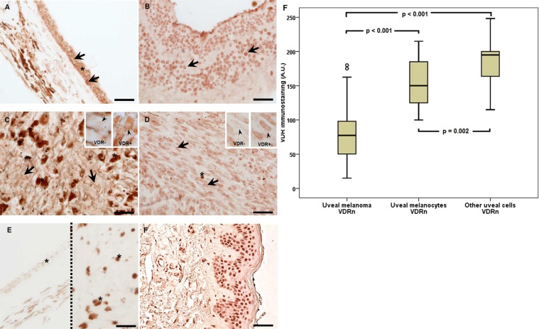
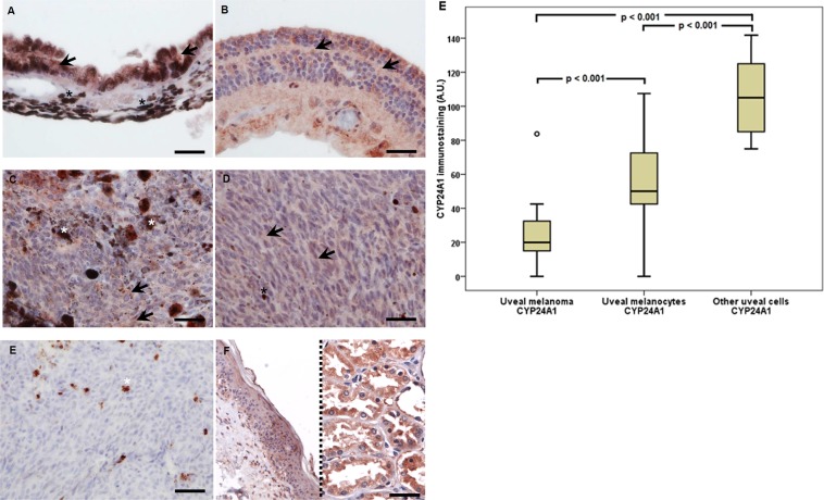
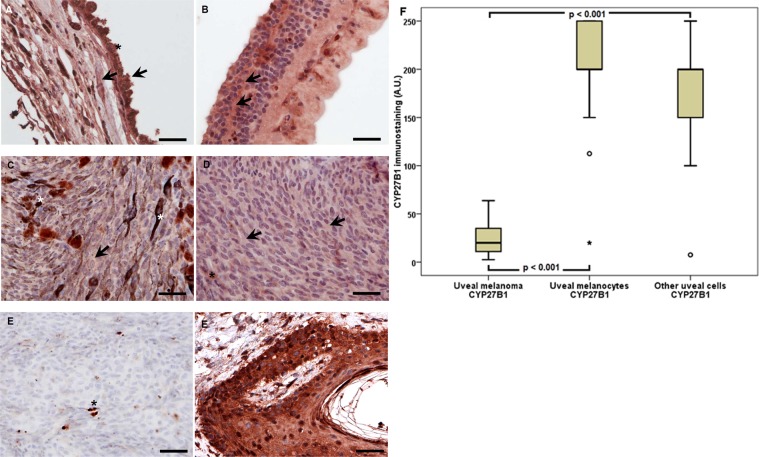
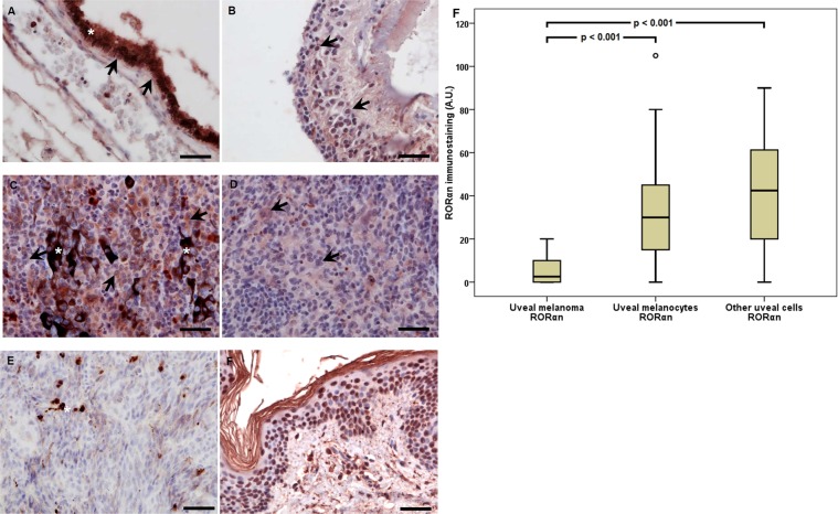
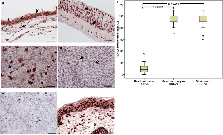
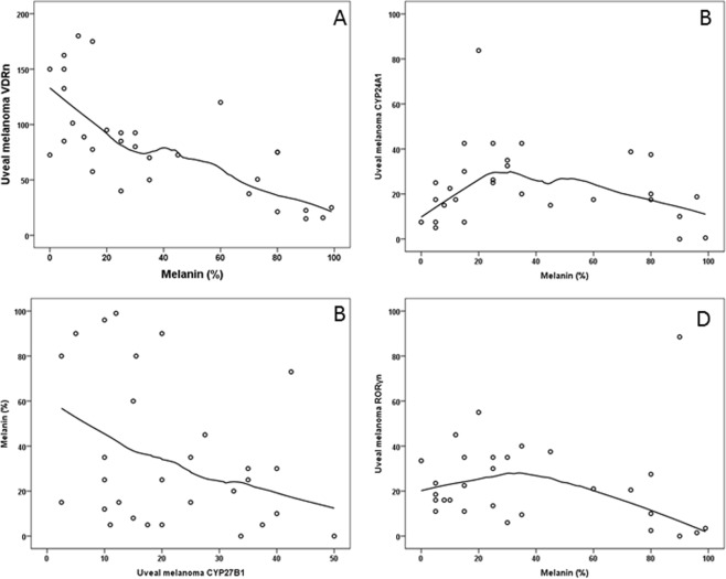
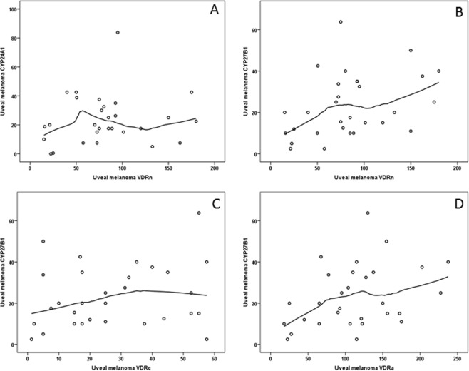
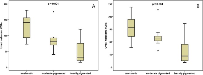
In recent years, a significant number of studies have investigated the preventive role of vitamin D in a number of different neoplasms. In this study, we analyze various components of the vitamin D signaling pathways in the human uveal tract and uveal melanoma, including analysis of the expression of vitamin D receptors (VDR), the activating and inactivating hydroxylases, respectively, CYP27B1 and CYP24A1, and the retinoic acid-related orphan receptors (ROR) α (RORα) and γ (RORγ) in these tissues. We further analyzed the expression of VDR, CYP27B1, CYP24A1, and ROR in relation to melanin levels, clinical stage and prognosis. Our study indicated that the uveal melanoma melanin level inversely correlated with VDR expression. We further showed that vitamin D is metabolized in uveal melanoma. This is significant because until now there has been no paper published, that would describe presence of VDR, hydroxylases CYP27B1 and CYP24A1, and RORα and RORγ in the human uveal tract and uveal melanomas. The outcomes of our research can contribute to the development of new diagnostic and therapeutic methods in uveal tract disorders, especially in uveal melanoma. The presented associations between vitamin D signaling elements and uveal melanoma in comparison to uveal tract encourage future clinical research with larger patients' population.


Conflict of interest statement

The authors declare no competing interests.

Figures

Figure 1
Figure 1
Representative VDR immunostaining in uveal pigmented cells (A), other cells (B), strongly pigmented (C) and slightly pigmented (D) melanomas and negative (E; two separate samples separated with dotted line) and positive (F) controls. Inserts present high magnification of negative (VDR−) and positive (VDR+) cell nuclei (as indicated by arrow heads). Arrows indicate nuclear VDR immunostaining, asterisks indicate melanin, scale bar: 50 μm. Mean nuclear VDR immunolabelling (A.U. – arbitrary units) in melanoma, normal melanocytes and other normal uveal cells (F) (statistically significant differences are denoted with the Wilcoxon signed-rank test).
Figure 2
Figure 2
Representative CYP24A1 immunostaining in uveal pigmented cells (A), other cells (B), strongly pigmented (C) and slightly pigmented (D) melanomas and negative (E) and positive (F) controls. For CYP24A1 in addition to normal skin (left) also kidney (right) samples served as positive controls. Arrows indicate CYP24A1 immunostaining, asterisks indicate melanin, scale bar: 50 μm. Mean CYP24A1 immunolabelling in melanoma, normal melanocytes and other normal uveal cells (F) (statistically significant differences are denoted with the Wilcoxon signed-rank test).
Figure 3
Figure 3
Representative CYP27B1 immunostaining in uveal pigmented cells (A), other cells (B), strongly pigmented (C) and slightly pigmented (D) melanomas and negative (E) and positive (F) controls. Arrows indicate CYP27B1 immunostaining, asterisks indicate melanin, scale bar: 50 μm. Mean CYP27B1 immunolabelling in melanoma, normal melanocytes and other normal uveal cells (F) (statistically significant differences are denoted with the Wilcoxon signed-rank test).
Figure 4
Figure 4
Representative RORα immunostaining in uveal pigmented cells (A), other cells (B), strongly pigmented (C) and slightly pigmented (D) melanomas and negative (E) and positive (F) controls. Arrows indicate RORα immunostaining, asterisks indicate melanin, scale bar: 50 μm. Mean RORα nuclear (RORαn) immunolabeling in melanoma, normal melanocytes, and other normal uveal cells (F) (statistically significant differences are denoted with the Wilcoxon signed-rank test).
Figure 5
Figure 5
Representative RORγ immunostaining in uveal pigmented cells (A), other cells (B), strongly pigmented (C) and slightly pigmented (D) melanomas and negative (E) and positive (F) controls. Arrows indicate RORγ immunostaining, asterisks indicate melanin, scale bar: 50 μm. Mean RORγ nuclear (RORγn) immunolabelling in uveal melanoma, normal melanocytes and other normal uveal cells (F) (statistically significant differences are denoted with the Wilcoxon signed-rank test).
Figure 6
Figure 6
The correlation of: VDRn immunostaining and the level of melanin (A); CYP24A1 immunostaining and melanin (B); CYP27B1immunostaining and melanin (C); RORγn immunostaining and melanin (D).
Figure 7
Figure 7
The correlation between VDRn immunostaining and CYP24A1 level (A). The correlation of the level of CYP27B1 to VDRn (B), VDRc (C) and VDRa (D).
Figure 8
Figure 8
Comparison of VDRn (A) and VDRa (B) expression with the clinical assessment of tumor pigmentation (the Wilcoxon signed-rank test).

References

    1. Chang AE, Karnell LH, Menck HR. The National Cancer Data Base Report on Cutaneous and Noncutaneous Melanoma. Cancer. 1998;83:1664–1678. doi: 10.1002/(SICI)1097-0142(19981015)83:83.0.CO;2-G. - DOI - PubMed
    1. Shields CL, et al. Metastasis of uveal melanoma millimeter-by-millimeter in 8033 consecutive eyes. Arch. Ophthalmol. 2009;127:989–998. doi: 10.1001/archophthalmol.2009.208. - DOI - PubMed
    1. Kaliki S, Shields CL. Uveal melanoma: Relatively rare but deadly cancer. Eye. 2017;31:241–257. doi: 10.1038/eye.2016.275. - DOI - PMC - PubMed
    1. Singh AD, Turell ME, Topham AK. Uveal melanoma: Trends in incidence, treatment, and survival. Ophthalmology. 2011;118:1881–1885. doi: 10.1016/j.ophtha.2011.01.040. - DOI - PubMed
    1. Virgili Gianni, Gatta Gemma, Ciccolallo Laura, Capocaccia Riccardo, Biggeri Annibale, Crocetti Emanuele, Lutz Jean-Michel, Paci Eugenio. Incidence of Uveal Melanoma in Europe. Ophthalmology. 2007;114(12):2309-2315.e2. doi: 10.1016/j.ophtha.2007.01.032. - DOI - PubMed

Publication types

Substances