. 2019 Jun 24;9(1):9073.
doi: 10.1038/s41598-019-45658-2.

Anaplasma phagocytophilum modifies tick cell microRNA expression and upregulates isc-mir-79 to facilitate infection by targeting the Roundabout protein 2 pathway

Affiliations

Anaplasma phagocytophilum modifies tick cell microRNA expression and upregulates isc-mir-79 to facilitate infection by targeting the Roundabout protein 2 pathway

Sara Artigas-Jerónimo et al. Sci Rep. .

Abstract

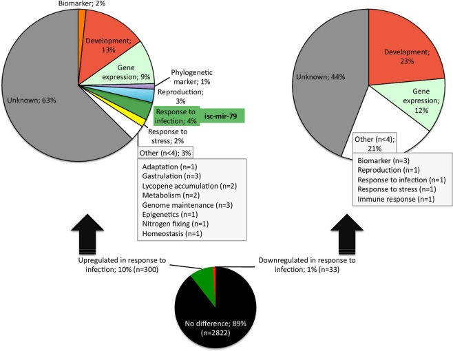
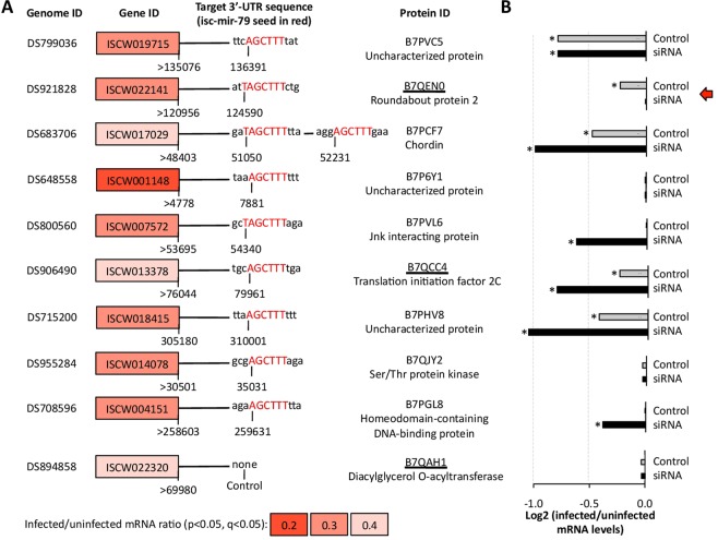
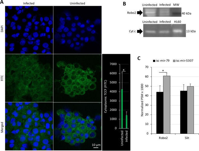
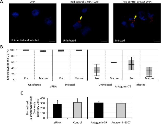
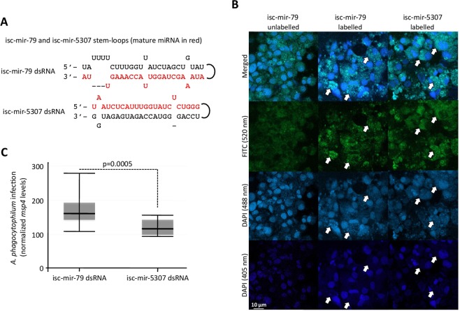
The microRNAs (miRNAs) are a class of small noncoding RNAs that have important regulatory roles in multicellular organisms including innate and adaptive immune pathways to control bacterial, parasite and viral infections, and pathogens could modify host miRNA profile to facilitate infection and multiplication. Therefore, understanding the function of host miRNAs in response to pathogen infection is relevant to characterize host-pathogen molecular interactions and to provide new targets for effective new interventions for the control infectious diseases. The objective of this study was to characterize the dynamics and functional significance of the miRNA response of the tick vector Ixodes scapularis in response to Anaplasma phagocytophilum infection, the causative agent of human and animal granulocytic anaplasmosis. To address this objective, the composition of tick miRNAs, functional annotation, and expression profiling was characterized using high throughout RNA sequencing in uninfected and A. phagocytophilum-infected I. scapularis ISE6 tick cells, a model for tick hemocytes involved in pathogen infection. The results provided new evidences on the role of tick miRNA during pathogen infection, and showed that A. phagocytophilum modifies I. scapularis tick cell miRNA profile and upregulates isc-mir-79 to facilitate infection by targeting the Roundabout protein 2 (Robo2) pathway. Furthermore, these results suggested new targets for interventions to control pathogen infection in ticks.


Conflict of interest statement

The authors declare no competing interests.

Figures

Figure 1
Figure 1
Characterization of the miRNA profile in response to A. phagocytophilum infection of tick ISE6 cells. (A) Total of 3,155 miRNAs were identified after miRNA-seq. Of them, 300 and 33 were upregulated and downregulated in response to A. phagocytophilum infection, respectively. Differentially expressed miRNAs (p < 0.05, q < 0.05) were functionally annotated based on the previously published information for homologous miRNAs in ticks and other species. Of the 16 miRNAs within the response to infection category and upregulated in response to infection, the isc-mir-79 (infected/uninfected ratio = 4.4; p = 0.002, q = 0.047) was selected for further analysis.
Figure 2
Figure 2
Validation of miRNA levels in response to A. phagocytophilum infection of tick ISE6 cells. (A) Forward primers were designed for selected miRNAs upregulated in response to infection, and used to characterize by qRT-PCR the levels of pre and mature isc-mir-79 (shown as example), isc-mir-185, isc-mir-7544, isc-mir-9771b, isc-mir-2951, isc-mir-B8, isc-mir-491, and isc-mir-5307 using the miScript universal reverse primer. The mature-isc-mir-5037, which was not differentially regulated in response to infection, was used as control miRNA. The miRNA levels were characterized in uninfected and A. phagocytophilum-infected tick cells and shown as infected/uninfected ratio (B) or Ave + S.D. for pre and mature isc-mir-79, and isc-mir-5307 levels (C). The miRNA levels were normalized against tick rps4. The normalized Ct values were compared between groups by Student’s t-test with unequal variance (*p < 0.05; N = 3 biological replicates).
Figure 3
Figure 3
Putative targets for isc-mir-79 in the mRNA of genes downregulated in response to A. phagocytophilum infection of tick ISE6 cells. (A) Putative isc-mir-79 targets were predicted using RNAhybrid (https://bibiserv2.cebitec.uni-bielefeld.de/rnahybrid). Biological process functional annotations were done using Blast2GO. Data for genes significantly downregulated (p < 0.05) in response to A. phagocytophilum infection of tick ISE6 cells were obtained from Villar et al.. Protein identity is shown, and the protein identified in the proteome of tick ISE6 cells (Villar et al.) is underlined. (B) Regulation of predicted isc-mir-79 target mRNAs in response to isc-mir-79 knockdown and A. phagocytophilum infection. Only genes that were downregulated in response to A. phagocytophilum infection of tick ISE6 cells were included. The mRNA levels were normalized against tick rps4, and normalized Ct values were represented as the log2 (infected to uninfected ratio) and compared between control uninfected and control infected (Control) or isc-mir-79 siRNA-treated infected (siRNA) tick cells by Student’s t-test with unequal variance (p > 0.05; N = 3 biological replicates). None of the genes were confirmed as putative mRNA targets for isc-mir-79.
Figure 4
Figure 4
Putative targets for isc-mir-79 in the 3′-UTR of genes downregulated in response to A. phagocytophilum infection of tick ISE6 cells. (A) Data for genes significantly downregulated (p < 0.05) in response to A. phagocytophilum infection of tick ISE6 cells were obtained from Villar et al.. Putative target sites (6–8 bp; seed sequence 5′-AGCTTTA-3′) for mature isc-mir-79 (3′-AUUGAAACCAUUGGAUCGAAAAUA-5′) were predicted using I. scapularis genome sequence of corresponding 3′-UTRs (60–5000 bp) as AGCTTT, AGCTTTA, TAGCTTT, TAGCTTTA (weaker to stronger prediction). Proteins identified in the proteome of tick ISE6 cells (Villar et al.) are underlined. (B) Regulation of predicted isc-mir-79 target genes in response to isc-mir-79 knockdown and A. phagocytophilum infection. The mRNA levels were normalized against tick rps4, and normalized Ct values were represented as the log2 (infected to uninfected ratio) and compared between control uninfected and control infected (Control) or isc-mir-79 siRNA-treated infected (siRNA) tick cells by Student’s t-test with unequal variance (*p < 0.05; N = 3 biological replicates). The identified putative target for isc-mir-79 (red arrow) corresponds to the gene significantly downregulated in response to infection but not after miRNA knockdown.
Figure 5
Figure 5
Protein levels of putative isc-mir-79 targets in tick ISE6 cells. (A) Representative images of immunofluorescence analysis of uninfected and A. phagocytophilum-infected ISE6 tick cells incubated with mouse anti-Robo antibodies. Goat anti-mouse IgG-FITC secondary antibodies were used to label Robo (green). Host cell nucleus was stained with DAPI (blue). Bar, 10 µm. The total cytoplasmic corrected cellular fluorescence (TCCF) was calculated as integrated density − (area of selected cell × mean fluorescence of background readings) and compared between infected and uninfected cells by Student’s t-test with unequal variance (*p = 0.001; N = 10 biological replicates). (B) Western-blot analysis of tick Robo2 and Cyt c (control protein not affected by A. phagocytophilum infection) levels in uninfected and A. phagocytophilum-infected ISE6 tick cells incubated with anti-Robo or anti-Cyt c antibodies. The position for tick Robo2 (predicted molecular weight, 42 kDa) and Cyt c (predicted molecular weight, 13 kDa) is shown with an arrow. For Cyt c, proteins from human HL60 cells were included as a positive control. (C) The isc-mir-79 and control isc-mir-5307 miRNA levels were increased in tick ISE6 cells with dsRNA miRNA mimics. Proteomics analysis was targeted at Robo2 (B7QEN0) and Slit (B7PL46) proteins identified as an isc-mir-79 3′-UTR target or not affected by this miRNA, respectively. The results of the normalized PSM x 1000 values were compared between groups by Chi2-test (*p < 0.05; N = 3 biological replicates).
Figure 6
Figure 6
Knockdown of isc-mir-79 and effect on A. phagocytophilum infection of tick ISE6 cells. (A) Representative images to confirm the delivery of siRNA into tick ISE6 cells. Cytocentrifuge preparations of ISE6 cells treated with an Accell red fluorescent non-targeting control siRNA (red, yellow arrows) were mounted using Prolong Gold antifade reagent with DAPI (blue). Bars, 10 µm. (B) The isc-mir-79 was knockdown using a isc-mir-79 miRNA-specific custom siRNA or isc-mir-79 and isc-mir-5307 miRNA-specific antisense oligonucleotide antagomirs. The miRNA levels were characterized in uninfected and A. phagocytophilum-infected tick cells and normalized against tick rps4 to calculate the knockdown percent with respect to medium-treated or isc-mir-5307 antagomir-treated cells for siRNA and Antagomir-79, respectively. (C) After treatment with siRNA or antagomirs, tick ISE6 cells were infected with A. phagocytophilum. DNA samples were analyzed by real-time qPCR using the A. phagocytophilum msp4 and normalized against tick 16 S rRNA. Normalized Ct values were compared between treated and control cells by Student’s t-test with unequal variance (p > 0.05; N = 3 biological replicates).
Figure 7
Figure 7
Increase of isc-mir-79 levels and effect on A. phagocytophilum infection of tick ISE6 cells. (A) The miRNA dsRNA mimics were constructed using RNA oligonucleotides for isc-mir-79 and isc-mir-5307 and their complementary sequences. (B) The delivery of miRNA mimics into tick ISE6 cells was confirmed using fluorescent labeled and unlabeled miRNA mimics. Emission peak wavelengths are for DAPI RNA (500 nm), DAPI DNA (460 nm) and FITC (519 nm). Therefore, fluorescence was detected at 405 nm (closer to DNA then RNA), 488 nm (closer to RNA than DNA) and 520 nm for FITC. White arrows show examples of regions with miRNA labeling and some RNA labeling but without DNA labeling, thus corresponding to miRNA mimics inside the cell. (C) Tick ISE6 cells were incubated with dsRNA, washed and then treated with fresh medium containing cell free A. phagocytophilum. DNA samples were analyzed by real-time qPCR using the A. phagocytophilum msp4 and normalized against tick 16 S rRNA. Normalized Ct values were compared between isc-mir-79 and isc-mir-5307 dsRNA treated cells by Student’s t-test with unequal variance (p = 0.0005; N = 4 biological replicates).
Figure 8
Figure 8
Proposed mechanism affected by isc-mir-79 to facilitate pathogen infection. (A,B) Tick ISE6 cells were incubated with siRNAs targeting Robo2 or Robo paralogs, washed and then treated with fresh medium containing cell free A. phagocytophilum ((A) high infection: 90–100% infected cells; B, low infection: 5–10% infected cells). Control cells were treated with siRNAs targeting the unrelated Rs86 gene. DNA samples were analyzed by real-time qPCR using the A. phagocytophilum msp4 and normalized against tick 16 S rRNA. Normalized Ct values were compared between Robo and Rs86 siRNAs treated cells by Chi test (*p < 0.0005; **p < 0.00005; N = 4 biological replicates). (C) Based on these results we speculated that A. phagocytophilum uses unknown type IV secretion system (T4SS) effectors or other mechanisms to promote isc-mir-79 overexpression and target Robo2 to suppress or diminish protective proinflammatory responses mediated by the Slit-Robo pathway to facilitate infection.

References

    1. Jongejan F, Uilenberg G. The global importance of ticks. Parasitology. 2004;129:S3–S14. doi: 10.1017/S0031182004005967. - DOI - PubMed
    1. de la Fuente J, Estrada-Peña A, Venzal JM, Kocan KM, Sonenshine DE. Overview: Ticks as vectors of pathogens that cause disease in humans and animals. Front. Biosci. 2008;13:6938–6946. doi: 10.2741/3200. - DOI - PubMed
    1. Ayllón N, et al. Systems biology of tissue-specific response to Anaplasma phagocytophilum reveals differentiated apoptosis in the tick vector Ixodes scapularis. PLoS Genet. 2015;11:e1005120. doi: 10.1371/journal.pgen.1005120. - DOI - PMC - PubMed
    1. Severo, M. S., Pedra, J. H. F., Ayllón, N., Kocan, K. M. & de la Fuente, J. Anaplasma. In: Tang YW, Sussman M, Liu D, Poxton I, Schwartzman J, editors. Molecular Medical Microbiology (2nd edition) 2033–2042 (Elsevier Academic Press, 2015).
    1. Truchan HK, et al. The pathogen-occupied vacuoles of Anaplasma phagocytophilum and Anaplasma marginale interact with the endoplasmic reticulum. Front. Cell. Infect. Microbiol. 2016;6:22. doi: 10.3389/fcimb.2016.00022. - DOI - PMC - PubMed

Publication types