. 2019 Jun 24;9(1):9206.
doi: 10.1038/s41598-019-45651-9.

The extracellular matrix proteoglycan lumican improves survival and counteracts cardiac dilatation and failure in mice subjected to pressure overload

Affiliations

The extracellular matrix proteoglycan lumican improves survival and counteracts cardiac dilatation and failure in mice subjected to pressure overload

Naiyereh Mohammadzadeh et al. Sci Rep. .

Abstract

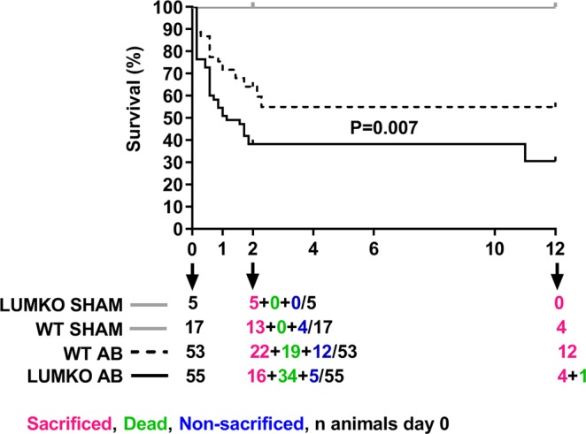
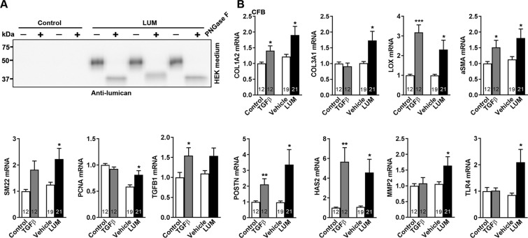
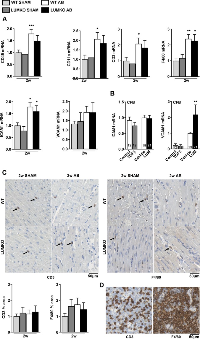
Left ventricular (LV) dilatation is a key step in transition to heart failure (HF) in response to pressure overload. Cardiac extracellular matrix (ECM) contains fibrillar collagens and proteoglycans, important for maintaining tissue integrity. Alterations in collagen production and cross-linking are associated with cardiac LV dilatation and HF. Lumican (LUM) is a collagen binding proteoglycan with increased expression in hearts of patients and mice with HF, however, its role in cardiac function remains poorly understood. To examine the role of LUM in pressure overload induced cardiac remodeling, we subjected LUM knock-out (LUMKO) mice to aortic banding (AB) and treated cultured cardiac fibroblasts (CFB) with LUM. LUMKO mice exhibited increased mortality 1-14 days post-AB. Echocardiography revealed increased LV dilatation, altered hypertrophic remodeling and exacerbated contractile dysfunction in surviving LUMKO 1-10w post-AB. LUMKO hearts showed reduced collagen expression and cross-linking post-AB. Transcriptional profiling of LUMKO hearts by RNA sequencing revealed 714 differentially expressed transcripts, with enrichment of cardiotoxicity, ECM and inflammatory pathways. CFB treated with LUM showed increased mRNAs for markers of myofibroblast differentiation, proliferation and expression of ECM molecules important for fibrosis, including collagens and collagen cross-linking enzyme lysyl oxidase. In conclusion, we report the novel finding that lack of LUM attenuates collagen cross-linking in the pressure-overloaded heart, leading to increased mortality, dilatation and contractile dysfunction in mice.


Conflict of interest statement

The authors declare no competing interests.

Figures

Figure 1
Figure 1
Increased mortality of LUMKO mice post-AB. Kaplan-Meier survival curves for lumican knock-out (LUMKO, n = 55) and wild-type (WT, n = 53) mice 12 weeks post-aortic banding (AB). We had no mortality in SHAM groups. At 2w, we sacrificed 5 LUMKO SHAM and 13 WT SHAM. The remainder 4 WT SHAM lived until 12w. However, we had significant mortality in AB groups. At 2w, in WT AB group (n = 53), we sacrificed 22 mice, 19 mice died up until 2w and 12 mice lived until 12w (22 + 19 + 12 = 53). At 2w, in LUMKO AB group, we sacrificed 16 mice, 34 mice died up until 2w and 5 mice lived until 12w (one died after 10w) (16 + 34 + 5 = 55). Differences were tested using Log-rank (Mantel–Cox) test, p = 0.007.
Figure 2
Figure 2
Increased left ventricular dilatation and exacerbated contractile dysfunction in LUMKO mice post-AB. (A) Serial echocardiography of lumican knock-out (LUMKO, n = 3–17) and wild-type (WT, n = 16–35) pre- and 1–10 weeks (w) post-aortic banding (AB), showing left ventricular (LV) internal diameter in diastole (LVIDd), and systole (LVIDs), and relative wall thickness (RWT %), and left atrial diameter (LAD) and fractional shortening (FS %). Representative 2D and M-mode echocardiograms are shown in A. (B) Representative Wheat Germ Agglutinate (WGA)-stained mid-ventricular sections and quantitative measurement of cardiomyocyte cross-sectional areas (CSA) in LUMKO and WT 2w and 12w post-SHAM and -AB (n SHAM 3–5, n AB 4–10, n = 2000–15000 cardiomyocytes). Scale bars 50 µm. (C) Heart and lung weights normalized to body weight (HW/BW and LW/BW, respectively) in LUMKO and WT 2w and 12w post-SHAM and -AB (n SHAM 3–12, n AB 11–20). The data are presented as mean ± SEM. Differences were tested using one-way ANOVA with Dunn’s post-hoc test vs. WT SHAM, ***p < 0.005; **p < 0.01; *p < 0.05, or one-way ANOVA with Tukey’s multiple comparisons test vs. WT AB, δp < 0.05; δδp < 0.01; δδδp < 0.005 (B,C 2w and 12w), or two-way ANOVA, δp < 0.05 (A).
Figure 3
Figure 3
Reduced collagen cross-linking and collagen expression in hearts of LUMKO mice post-AB. Lumican knock-out (LUMKO) and wild-type (WT) mice were subjected to aortic banding (AB) for 12 weeks (w). (A,B) Representative images of mid-ventricular histology sections 2w and 12w post-SHAM and-AB, stained with Picrosirious Red visualizing fibrillar collagens (A, non-polarized light, bright field) and collagen cross-linking (B, polarized light). Fibrotic area = area of red staining/total area in %. Collagen crosslinking area = area of orange/green (cross-linked collagens)/total area in % (n SHAM 3–4, n AB 4–10). Scale bars 50 µm. (C,D) Relative left ventricular (LV) mRNA levels of fibrillar collagens I and III (COL1A2 and COL3A1), and myofibroblast differentiation marker αSMA and SM22 (n SHAM 4–14, n AB 3–22). mRNA expression was normalized to expression of ribosomal protein L32 (RPL32). (E) Quantitative protein levels of the myofibroblast differentiation marker alpha-smooth muscle actin (αSMA) in LVs of LUMKO and WT mice 2w and 12w post-SHAM and-AB (2w: n WT SHAM = 9, n WT AB = 13, n LUMKO SHAM = 9, n LUMKO AB = 14 and 12w: n WT SHAM = 4, n WT AB = 12, n LUMKO AB = 3). Cultured cardiac fibroblasts (CFB) were used as positive control. Coomassie staining was used as loading control. Immunoblots are presented in Fig. S5 (2w) and Fig. S6 (12w). The data are presented as mean ± SEM. Differences were tested using one-way ANOVA with Dunn’s post-hoc test vs. WT SHAM, *p < 0.05; **p < 0.01; ***p < 0.005; or an unpaired t-test vs. WT AB, δp < 0.01.
Figure 4
Figure 4
Increased mRNA expression of myofibroblast markers and ECM components including collagens in cardiac fibroblasts treated with LUM. Cultured cardiac fibroblasts from neonatal rats (n = 3 cell isolations) were treated with LUM or vehicle conditioned medium for 24 h. Non-treated cells (control) and cells treated with the pro-fibrotic growth factor transforming growth factor (TGF)β1 served as controls. (A) LUM was produced by transfection of human endothelial kidney (HEK)293 cells with human LUM. LUM was secreted into the cell medium as 50 kDa glycosylated proteoglycan (Fig. S8). PNGaseF treatment results in a 38 kDa deglycosylated core protein (Fig. S8). (B) mRNA expression of fibrillar collagens I and III (COL1A2 and COL3A1), the collagen cross-linking enzyme lysyl oxidase (LOX), the myofibroblast signature genes αSMA and SM22, the proliferating cell nuclear antigen (PCNA), TGFβ1 and the extracellular matrix components periostin (POSTN), hyaluronan synthase 2 (HAS2), matrix metalloproteinase 2 (MMP-2) and toll-like receptor-4 (TLR-4). mRNA expression was normalized to expression of ribosomal protein L32 (RPL32). Data are presented as mean ± SEM. Differences were tested using an unpaired t-test vs. Vehicle, *p < 0.05, or vs. Control, ***p ≤ 0.005; **p ≤ 0.01.
Figure 5
Figure 5
No alteration of immune cell infiltration in LUMKO mice post-AB. (A) left ventricular (LV) mRNA expression of immune cell surface markers (leukocytes, CD45 (encoding cluster of differentiation 45) and CD11a (encoding cluster of differentiation 11), T-cells, CD3 (encoding cluster of differentiation 3), and macrophages, F4/80 (encoding adhesion G protein-coupled receptor E1) and immune cell adhesion molecules (ICAM1 and VCAM1, encoding intercellular adhesion molecule-1 and vascular cell adhesion molecule 1, respectively) in LUMKO and WT mice 2w post-SHAM and -AB operations (n SHAM 1–5, n AB 9–10). (B) mRNA expression of ICAM1 and VCAM1 in cultured neonatal rat cardiac fibroblasts (n = 3 cell isolations) treated with LUM or vehicle conditioned medium for 24 h. Non-treated cells (control) and cells treated with the pro-fibrotic growth factor transforming growth factor (TGF) β1 served as controls. mRNA expression was normalized to expression of ribosomal protein L32 (RPL32). (C) Representative immuno-stained mid-ventricular sections and quantitative measurement of CD3 (T-lymphocyte) and F4/80 (macrophage) infiltration markers in LUMKO and WT 2w post-SHAM and -AB (n WT SHAM = 4, n WT AB = 6, n LUMKO SHAM = 3, n LUMKO AB = 8). Adult WT mouse spleen sections were stained for CD3 and F4/80 as positive controls (D). Scale bars 50 µm. Data are presented as mean ± SEM. Differences were tested using one-way ANOVA with Dunn’s post-hoc test vs. WT SHAM (A), or an unpaired t-test vs. WT SHAM (A CD45) or vs. Control (B), ***p < 0.005; **p < 0.01; *p < 0.05.
Figure 6
Figure 6
RNA-sequencing revealed novel LUM-dependent molecular mechanisms associated with cardiac remodeling. Lumican knock-out (LUMKO) and wild-type (WT) mice were subjected to aortic banding (AB) and left ventricles (LV) harvested at 2 weeks (w) post-AB for RNA-sequencing of pools of n = 3. 714 differentially expressed (DE) transcripts were identified, i.e. 526 > 1.33-fold up- and 188 < 0.75-fold down-regulated, (p < 0.001), please see Suppl. File. (A) Top 10 down-regulated (except LUM itself, which was not expressed) and top 10 up-regulated transcripts in LUMKO vs. WT. (B) Cultured cardiac fibroblasts from neonatal rats (n = 3 cell isolations) were treated with LUM or vehicle conditioned medium for 24 h. Non-treated cells (control) and cells treated with the pro-fibrotic growth factor transforming growth factor (TGF) β1 served as controls. mRNA expression of spondin-2 (SPON2) and growth differentiation factor 15 (GDF-15) identified in (A); and mRNA expression of miR-21-5p and -3p, respectively, predicted in (C). Expression was normalized to expression of ribosomal protein L32 (RPL32) or U6. (C) Ingenuity pathway analysis (IPA) prediction of upstream regulators of DE transcripts in LUMKO vs. WT LVs 2w post-AB (Z-score >2 = activated, <2 = inactivated). The data are presented as mean ± SEM. Differences were tested using an unpaired t-test vs. Vehicle or Control, *p < 0.05; ***p < 0.005.

Similar articles

Cited by

References

    1. Grossman W, Paulus WJ. Myocardial stress and hypertrophy: a complex interface between biophysics and cardiac remodeling. The Journal of clinical investigation. 2013;123:3701–3703. doi: 10.1172/jci69830. - DOI - PMC - PubMed
    1. Kehat I, Molkentin JD. Molecular pathways underlying cardiac remodeling during pathophysiological stimulation. Circulation. 2010;122:2727–2735. doi: 10.1161/circulationaha.110.942268. - DOI - PMC - PubMed
    1. Berk BC, Fujiwara K, Lehoux S. ECM remodeling in hypertensive heart disease. The Journal of clinical investigation. 2007;117:568–575. doi: 10.1172/jci31044. - DOI - PMC - PubMed
    1. Badenhorst D, et al. Cross-linking influences the impact of quantitative changes in myocardial collagen on cardiac stiffness and remodelling in hypertension in rats. Cardiovascular research. 2003;57:632–641. doi: 10.1016/S0008-6363(02)00733-2. - DOI - PubMed
    1. Lopez B, et al. Impact of treatment on myocardial lysyl oxidase expression and collagen cross-linking in patients with heart failure. Hypertension. 2009;53:236–242. doi: 10.1161/hypertensionaha.108.125278. - DOI - PubMed

Publication types