

Claudin-18 expression in oesophagogastric adenocarcinomas: a tissue microarray study of 523 molecularly profiled cases
- PMID: 31235864
- PMCID: PMC6738069
- DOI: 10.1038/s41416-019-0508-4
Claudin-18 expression in oesophagogastric adenocarcinomas: a tissue microarray study of 523 molecularly profiled cases
Abstract
Background: Claudin-18 (CLDN18) is a highly specific tight junction protein of the gastric mucosa. An isoform of CLDN18, the Claudin 18.2, has recently emerged as an innovative drug target for metastatic gastric cancer.
Methods: We investigated the immunohistochemical profile of CLDN18, p53, p16, E-cadherin, MSH2, MSH6, MLH1, PSM2, HER2, and PDL-1 in a large series of 523 primary gastric carcinomas (GCs; n = 408) and gastro-oesophageal carcinomas (GECs; n = 115) and 135 matched and synchronous nodal metastases. The status of HER2 and EBER by means of chromogenic in situ hybridisation (CISH) was also evaluated.
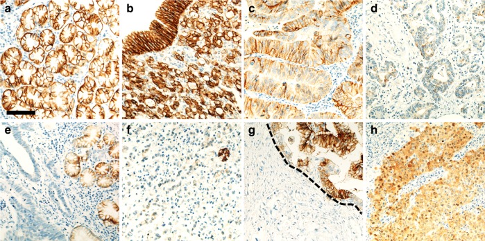
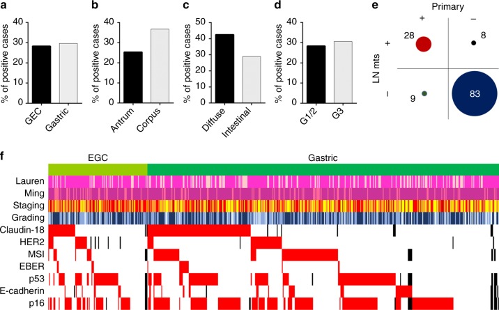
Results: High membranous CLDN18 expression was present in 150/510 (29.4%) primary cases and in 45/132 (34.1%) metastases. An abnormal expression (i.e. nuclear and/or cytoplasmic) was observed in 115 (22.5%) primary cases and in 33 (25.0%) metastases. A 38.8% of the cases showed significant CLDN18 intratumoural variability among the different tissue microarray cores obtained from the same tumour. Positive membrane CLDN18 expression was statistically associated with non-antral GCs (p = 0.016), Lauren diffuse type (p = 0.009), and with EBV-associated cancers (p < 0.001).
Conclusions: CLDN18 is frequently expressed in gastric and gastro-oesophageal cancers; further studies should investigate the prognostic significance of CLDN18 heterogeneity in order to implement its test into clinical practice.
Conflict of interest statement
F.L. had roles as consultant or advisor for Roche, Bayer, Amgen and Genentech. S.L. had roles as consultant or advisor for Amgen, Bayer, Merck Serono and Lilly. She received research funding from Amgen and Merck Serono and is part of the speaker’s bureau of Lilly and BMS. N.V. received honoraria for lectures from Merck Serono, Bayer, Eli Lilly and Pfizer. The other authors declare that they have no conflict of interest.
Figures
References
-
- Mathiak M, Warneke VS, Behrens HM, Haag J, Boger C, Kruger S, et al. Clinicopathologic characteristics of microsatellite instable gastric carcinomas revisited: urgent need for standardization. Appl. Immunohistochem. Mol. Morphol. 2017;25:12–24. doi: 10.1097/PAI.0000000000000264. - DOI - PMC - PubMed
Publication types
MeSH terms
Substances
Grants and funding
LinkOut - more resources
Full Text Sources
Other Literature Sources
Medical
Research Materials
Miscellaneous
