. 2019 Jul;26(7):619-627.
doi: 10.1038/s41594-019-0248-4. Epub 2019 Jun 24.

Cryo-EM structures of four polymorphic TDP-43 amyloid cores

Affiliations

Cryo-EM structures of four polymorphic TDP-43 amyloid cores

Qin Cao et al. Nat Struct Mol Biol. 2019 Jul.

Abstract

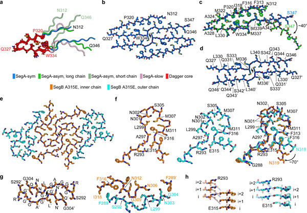
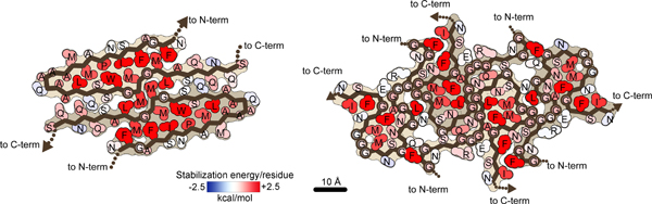
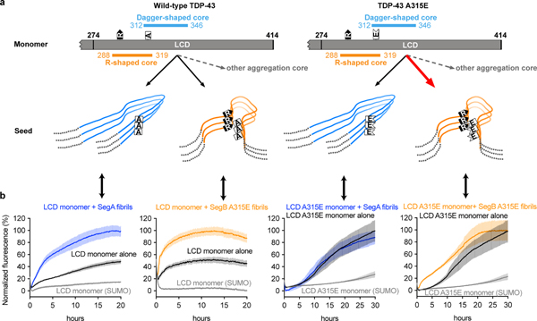
The DNA and RNA processing protein TDP-43 undergoes both functional and pathogenic aggregation. Functional TDP-43 aggregates form reversible, transient species such as nuclear bodies, stress granules, and myo-granules. Pathogenic, irreversible TDP-43 aggregates form in amyotrophic lateral sclerosis and other neurodegenerative conditions. Here we find the features of TDP-43 fibrils that confer both reversibility and irreversibility by determining structures of two segments reported to be the pathogenic cores of human TDP-43 aggregation: SegA (residues 311-360), which forms three polymorphs, all with dagger-shaped folds; and SegB A315E (residues 286-331 containing the amyotrophic lateral sclerosis hereditary mutation A315E), which forms R-shaped folds. Energetic analysis suggests that the dagger-shaped polymorphs represent irreversible fibril structures, whereas the SegB polymorph may participate in both reversible and irreversible fibrils. Our structures reveal the polymorphic nature of TDP-43 and suggest how the A315E mutation converts the R-shaped polymorph to an irreversible form that enhances pathology.


Figures

Figure 1
Figure 1. Cryo-EM Structure of TDP-43 polymorphic fibrils.
a, Schematic of full-length TDP-43. SegA (residues 311–360) and SegB (residues 286–331) identified for structural determination are shown as gray bars, respectively above and below the low-complexity domain (LCD). The color bars show the range of residues visualized in the structure of each polymorph. Panels b-e: (left) fibril reconstructions showing left-handed twist and pitch; (middle) density and atomic model of one cross-sectional layer of each fibril; (right) schematic model showing protein chain (blue) and residues (hydrophobic in yellow, polar in green, glycine in pink, glutamate in red, and arginine in blue).
Figure 2
Figure 2. Structure of the dagger-shaped fold and the R-shaped fold.
a,Superposition of the dagger-shaped folds (residue 312–346) in three SegA polymorphic fibrils. The core region of the dagger-shaped folds (residue 320–334) is colored red with side chains shown, and the rest of the dagger-shaped fold is colored according to the key. b, Atomic model of SegA-sym. c, Pairwise superposition of the dagger-shaped fold of SegA-sym vs. SegA-asym long chain. d, Continuous dimer interface of SegA-sym. e, Atomic model of SegB A315E. Each of the four chains is R-shaped, with a pseudo-21 axis relating the two pairs. The inner two chains are identical as are the two outer chains. f, Structures of the inner (left) and outer (middle) chains and their superposition (right). Notice the proximity of R293 to the pathogenic variant E315 which replaces A315 in the reference sequence. g, View parallel to the fibril axis showing the symmetric interface between the two inner chains (left) and the asymmetric interface between the inner and outer chains (right). h, View perpendicular to the fibril showing the inter-layer interaction of Arg293 with Glu315 on the inner chain (left) and outer chain (right). (Detailed alignment parameters and RMSD values listed in Supplementary Table 2).
Figure 3
Figure 3. Calculated solvation energy maps for TDP-43 fibrils based on atomic solvation parameters.
Solvation energy maps of SegA-sym (left) and SegB A315E (right). Residues are colored from unfavorable (blue, −2.5 kcal/mol) to favorable stabilization energy (red, 2.5 kcal/mol). Notice that both the dagger-shaped fold (SegA-sym) and the R-shaped fold (SegB A315E) show stable cores.
Figure 4
Figure 4. Speculative model of TDP-43 amyloid aggregation based on fibril structures reported here.
a, (Left) Monomeric TDP-43 can form amyloid fibrils with either dagger-shaped or R-shaped cores (or perhaps other so far unidentified cores). The ALS hereditary A315E mutation accelerates the R-shaped core through electrostatic interaction of Glu315 with Arg293. b, Experiments seeding TDP-43 fibril formation support the model of Panel A. Notice that both SegA and SegB A315E fibrils seed LCD monomer (far left and middle left). In contrast, only SegB A315E fibrils seed LCD A315E monomer whereas SegA fibrils barely seed LCD A315E monomer (far right and middle right). Data are shown as mean ± s.d., n=4 independent experiments.

References

    1. Anfinsen CB Principles that govern the folding of protein chains. Science 181, 223–230 (1973). - PubMed
    1. Li B. et al. Cryo-EM of full-length α-synuclein reveals fibril polymorphs with a common structural kernel. Nat. Commun. 9, 3609 (2018). - PMC - PubMed
    1. Zhang W. et al. Heparin-induced tau filaments are polymorphic and differ from those in Alzheimer’s and Pick’s disease. (2018). doi:10.1101/468892 - DOI - PMC - PubMed
    1. Neumann M. et al. Ubiquitinated TDP-43 in Frontotemporal Lobar Degeneration and Amyotrophic Lateral Sclerosis. Science 314, 130–133 (2006). - PubMed
    1. Hasegawa M. et al. Phosphorylated TDP-43 in frontotemporal lobar degeneration and amyotrophic lateral sclerosis. Ann. Neurol. 64, 60–70 (2008). - PMC - PubMed

METHODS-ONLY REFERENCES

    1. Suloway C. et al. Automated molecular microscopy: the new Leginon system. J. Struct. Biol. 151, 41–60 (2005). - PubMed
    1. Grant T. & Grigorieff N. Automatic estimation and correction of anisotropic magnification distortion in electron microscopes. J. Struct. Biol. 192, 204–208 (2015). - PMC - PubMed
    1. Rohou A. & Grigorieff N. CTFFIND4: Fast and accurate defocus estimation from electron micrographs. J. Struct. Biol. 192, 216–221 (2015). - PMC - PubMed
    1. Grant T. & Grigorieff N. Measuring the optimal exposure for single particle cryo-EM using a 2.6 Å reconstruction of rotavirus VP6. eLife 4, e06980 (2015). - PMC - PubMed
    1. Tang G. et al. EMAN2: an extensible image processing suite for electron microscopy. J. Struct. Biol. 157, 38–46 (2007). - PubMed

Publication types