

Colonic malignant peripheral nerve sheath tumour in a cat
- PMID: 31236282
- PMCID: PMC6572897
- DOI: 10.1177/2055116919849979
Colonic malignant peripheral nerve sheath tumour in a cat
Abstract
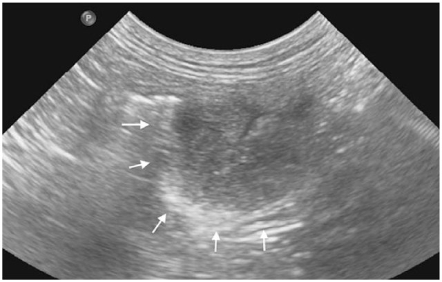
Case summary: A 14-year-old male neutered domestic mediumhair cat presented with a 4 month history of inappetence and weight loss. Pertinent abnormalities on haematology and biochemistry included a mild microcytic regenerative anaemia (packed cell volume [PCV] 24% [reference interval (RI) 30-45%], mean cell volume 30.8 fl [RI 40-45 fl], absolute reticulocyte count 326.8 × 10¹²) and increased alkaline phosphatase activity (76 IU/l; RI <50 IU/l). Abdominal ultrasound and CT scan revealed masses in the transverse colon (2.0 cm × 1.2 cm) and right medial liver lobe (5.0 cm diameter). Thoracic radiographs were unremarkable. Right medial liver lobe resection and colectomy were performed. Immunohistochemistry was positive for S-100 protein, vimentin and glial fibrillary acidic protein, very weakly positive for c-kit and negative for muscle-specific actin and CD18, consistent with a colonic malignant peripheral nerve sheath tumour (MPNST) with a hepatic metastasis. Postoperative treatment with metronomic cyclophosphamide was well tolerated. Eighteen months postoperatively the cat re-presented after 3 days of progressive lethargy and inappetence. Haematology revealed a marked non- or pre-regenerative anaemia (PCV 10%). Coagulation times were prolonged (prothrombin time 39 s [RI 15-22 s] and activated partial thromboplastin time >300 s [RI 65-119 s]). Abdominal ultrasound identified multiple renal and hepatic nodules. Euthanasia was performed and post-mortem examination confirmed metastasis of the MPNST.
Relevance and novel information: This report describes the treatment of a metastatic colonic peripheral nerve sheath tumour in a cat. Feline visceral MPNSTs are rare and little is known about prognosis or optimal treatment.
Keywords: Peripheral nerve sheath tumour; cyclophosphamide; metastasis; metronomic chemotherapy.
Conflict of interest statement
Conflict of interest: The authors declared no potential conflicts of interest with respect to the research, authorship, and/or publication of this article.
Figures


References
-
- Schulman FY, Johnson TO, Facemire PR, et al. Feline peripheral nerve sheath tumors: histologic, immunohistochemical, and clinicopathologic correlation (59 tumors in 53 cats). Vet Pathol 2009; 46: 1166–1180. - PubMed
-
- Stoll A, Suárez-Bonnet A, Summers B, et al. Malignant cutaneous peripheral nerve sheath tumour with rhabdomyosarcomatous differentiation (Triton tumour) in a domestic cat. J Comp Pathol 2018; 165: 1–5. - PubMed
-
- Hoffman A, Blocker T, Dubielzig R, et al. Feline periocular peripheral nerve sheath tumor: a case series. Vet Ophthalmol 2005; 8: 153–158. - PubMed
-
- Newkirk KM, Rohrbach BW. A retrospective study of eyelid tumors from 43 cats. Vet Pathol 2009; 46: 916–927. - PubMed
Publication types
LinkOut - more resources
Full Text Sources
Molecular Biology Databases
Miscellaneous