The surgical anatomy of soft tissue layers in the mastoid region
- PMID: 31236472
- PMCID: PMC6580058
- DOI: 10.1002/lio2.271
The surgical anatomy of soft tissue layers in the mastoid region
Abstract
Background: An understanding of the soft tissue layers in the mastoid region has become important for otologic reconstructive surgery. The objective of this study was to clarify the surgical anatomy of the soft tissue layers in the mastoid region and reveal its clinical significance.
Methods: Cadaveric study.
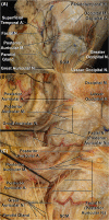
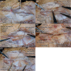
Results: Our dissections showed the soft tissue layers consisting of skin, subcutaneous layer, superficial and deep mastoid fasciae, and periosteum. The superficial mastoid fascia was continuous with the temporoparietal fascia cranially and the superficial cervical fascia caudally. The deep mastoid fascia could be clearly separated from the superficial mastoid fascia and has continuity to the loose alveolar layer in the temporoparietal region. However, it caudally fused with the fascia and ligament of the sternocleidomastoid.
Conclusions: A comprehensive understanding of soft tissue layers would improve otologic reconstructive surgery.
Level of evidence: NA.
Keywords: Temporal bone surgery; cadaveric study; fascia; otology; surgical anatomy.
Figures


Similar articles
-
Naming the soft tissue layers of the temporoparietal region: unifying anatomic terminology across surgical disciplines.Neurosurgery. 2010 Sep;67(3 Suppl Operative):ons120-9; discussion ons129-30. doi: 10.1227/01.NEU.0000383132.34056.61. Neurosurgery. 2010. PMID: 20679939 Review.
-
Anatomy of the orbital fasciae and the third eyelid in dogs.Am J Vet Res. 1990 Feb;51(2):260-3. Am J Vet Res. 1990. PMID: 2301837
-
Temporal branch of the facial nerve and its relationship to fascial layers.Arch Facial Plast Surg. 2010 Jan-Feb;12(1):16-23. doi: 10.1001/archfacial.2009.96. Arch Facial Plast Surg. 2010. PMID: 20083736
-
Superficial mastoid fascia as an accessible donor for various augmentations in Asian rhinoplasty.J Plast Reconstr Aesthet Surg. 2012 Aug;65(8):1035-40. doi: 10.1016/j.bjps.2012.03.002. Epub 2012 Mar 30. J Plast Reconstr Aesthet Surg. 2012. PMID: 22465595
-
The fascia: the forgotten structure.Ital J Anat Embryol. 2011;116(3):127-38. Ital J Anat Embryol. 2011. PMID: 22852442 Review.
Cited by
-
Biomedical application of materials for external auditory canal: History, challenges, and clinical prospects.Bioact Mater. 2024 May 25;39:317-335. doi: 10.1016/j.bioactmat.2024.05.035. eCollection 2024 Sep. Bioact Mater. 2024. PMID: 38827173 Free PMC article. Review.
-
"Benefits of the pedicled osteoplastic flap as a surgical approach of mastoidectomy in cochlear implant surgery".Eur Arch Otorhinolaryngol. 2022 May;279(5):2259-2268. doi: 10.1007/s00405-021-06907-1. Epub 2021 Jun 10. Eur Arch Otorhinolaryngol. 2022. PMID: 34110455 Free PMC article. Clinical Trial.
References
-
- Cho JM, Jeong JH, Woo KV, Lee YH. Versatility of retroauricular mastoid donor site: a convenient valuable warehouse of various free graft tissues in cosmetic and reconstructive surgery. J Craniofac Surg 2013;24(5):e486–e490. - PubMed
-
- Choi JW, Oh TS, Lee TJ, Kim TG. Inferiorly based postauricular periosteofascial flap for reconstruction of intractable small postauricular defects. J Craniofac Surg 2009;20(6):2091–2094. - PubMed
-
- Datta G, Carlucci S. Reconstruction of the retroauricular fold by 'nonpedicled' superficial mastoid fascia: details of anatomy and surgical technique. J Plast Reconstr Aesthet Surg 2008;61(suppl 1):S92–S97. - PubMed
-
- Dogan T, Aydin HU. Mastoid fascia tissue as a graft for restoration of nasal contour deformities. J Craniofac Surg 2012;23(4):e314–e316. - PubMed
-
- Hong ST, Kim DW, Yoon ES, Kim HY, Dhong ES. Superficial mastoid fascia as an accessible donor for various augmentations in Asian rhinoplasty. J Plast Reconstr Aesthet Surg 2012;65(8):1035–1040. - PubMed
LinkOut - more resources
Full Text Sources
Research Materials
