Review
. 2019 Aug;45(8):1061-1071.
doi: 10.1007/s00134-019-05664-4. Epub 2019 Jun 24.

Expiratory muscle dysfunction in critically ill patients: towards improved understanding

Affiliations
Review

Expiratory muscle dysfunction in critically ill patients: towards improved understanding

Zhong-Hua Shi et al. Intensive Care Med. 2019 Aug.

Abstract

Introduction: This narrative review summarizes current knowledge on the physiology and pathophysiology of expiratory muscle function in ICU patients, as shared by academic professionals from multidisciplinary, multinational backgrounds, who include clinicians, clinical physiologists and basic physiologists.

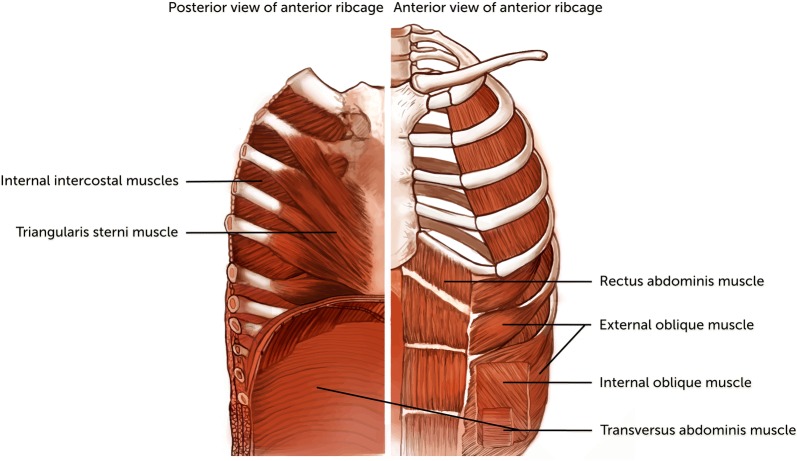
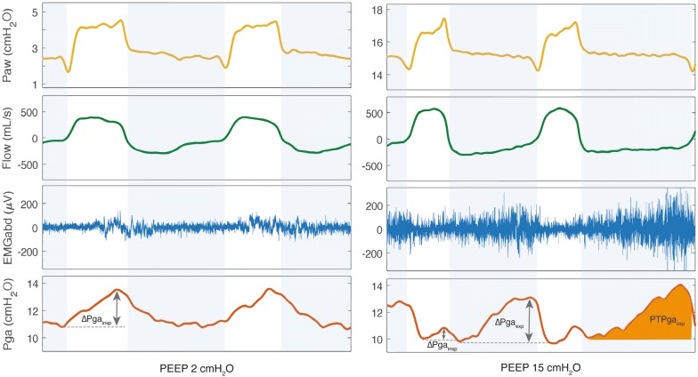
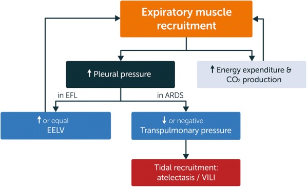
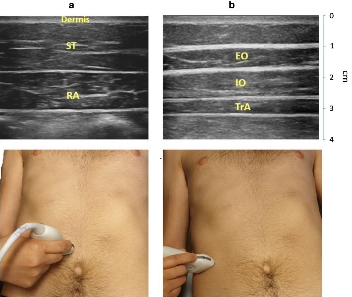
Results: The expiratory muscles, which include the abdominal wall muscles and some of the rib cage muscles, are an important component of the respiratory muscle pump and are recruited in the presence of high respiratory load or low inspiratory muscle capacity. Recruitment of the expiratory muscles may have beneficial effects, including reduction in end-expiratory lung volume, reduction in transpulmonary pressure and increased inspiratory muscle capacity. However, severe weakness of the expiratory muscles may develop in ICU patients and is associated with worse outcomes, including difficult ventilator weaning and impaired airway clearance. Several techniques are available to assess expiratory muscle function in the critically ill patient, including gastric pressure and ultrasound.

Conclusion: The expiratory muscles are the "neglected component" of the respiratory muscle pump. Expiratory muscles are frequently recruited in critically ill ventilated patients, but a fundamental understanding of expiratory muscle function is still lacking in these patients.

Keywords: Acute respiratory failure; Expiratory muscles; Mechanical ventilation; Respiratory muscle monitoring; Respiratory muscle weakness.


Figures

Fig. 1
Fig. 1
The expiratory muscles of the respiratory muscle pump. The respiratory muscle pump is a complex organ that involves a large number of muscles that contribute to inspiration or expiration. This figure schematically demonstrates the expiratory muscles. With the exception of the diaphragm, other inspiratory muscles are not shown
Fig. 2
Fig. 2
Physiology of expiratory muscle recruitment. Schematic illustration of the causes and consequences of expiratory muscle recruitment under physiological (healthy) conditions. All the consequences of expiratory muscle recruitment occur during expiration, except for the increased inspiratory muscle capacity (which occurs during the subsequent inspiration). See main text for explanation. EELV end-expiratory lung volume, PEEPi intrinsic positive end-expiratory pressure, PEEPe external positive end-expiratory pressure
Fig. 3
Fig. 3
Activation of the abdominal muscles during high PEEP. Tracing of airway pressure (Paw), flow, EMG of the abdominal muscles (EMGabd) and gastric pressure (Pga) obtained from a healthy subject during non-invasive ventilation with PEEP levels of 2 cmH2O (left) and 15 cmH2O (right). At 2 cmH2O of PEEP there is no evidence of activation of the abdominal wall muscles (no EMGabd activity during expiration and no rise in Pga during expiration), however at 15 cmH2O of PEEP, the abdominal muscles are recruited during the expiratory phase, as shown by the presence of EMGabd activity during expiration and the rise in Pga during expiration. White column: inspiration; blue column: expiration. In the Pga tracing obtained during PEEP 15 cmH2O calculation of parameters to estimate expiratory muscle activity are shown: increase in gastric pressure during expiration (ΔPgaexp); and the gastric pressure–time product during expiration (PTPgaexp) represented by the orange area. EMGabd electromyography of abdominal wall muscles, Paw airway pressure, PEEP positive end-expiratory pressure, Pga gastric pressure, PTPgaexp gastric pressure-time product during expiration
Fig. 4
Fig. 4
Pathophysiology of expiratory muscle recruitment. Schematic illustration of the pathophysiological consequences of expiratory muscle recruitment in critically ill patients. The depicted relationships are mostly hypothetical due to the low number of studies on expiratory muscle function in ICU patients. The elevated pleural pressure caused by expiratory muscle recruitment might lead to dynamic airway collapse, especially in patients who already have expiratory flow limitation (EFL). This leads to an equal or increased end-expiratory lung volume (EELV). On the other hand, elevated pleural pressure might lead to negative expiratory transpulmonary pressures, especially in diseases with an increased lung elastance such as in ARDS, which in turn leads to atelectasis and tidal recruitment. EFL expiratory flow limitation, ARDS acute respiratory distress syndrome, VILI ventilator-induced lung injury
Fig. 5
Fig. 5
Role of expiratory muscle recruitment in the development of expiratory flow limitation (EFL). Schematic and simplified illustration demonstrating the role of expiratory muscle activation in EFL. ac With activation of the expiratory muscles the abdominal pressure increases, also increasing pleural pressure during expiration. This decreases the transluminal pressure resulting in partial airway collapse and therefore EFL. With higher expiratory muscle pressure the flow-limiting site, or choking point, moves towards the alveoli. Note that gravitational forces are not considered in this illustration. Pab abdominal pressure, Palv alveolar pressure, Pao airway opening pressure, Ppl pleural pressure
Fig. 6
Fig. 6
Ultrasound image of the abdominal muscles. Left: ultrasound image of the rectus abdominis muscle (RA) (top), obtained with the probe placed 2–3 cm above the umbilicus and 2–3 cm from the midline (bottom). Right: ultrasound image of the external oblique muscle (EO), internal oblique muscle (IO) and transversus abdominis muscle (TrA) (top), obtained with the probe placed midway between the costal margin and the iliac crest, along the anterior axillary line (bottom)

References

    1. De Troyer A, Boriek AM. Mechanics of the respiratory muscles. Compr Physiol. 2011;1:1273–1300. - PubMed
    1. Dres M, Goligher EC, Heunks LMA, Brochard LJ. Critical illness-associated diaphragm weakness. Intensive Care Med. 2017;43:1441–1452. doi: 10.1007/s00134-017-4928-4. - DOI - PubMed
    1. Goligher EC, Fan E, Herridge MS, Murray A, Vorona S, Brace D, Rittayamai N, Lanys A, Tomlinson G, Singh JM, Bolz SS, Rubenfeld GD, Kavanagh BP, Brochard LJ, Ferguson ND. Evolution of diaphragm thickness during mechanical ventilation. impact of inspiratory effort. Am J Respir Crit Care Med. 2015;192:1080–1088. doi: 10.1164/rccm.201503-0620OC. - DOI - PubMed
    1. Levine S, Nguyen T, Taylor N, Friscia ME, Budak MT, Rothenberg P, Zhu J, Sachdeva R, Sonnad S, Kaiser LR, Rubinstein NA, Powers SK, Shrager JB. Rapid disuse atrophy of diaphragm fibers in mechanically ventilated humans. N Engl J Med. 2008;358:1327–1335. doi: 10.1056/NEJMoa070447. - DOI - PubMed
    1. Jaber S, Petrof BJ, Jung B, Chanques G, Berthet JP, Rabuel C, Bouyabrine H, Courouble P, Koechlin-Ramonatxo C, Sebbane M, Similowski T, Scheuermann V, Mebazaa A, Capdevila X, Mornet D, Mercier J, Lacampagne A, Philips A, Matecki S. Rapidly progressive diaphragmatic weakness and injury during mechanical ventilation in humans. Am J Respir Crit Care Med. 2011;183:364–371. doi: 10.1164/rccm.201004-0670OC. - DOI - PubMed

MeSH terms