Review
. 2020 Feb;33(1):99-110.
doi: 10.1007/s10278-019-00239-7.

Comprehensive Review of 3D Segmentation Software Tools for MRI Usable for Pelvic Surgery Planning

Affiliations
Review

Comprehensive Review of 3D Segmentation Software Tools for MRI Usable for Pelvic Surgery Planning

Alessio Virzì et al. J Digit Imaging. 2020 Feb.

Abstract

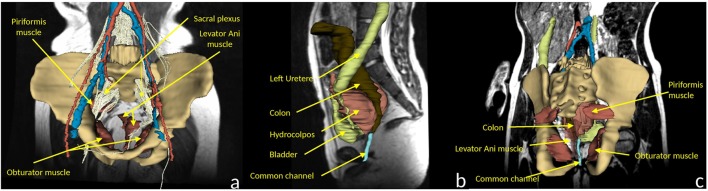
Patient-specific 3D modeling is the first step towards image-guided surgery, the actual revolution in surgical care. Pediatric and adolescent patients with rare tumors and malformations should highly benefit from these latest technological innovations, allowing personalized tailored surgery. This study focused on the pelvic region, located at the crossroads of the urinary, digestive, and genital channels with important vascular and nervous structures. The aim of this study was to evaluate the performances of different software tools to obtain patient-specific 3D models, through segmentation of magnetic resonance images (MRI), the reference for pediatric pelvis examination. Twelve software tools freely available on the Internet and two commercial software tools were evaluated using T2-w MRI and diffusion-weighted MRI images. The software tools were rated according to eight criteria, evaluated by three different users: automatization degree, segmentation time, usability, 3D visualization, presence of image registration tools, tractography tools, supported OS, and potential extension (i.e., plugins). A ranking of software tools for 3D modeling of MRI medical images, according to the set of predefined criteria, was given. This ranking allowed us to elaborate guidelines for the choice of software tools for pelvic surgical planning in pediatric patients. The best-ranked software tools were Myrian Studio, ITK-SNAP, and 3D Slicer, the latter being especially appropriate if nerve fibers should be included in the 3D patient model. To conclude, this study proposed a comprehensive review of software tools for 3D modeling of the pelvis according to a set of eight criteria and delivered specific conclusions for pediatric and adolescent patients that can be directly applied to clinical practice.

Keywords: 3D modeling; MRI; Pelvic surgery; Segmentation software.


Figures

Fig. 1
Fig. 1
Example of (a) coronal, (b) sagittal, and (c) axial slices of one of the 3D T2-w MRI used in this study
Fig. 2
Fig. 2
Example of 3D pelvic reconstructions obtained through segmentation of the T2-w MRI volume, using (a) 3D Slicer, (b) ITK-SNAP, and (c) Myrian Studio
Fig. 3
Fig. 3
Example of organ segmentations in a few MRI coronal slices. From top to bottom: (a) rectum, (b) bones, (c) bladder, (d) veins (blue) and arteries (red), (e) prostate, and (f) uterus
Fig. 4
Fig. 4
3D modeling of a 7-year-old patient with a rhabdomyosarcoma of the right obturator muscle
Fig. 5
Fig. 5
3D modeling of a 9-year-old patient with a non-repaired cloacal malformation (with a colostomy), a sacral agenesia, and no dysraphism. a Coronal view integrating the sacral tractogram. Note the asymmetry of levator ani muscles. b Left sagittal view. Note the compression by the hydrocolpos on the left ureter. c Coronal posterior view, showing the muscular system and the common channel in PSARP position

References

    1. Lo Presti G, Carbone M, Ciriaci D, Aramini D, Ferrari M, Ferrari V. Assessment of DICOM viewers capable of loading patient-specific 3D models obtained by different segmentation platforms in the operating room. J Digit Imaging. 2015;28(5):518–527. doi: 10.1007/s10278-015-9786-4. - DOI - PMC - PubMed
    1. Ferrari V, Carbone M, Cappelli C, Boni L, Melfi F, Ferrari M, Mosca F, Pietrabissa A. Value of multidetector computed tomography image segmentation for preoperative planning in general surgery. Surg Endosc. 2012;26(3):616–626. doi: 10.1007/s00464-011-1920-x. - DOI - PMC - PubMed
    1. Azagury DE, Dua MM, Barrese JC, Henderson JM, Buchs NC, Ris F, Cloyd JM, Martinie JB, Razzaque S, Nicolau S, Soler L, Marescaux J, Visser BC. Image-guided surgery. Curr Probl Surg. 2015;52(12):476–520. doi: 10.1067/j.cpsurg.2015.10.001. - DOI - PubMed
    1. Haak D, Page C-E, Deserno TM. A survey of DICOM viewer software to integrate clinical research and medical imaging. J Digit Imaging. 2016;29(2):206–215. doi: 10.1007/s10278-015-9833-1. - DOI - PMC - PubMed
    1. Valeri G, Mazza FA, Maggi S, Aramini D, La Riccia L, Mazzoni G, Giovagnoni A. Open source software in a practical approach for post processing of radiologic images. Radiol Med. 2015;120(3):309–323. doi: 10.1007/s11547-014-0437-5. - DOI - PubMed

LinkOut - more resources