. 2019 Sep;12(5):1003-1013.
doi: 10.1111/1751-7915.13455. Epub 2019 Jun 25.

Cell density-dependent auto-inducible promoters for expression of recombinant proteins in Pseudomonas putida

Affiliations

Cell density-dependent auto-inducible promoters for expression of recombinant proteins in Pseudomonas putida

Annika Meyers et al. Microb Biotechnol. 2019 Sep.

Abstract

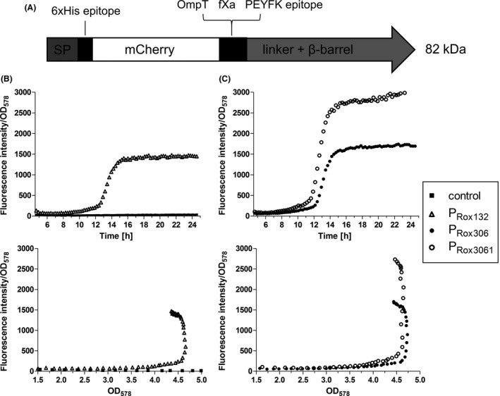
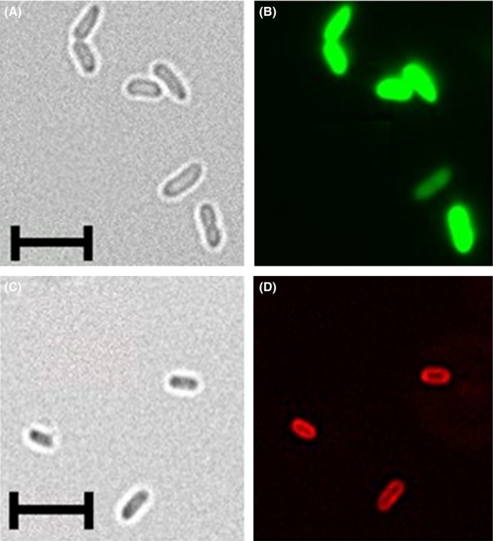
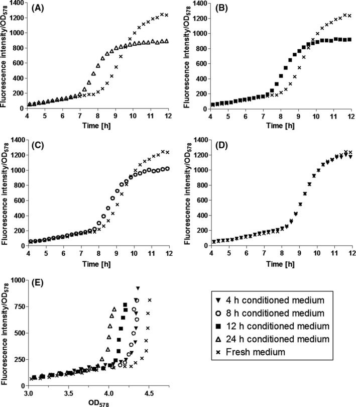
Inducible promoters such as Plac are of limited usability for industrial protein production with Pseudomonas putida. We therefore utilized cell density-dependent auto-inducible promoters for recombinant gene expression in P. putida KT2440 based on the RoxS/RoxR Quorum Sensing (QS) system of the bacterium. To this end, genetic regions upstream of the RoxS/RoxR-regulated genes ddcA (PR ox132 ) and PP_3332 (PR ox306 ) were inserted into plasmids that mediated the expression of superfolder green fluorescent protein (sfGFP) and surface displayed mCherry, confirming their promoter functionalities. Mutation of the Pribnow box of PR ox306 to the σ70 consensus sequence (PR ox3061 ) resulted in a more than threefold increase of sfGFP production. All three promoters caused cell density-dependent expression, starting transcription at optical densities (OD578 ) of approximately 1.0 (PR ox132 , PR ox306 ) or 0.7 (PR ox3061 ) as determined by RT-qPCR. The QS dependency of PR ox306 was further shown by cultivating P. putida in media that had already been used for cultivation and thus contained bacterial signal molecules. The longer P. putida had grown in these media before, the earlier protein expression in freshly inoculated P. putida appeared with PR ox306 . This confirmed previous findings that a bacterial compound accumulates within the culture and induces protein expression.


Conflict of interest statement

None declared.

Figures

Figure 1
Figure 1
A. Genetic origins of PRox promoters. Top panel: PRox132 is part of the upstream genetic region of ddcA (light grey). It starts with the RoxR recognition element (Rox RE), the regulatory regions (−35 and −10) and a ribosome binding site (RBS), which is located in the 5′ untranslated region (white). Bottom panel: PRox306 consists of the whole intergenic region between P. putida KT2440 genes PP_3332 and PP_3331. B. DNA sequences of PRox132, PRox306 and PRox3061. The −35 and −10 regions predicted by BPROM (Solovyev and Salamov, 2011) are depicted in bold. Experimentally determined transcriptional start sites are marked with + 1. In PRox3061, the −10 region was mutated to the consensus sequence of the major σ‐factor of the σ70 family, TATAAT. The initiation codon of the regulated gene is underlined. The sequence of the Rox RE is shaded, the putative RBS sequence depicted in italics.
Figure 2
Figure 2
Expression of sfGFP by P. putida (A) under control of PRox132, PRox306 and PRox3061 and (B) under control of the constitutive promoter PGAP and the inducible promoter PBAD. C. Comparison of sfGFP expression under control of PRox3061 in LB and MOPS medium. D. Quantitative comparison of sfGFP accumulation within the cells with different promoters. P. putida cells containing plasmids for expression of sfGFP under control of the different promoters were cultivated in 500 ml shake flasks in LB or MOPS medium at 30°C, and OD 578 and fluorescence intensity (FI, excitation: 485 nm, emission: 510 nm) were monitored. After reaching stationary phase, the cells were lysed, their proteins separated by SDSPAGE and sfGFP detected by means of anti‐sfGFP and secondary HRP‐conjugated antibodies. The detected band intensities were quantified and provided relative to the band intensity of PRox3061. The data are derived from one representative experiment, with the mean of three technical replicates shown. The error bars represent the standard deviation.
Figure 3
Figure 3
A. Scheme of the unprocessed MATEmCherry fusion protein. After cleavage of the signal peptide (SP), the protein consists of a 6xHis epitope, the passenger mCherry, OmpT and fXa cleavage sites, a PEYFK epitope and the EhaA linker and β‐barrel (Sichwart et al., 2015). B,C. Expression of MATEmCherry under control of PRox132, PRox306 and PRox3061. P. putida without plasmid and with plasmid pPRox132MATEmCherry (B), pPRox306MATEmCherry and pPRox3061MATEmCherry (C) were cultivated in LB medium in a 24‐well MTP at 30°C. OD 578 and fluorescence intensity (FI, excitation: 580 nm, emission: 620 nm) were monitored. These data are mean values of biological triplicates. Error bars are not visible due to small standard deviations that are covered by the icons.
Figure 4
Figure 4
Analysis of P. putida pPRox3061‐sfGFP (A,B), and pPRox132MATEmCherry (C,D) via fluorescence microscopy. The strains were cultivated at 30°C, 200 rpm in LB medium for 8 h and 24 h respectively. 2.5 × 106 cells were washed three times with PBS and fixed on a microscope slide with DABCO/Mowiol. The samples were analysed with the 100× oil immersion lens of a BZ‐9000 fluorescence microscope (Keyence, Neu‐Isenburg, Germany). A,C. Brightfield pictures. (B) GFP‐filter, excitation 472/30 nm, emmission 593/40 nm. D. TexasRed‐filter, excitation 560/40 nm, emmission 630/75 nm. The length of the scales corresponds to 5 μm.
Figure 5
Figure 5
RTqPCR analysis of MATEmCherry expression under control of PBG35 (A), PRox132 (B), PRox306 (C) and PRox3061 (D). P. putida cells containing plasmids for expression of MATEmCherry under control of different promoters were cultivated in LB medium at 30°C, 200 rpm. At different time points, 2.5 × 108 cells were removed from the cultures and the total amount of RNA was isolated. 1000 ng of total RNA was reversely transcribed to cDNA. 50 ng cDNA was amplified and analysed with specific primers in a qPCR cycler. The gene expression of MATEmCherry was determined relative to the gene expression of the reference gene rpoD (Fujita et al., 1995). The measured Cq values were analysed by applying a model of Pfaffl (2001). All RTqPCR analyses were performed as biological triplicates, each of them conducted as technical triplicates. Error bars indicate the standard deviation.
Figure 6
Figure 6
Influence of conditioned medium (CM) on expression of MATEmCherry. P. putida pPRox306MATE‐mCherry was cultivated in LB medium to an OD 578 = 0.8. Five millilitres of the cell suspension were harvested and suspended in mixtures of 4 ml fresh LB and 1 ml of different CM. The cells were then cultivated in a 24‐well MTP for another 12 h while monitoring OD 578 and fluorescence intensity (FI). A. 24 h CM, (B) 12 h CM, (C) 8 h CM, (D) 4 h CM. These data are mean values of biological triplicates. Error bars are not visible due to small standard deviations that are covered by the icons.

References

    1. Anderson, J.C. , Clarke, E.J. , Arkin, A.P. , and Voigt, C.A. (2006) Environmentally controlled invasion of cancer cells by engineered bacteria. J Mol Biol 355: 619–627. - PubMed
    1. Briand, L. , Marcion, G. , Kriznik, A. , Heydel, J.M. , Artur, Y. , Garrido, C. , et al (2016) A self‐inducible heterologous protein expression system in Escherichia coli . Sci Rep 6: 33037. - PMC - PubMed
    1. Cao, L. , Wang, Q. , Zhang, J. , Li, C. , Yan, X. , Lou, X. , et al (2012) Construction of a stable genetically engineered rhamnolipid‐producing microorganism for remediation of pyrene‐contaminated soil. World J Microb Biot 28: 2783–2790. - PubMed
    1. Cha, M. , Lee, N. , Kim, M. , Kim, M. , and Lee, S. (2008) Heterologous production of Pseudomonas aeruginosa EMS1 biosurfactant in Pseudomonas putida . Bioresource Technol 99: 2192–2199. - PubMed
    1. Conway, T. , Sewell, G.W. , and Ingram, L.O. (1987) Glyceraldehyde‐3‐phosphate dehydrogenase gene from Zymomonas mobilis: cloning, sequencing, and identification of promoter region. J Bacteriol 169: 5653–5662. - PMC - PubMed

Publication types

MeSH terms

LinkOut - more resources