. 2019;8(1):946-958.
doi: 10.1080/22221751.2019.1632676.

Immunization with a recombinant antigen composed of conserved blocks from TSA56 provides broad genotype protection against scrub typhus

Affiliations

Immunization with a recombinant antigen composed of conserved blocks from TSA56 provides broad genotype protection against scrub typhus

Hong-Il Kim et al. Emerg Microbes Infect. 2019.

Abstract

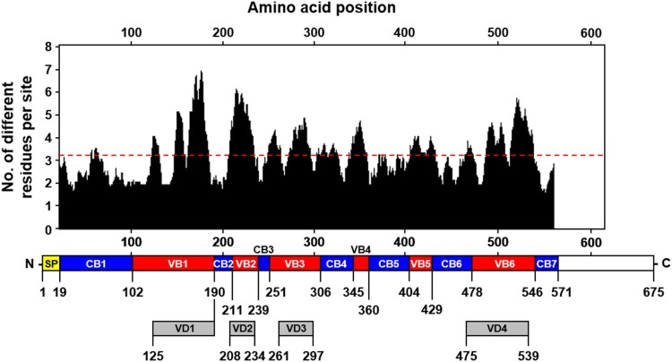
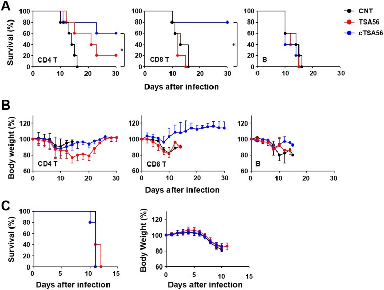
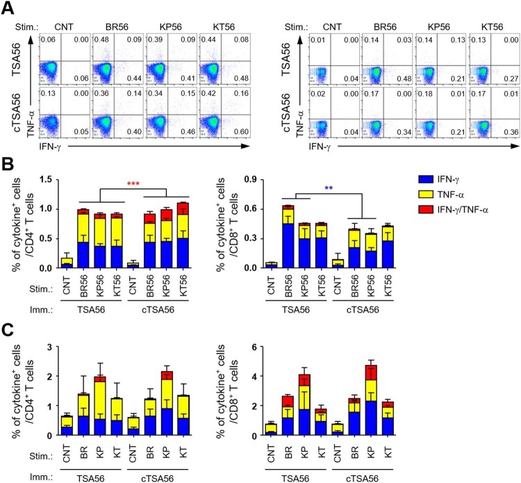
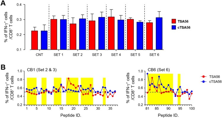
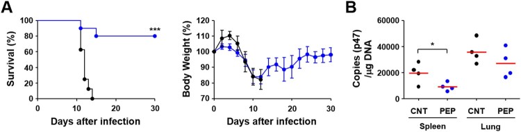
Scrub typhus is an acute febrile disease caused by Orientia tsutsugamushi infection. Despite the wide range of approaches explored during the last seventy years, an effective prophylactic vaccine is not yet available. Here, we developed a novel recombinant antigen derived from conserved regions of 56 kDa type-specific antigen (TSA56), a major outer membrane protein responsible for genetic heterogeneity and antigenicity, and evaluated it as a protective vaccine antigen. Our findings demonstrate that immunization with conserved blocks of TSA56 (cTSA56) not only provides protective immunity against lethal challenges with the homologous genotype, but also confers significantly better protection against heterologous genotypes than TSA56. Adoptive transfer of CD4+ or CD8+ T cells from immunized mice provided significantly enhanced protection against lethal challenge, whereas immune B cells failed to do so, indicating that cellular immunity against the conserved epitopes plays a protective role. Moreover, immunization with a 10-mer peptide mixture, screened from CD8+ T cell epitopes within the conserved region of TSA56, provided enhanced protection against lethal challenge with O. tsutsugamushi. Therefore, this novel recombinant antigen is a promising candidate for scrub typhus vaccine against a wide range of O. tsutsugamushi genotypes.

Keywords: CD8 T cell; Scrub typhus; TSA56; conserved blocks; vaccine.


Figures

Figure 1.
Figure 1.
Characterization of conserved and variable blocks in aligned TSA56 sequences. The upper plot shows the moving mean, for a window of 10 amino acid residues, of the number of different amino acids observed at a position in the amino acid alignment using 206 tsa56 genes (list available at ref. 3). The overall average of sequence variation is noted by the red dashed line. The location of conserved (CB1 ∼ 7, blue) and variable blocks (VB1 ∼ 6, red) annotated in this study and the variable domains (VD1 ∼ 4, grey) defined in a previous work (ref. 12) is shown below the graph. SP, signal peptide.
Figure 2.
Figure 2.
Pairwise comparison of sequence similarity and phylogenetic distance of CBs and VBs in TSA56 sequences. Amino acid similarity of each pair of blocks in TSA56 sequences was measured using MatGAT 2.1. Cophenetic distance, which represents phylogenetic distance between two sequences, was calculated by Ape-package. A pair of sequences, represented by each dot, belonging to different genotypes was chosen randomly for 1000 times and plotted.
Figure 3.
Figure 3.
Characterization of purified TSA56 and cTSA56 antigen. (A) Schematic representation of the recombinant TSA56 and cTSA56 used in this study. The amino acid positions of conserved (CB, blue) and variable (VB, red) blocks were indicated. SP: signal peptide. His: 6 × histidine tag. (B) Purified TSA56 and cTSA56 were was resolved by SDS-PAGE, stained with Coomassie blue (left panel), and immunoblotted with anti-His tag antibody (middle panel) or antisera collected from mice recovered from infection with O. tsutsugamushi Boryong genotype (right panel). (C) Antibody titres against the indicated antigens were measured by ELISA. Mice (n = 3/group) were immunized with TSA56 or cTSA56 three times at two week intervals and sera were collected at one week after the third immunization. (D) IgG2c and IgG1 ratio of the immune sera. Error bar, mean ± S.D.
Figure 4.
Figure 4.
Protective role of cTSA56 against heterologous genotype infection. Mice (n = 5/group) were immunized with the indicated antigens and challenged i.p. with 100 × LD50 of O. tsutsugamushi. Mice were immunized with antigens from the Boryong genotype and challenged with the indicated genotypes. Survival rate (A) and body weight change (B) of mice were observed for 30 d after infection. CNT, mock-immunized; *p < .05; **p < .01. (C) Bacterial loads in the spleens of mice (n = 4/group) infected with Boryong or Kato genotype were assessed by qRT-PCR using primer sets detecting the p47 gene of O. tsutsugamushi. The infected tissues were collected at 7 days after infection. Black circle, mock-immunized; red circle, TSA56-immunized; blue circle, cTSA56-immunized. Red line, mean; *p < .05; **p < .01.
Figure 5.
Figure 5.
Protective role of immune T cells against heterologous Karp genotype infection. Survival rate (A) and body weight change (B) of mice receiving adoptively transferred indicated populations of splenocytes by i.v. were assessed after challenge with 100 × LD50 of O. tsutsugamushi Karp genotype. Indicated immune cells purified from splenocytes of mock-immunized (CNT, black circles) or antigen-vaccinated mice (red, TSA56; blue, cTSA56) were transferred to naïve mice (n = 5/group) at one day before the lethal challenge. *p < .05; error bar, mean ± S.D. (C) Pooled sera collected from naïve mice (black circle) or mice immunized with TSA56 (red circle) or cTSA56 (blue circle) were injected to naïve recipient mice (150 μl/mouse) by i.p. and then, they were challenged i.p. with a lethal dose (100 × LD50) of the O. tsutsugamushi Karp genotype. Survival rate (left panel) and body weight change (right panel) of mice receiving pooled sera were assessed.
Figure 6.
Figure 6.
Comparison of antigen-specific T cell responses in mice immunized with TSA56 or cTSA56. (A) Splenocytes were collected from mice at two weeks after the third immunization with the indicated antigen and production of IFN-γ and/or TNF-α by CD4+ T (left panels) or CD8+ T (right panels) cells were analysed by flow cytometry after stimulation with the indicated antigen (CNT, unstimulated; BR56, TSA56 from Boryong; KP56, TSA56 from Karp; KT56, TSA56 from Kato genotype). Representative flow cytometric results are presented. (B) The percentile of cytokine positive cells among CD4+ or CD8+ T cell subsets are summarized. Stim., stimulated antigen (the same as in A); Imm., Immunized antigen. Data represent mean + S.D. from duplicate assays with three mice per group. **p < .01; ***p < .001. (C) Splenocytes were collected from mice infected with live O. tsutsugamushi (4 m.o.i.) of the indicated genotype (CNT, uninfected; BR, Boryong; KP, Karp; KT, Kato) for 4 h and further incubated in the presence of tetracycline for one day. Then, cells were analysed by flow cytometry. The percentile of cytokine positive cells among CD4+ or CD8+ T cell subsets are summarized. Stim., stimulated antigen; Imm., Immunized antigen. Data represent mean + S.D. from duplicate assays with three mice per group. Blue box, IFN-γ-positive; yellow box, TNF-α-positve; red box, IFN-γ and TNF-α-positive.
Figure 7.
Figure 7.
Screening of CD8+ T cell epitopes using a 10-mer peptide library derived from CBs of TSA56. (A) Splenocytes were collected from mice at two weeks after the third immunization with the indicated antigen and production of IFN-γ in CD8+ T cells were analysed by flow cytometry after stimulation with the indicated sets of peptides. Information of peptide sequences and peptide sets is summarized in Supplementary Table 4. The percentile of cytokine positive cells among CD8+ T cells are summarized. Data represent mean + S.D. from duplicate assays with three mice per group. CNT, unstimulated. (B) Immune splenocytes were stimulated with the indicated peptide and production of IFN-γ in CD8+ T cells were analysed by flow cytometry. The percentile of cytokine positive cells among CD8+ T cells are summarized. Dashed line indicates the baseline frequency (mean + 3 × S.D.) of unstimulated controls in three sets of immune splenocytes. Overlapping peptides showing higher levels of IFN-γ positive CD8+ T cells than the baseline frequency are highlighted in yellow.
Figure 8.
Figure 8.
Protection against O. tsutsugamushi Karp infection in mice immunized with CD8+ T cell epitope peptides. (A) Survival rate (upper) and body weight change (lower) of mice (n = 5/group) immunized with 39 peptides selected in Figure 5. Mice were immunized three times at two week intervals and challenged i.v. with 100 × LD50 of O. tsutsugamushi Karp genotype. ***p < .001. (B) Bacterial loads in the spleens and lungs of infected mice (n = 4/group) were assessed by qRT-PCR using primer sets detecting the p47 gene of O. tsutsugamushi. The infected tissues were collected at 7 days after infection. Black circle, mock-immunized; blue circle, peptide-immunized. Red line, mean; *p < .05.

Similar articles

Cited by

References

    1. Diaz FE, Abarca K, Kalergis AM.. An update on host-pathogen interplay and modulation of immune responses during Orientia tsutsugamushi infection. Clin Microbiol Rev. 2018 Apr;31(2). doi: 10.1128/CMR.00076-17 - DOI - PMC - PubMed
    1. Taylor AJ, Paris DH, Newton PN.. A systematic review of mortality from untreated scrub typhus (Orientia tsutsugamushi). PLoS Negl Trop Dis. 2015 Aug;9(8):e0003971. doi: 10.1371/journal.pntd.0003971 - DOI - PMC - PubMed
    1. Kim G, Ha NY, Min CK, et al. . Diversification of Orientia tsutsugamushi genotypes by intragenic recombination and their potential expansion in endemic areas. Plos Neglect Trop D. 2017 Mar;11(3). doi: 10.1371/journal.pntd.0005408 - DOI - PMC - PubMed
    1. Walker DH.Scrub typhus – scientific neglect, ever-widening impact. New Engl J Med. 2016 Sep 8;375(10):913–915. doi: 10.1056/NEJMp1608499 - DOI - PubMed
    1. Valbuena G, Walker DH.. Approaches to vaccines against Orientia tsutsugamushi. Front Cell Infect Microbiol. 2013;2:170. doi: 10.3389/fcimb.2012.00170 - DOI - PMC - PubMed

MeSH terms

Substances