Review
. 2019 Jun 24;20(12):3076.
doi: 10.3390/ijms20123076.

Role of Hedgehog Signaling in Vasculature Development, Differentiation, and Maintenance

Affiliations
Review

Role of Hedgehog Signaling in Vasculature Development, Differentiation, and Maintenance

Candice Chapouly et al. Int J Mol Sci. .

Abstract

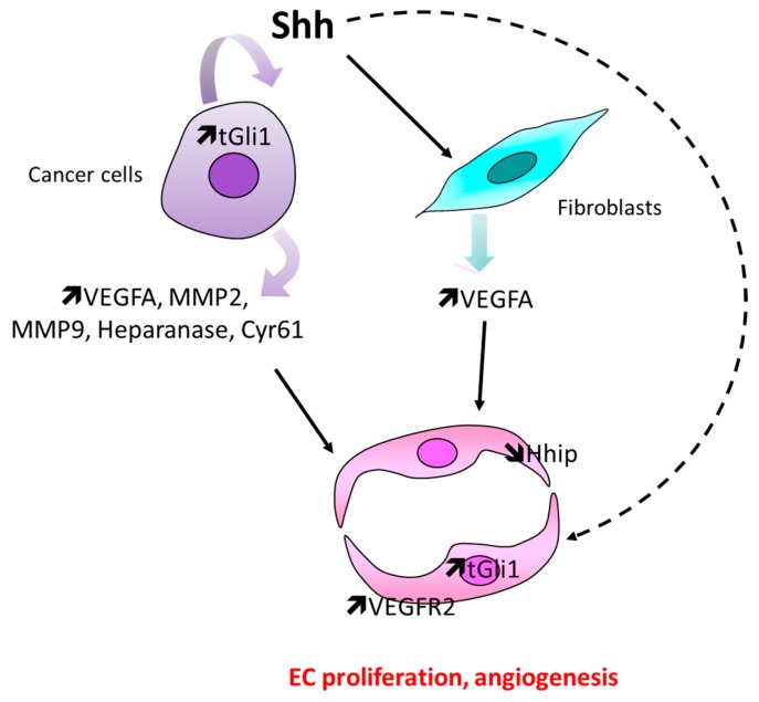
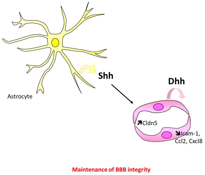
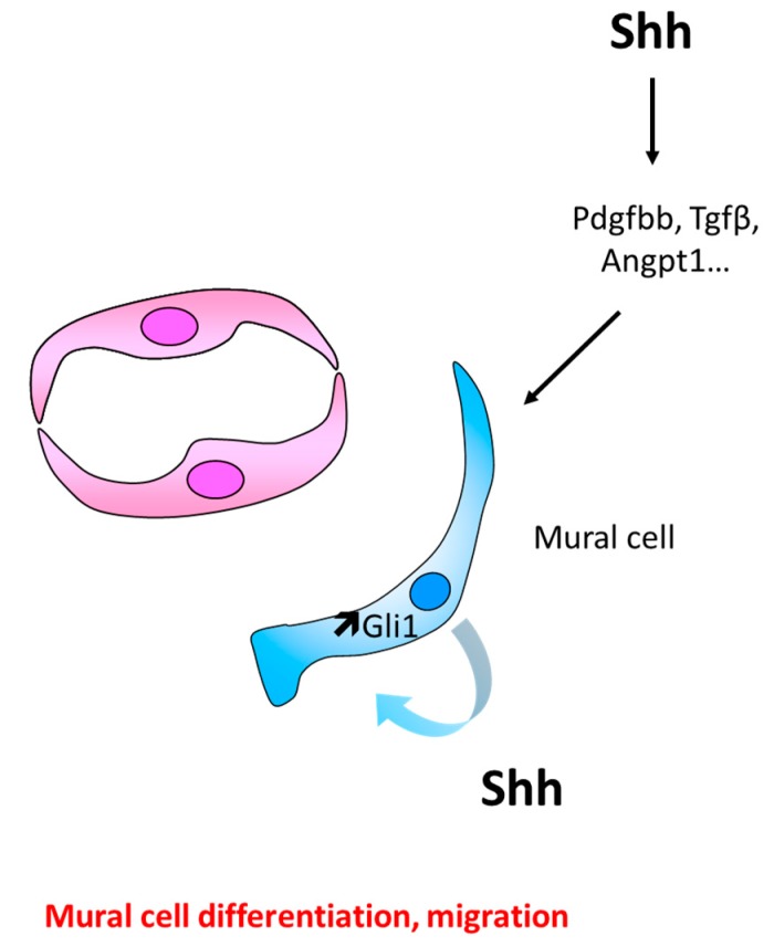
The role of Hedgehog (Hh) signaling in vascular biology has first been highlighted in embryos by Pepicelli et al. in 1998 and Rowitch et al. in 1999. Since then, the proangiogenic role of the Hh ligands has been confirmed in adults, especially under pathologic conditions. More recently, the Hh signaling has been proposed to improve vascular integrity especially at the blood-brain barrier (BBB). However, molecular and cellular mechanisms underlying the role of the Hh signaling in vascular biology remain poorly understood and conflicting results have been reported. As a matter of fact, in several settings, it is currently not clear whether Hh ligands promote vessel integrity and quiescence or destabilize vessels to promote angiogenesis. The present review relates the current knowledge regarding the role of the Hh signaling in vasculature development, maturation and maintenance, discusses the underlying proposed mechanisms and highlights controversial data which may serve as a guideline for future research. Most importantly, fully understanding such mechanisms is critical for the development of safe and efficient therapies to target the Hh signaling in both cancer and cardiovascular/cerebrovascular diseases.

Keywords: Hedgehog; angiogenesis; blood–brain barrier; endothelium; vasculogenesis.


Conflict of interest statement

The authors declare no conflicts of interest.

Figures

Figure 1
Figure 1
Shh post-transcriptional modification and secretion. Shh is synthetized as a full-length, 45 kDa protein. An autocatalytic reaction removes the carboxy-terminal domain and attaches a cholesterol moiety to the newly exposed carboxy-terminus. Then, Hhat catalyzes the addition of a palmitate to the amino-terminus [23]. Secretion and solubility of Shh depends on Disp1 and Scube2.
Figure 2
Figure 2
(A) Hh canonical signaling. In the absence of Hh ligands, Smo is inhibited by Ptch1 and Gli transcription factors are associated with SUFU negative regulator of hedgehog signaling (Sufu) and kinesin family member 7 (Kif7). This last complex promotes Gli3 and Gli2 phosphorylation by cAMP dependent protein kinase (PKA), casein kinase 1 (CK1), and glycogen synthase kinase 3 beta (GSK3. Once phosphorylated, Gli2 and Gli3 are processed by speckle type BTB/POZ protein (Spop)/cullin 3 (Cul3) ubiquitin ligase complex to generate Gli2R and Gli3R (repressor forms) respectively. Hh ligands binding to Ptch1 leads to Smo activation, which prevents Gli2 and Gli3 cleavage. Full-length Gli2 and Gli3 may then translocate to the nucleus and activates transcription. (B) Hh noncanonical signaling. Hh binding to Ptch1 or Cdon may, independently on Smo, promote cell survival or proliferation by modulating Caspase 9 (Casp9) or Cyclin D1 (CcnD1) activity, respectively. This is what is called type I noncanonical signaling. Alternatively, Hh ligands may activate PI3K/Akt, RhoA/ROCK or AMPK, via Smo, but independently on Gli transcription factors. This is what is called type II noncanonical signaling.
Figure 3
Figure 3
Schema representing the main cellular events involved in Hh-induced vasculogenesis and primary vascular plexus remodeling. Hh ligands promote EC differentiation indirectly via BMP4 upregulation in mesenchymal cells, while vascular remodeling, i.e., branching and pericyte recruitment, depends on Vegfa and/or Angpt1.
Figure 4
Figure 4
Schema representing the main cellular events involved in Hh-induced arterial differentiation. Briefly, Shh produced by the notochord upregulates Vegfa in somites, which, in turn, increases Notch signaling in ECs, and subsequently promotes the expression of the arterial marker EphrinB2.
Figure 5
Figure 5
Schema representing the main cellular events underlying the proangiogenic effect of Shh therapy in the setting of ischemia. When administered ectopically in ischemic tissues, Shh promotes angiogenesis indirectly by upregulating Vegfa, Cxcl12, and Angpt1 in fibroblasts and by recruiting bone marrow-derived proangiogenic cells.
Figure 6
Figure 6
Schema representing the main cellular events involved in Hh-induced tumor angiogenesis. In tumors, Shh, which is mainly produced by cancer cells, may promote angiogenesis either by increasing proangiogenic factor expression in cancer cells themselves, by promoting Vegfa expression in stromal fibroblast, or by promoting EC proliferation directly through Gli1 upregulation.
Figure 7
Figure 7
Schema representing the main cellular events underlying Hh maintenance of BBB integrity. Activation of Hh signaling in brain ECs promotes thigh junction integrity by increasing Cldn5 expression and BBB immune quiescence by downregulating Icam1, Ccl2, and Cxcl8. Brain ECs may either respond to Dhh, which is produced by EC themselves in physiological conditions, or to Shh, which is produced by astrocytes in certain pathological conditions.
Figure 8
Figure 8
Schema representing the main cellular events involved in Hh-induced mural cell recruitment and differentiation. Shh may either promote mural cell differentiation and migration indirectly by upregulating Angpt1, Pdgfbb, or Tgfβ in unidentified cells or by upregulating Gli1 directly in ECs.

Similar articles

Cited by

References

    1. Nusslein-Volhard C., Wieschaus E. Mutations affecting segment number and polarity in Drosophila. Nature. 1980;287:795–801. doi: 10.1038/287795a0. - DOI - PubMed
    1. Bitgood M.J., McMahon A.P. Hedgehog and Bmp genes are coexpressed at many diverse sites of cell-cell interaction in the mouse embryo. Dev. Biol. 1995;172:126–138. doi: 10.1006/dbio.1995.0010. - DOI - PubMed
    1. Ramalho-Santos M., Melton D.A., McMahon A.P. Hedgehog signals regulate multiple aspects of gastrointestinal development. Development. 2000;127:2763–2772. - PubMed
    1. St-Jacques B., Hammerschmidt M., McMahon A.P. Indian hedgehog signaling regulates proliferation and differentiation of chondrocytes and is essential for bone formation. Genes Dev. 1999;13:2072–2086. doi: 10.1101/gad.13.16.2072. - DOI - PMC - PubMed
    1. Bitgood M.J., Shen L., McMahon A.P. Sertoli cell signaling by Desert hedgehog regulates the male germline. Curr. Biol. 1996;6:298–304. doi: 10.1016/S0960-9822(02)00480-3. - DOI - PubMed