

Photoacoustic/Ultrasound/Optical Coherence Tomography Evaluation of Melanoma Lesion and Healthy Skin in a Swine Model
- PMID: 31238540
- PMCID: PMC6630987
- DOI: 10.3390/s19122815
Photoacoustic/Ultrasound/Optical Coherence Tomography Evaluation of Melanoma Lesion and Healthy Skin in a Swine Model
Abstract
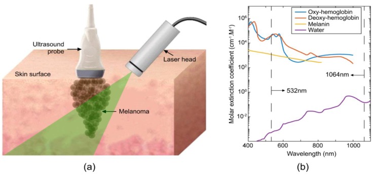
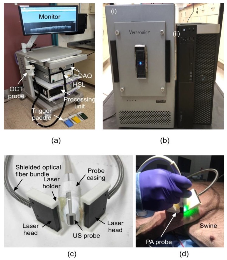
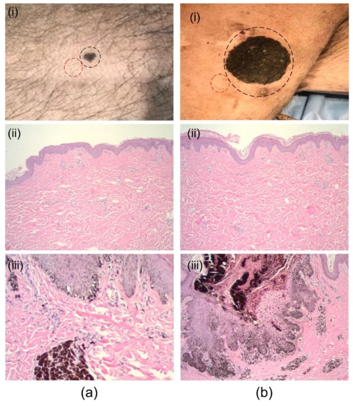
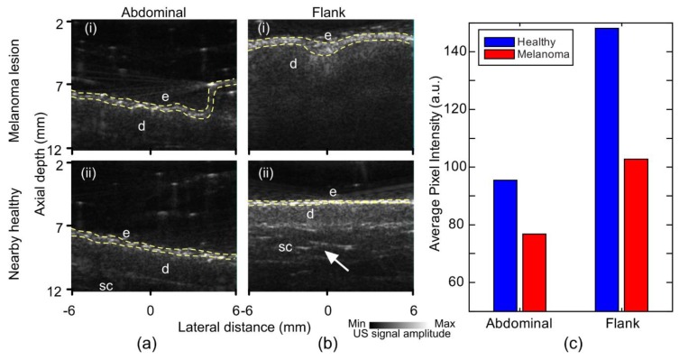
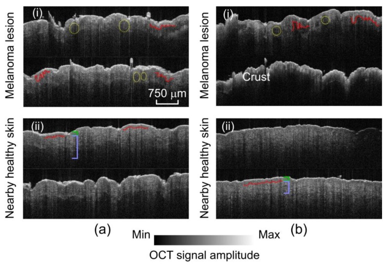
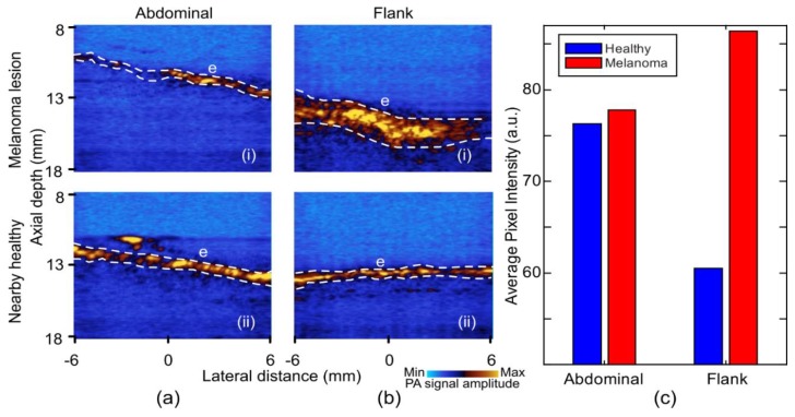
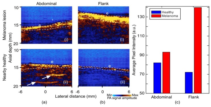
The marked increase in the incidence of melanoma coupled with the rapid drop in the survival rate after metastasis has promoted the investigation into improved diagnostic methods for melanoma. High-frequency ultrasound (US), optical coherence tomography (OCT), and photoacoustic imaging (PAI) are three potential modalities that can assist a dermatologist by providing extra information beyond dermoscopic features. In this study, we imaged a swine model with spontaneous melanoma using these modalities and compared the images with images of nearby healthy skin. Histology images were used for validation.
Keywords: cancer imaging; melanoma; optical coherence tomography; photoacoustic imaging; skin imaging; swine melanoma model; ultrasound.
Conflict of interest statement
The authors declare no conflict of interest.
Figures
References
-
- National Cancer Institute Surveillance, Epidemiology, and End Results Program . Cancer Stat Facts. National Institutes of Health; Washington, DC, USA: 2016. Cancer Stat Facts: Melanoma of the Skin.
-
- American Cancer Society . Cancer Facts & Figures 2018. American Cancer Society; Atlanta, GA, USA: 2018.
MeSH terms
Grants and funding
LinkOut - more resources
Full Text Sources
Medical
