. 2019 Jun 28;9(6):190066.
doi: 10.1098/rsob.190066. Epub 2019 Jun 26.

Structure of F1-ATPase from the obligate anaerobe Fusobacterium nucleatum

Affiliations

Structure of F1-ATPase from the obligate anaerobe Fusobacterium nucleatum

Jessica Petri et al. Open Biol. .

Abstract

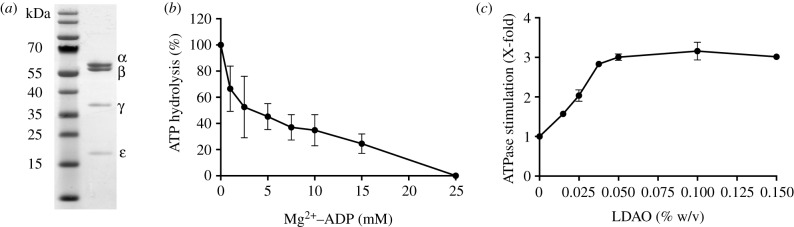
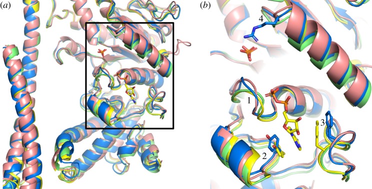
The crystal structure of the F1-catalytic domain of the adenosine triphosphate (ATP) synthase has been determined from the pathogenic anaerobic bacterium Fusobacterium nucleatum. The enzyme can hydrolyse ATP but is partially inhibited. The structure is similar to those of the F1-ATPases from Caldalkalibacillus thermarum, which is more strongly inhibited in ATP hydrolysis, and in Mycobacterium smegmatis, which has a very low ATP hydrolytic activity. The βE-subunits in all three enzymes are in the conventional 'open' state, and in the case of C. thermarum and M. smegmatis, they are occupied by an ADP and phosphate (or sulfate), but in F. nucleatum, the occupancy by ADP appears to be partial. It is likely that the hydrolytic activity of the F. nucleatum enzyme is regulated by the concentration of ADP, as in mitochondria.

Keywords: ATP hydrolysis; Fusobacterium nucleatum; catalytic F1-ATPase; pathogen; regulation; structure.


Conflict of interest statement

The authors declare no competing interests.

Figures

Figure 1.
Figure 1.
Characterization of F1-ATPase from F. nucleatum. (a) The subunits (1.75 µg of enzyme) were separated by SDS–PAGE and stained with Coomassie G-250 dye. Their identities (right-hand side) were verified by mass-mapping of tryptic peptides. The positions of molecular mass markers are indicated on the left. (b) The effect of ADP on ATP hydrolysis at an Mg2+ : ADP ratio of 2 : 1 was assayed by the release of inorganic phosphate. A specific activity of 3.5 U mg−1 was set to 100%. (c) The effect of LDAO on ATP hydrolysis was measured with an ATP-regenerating assay. A value of 1 corresponds to a specific activity of 3.8 U mg−1. Error bars represent the standard deviation of the mean from a biological triplicate. Where no error bars are shown, they are smaller than the diameter of the data points.
Figure 2.
Figure 2.
Structure of F1-ATPase from F. nucleatum. (a) Side view of the structure of molecule 1 in ribbon representation with the α-, β-, γ- and ɛ-subunits in red, yellow, blue and green, and bound nucleotides in a black space-filling representation. The green spheres represent Mg2+ ions. (b, c) Comparison of the structure of the F1-ATPase from F. nucleatum (6q45; red) with the structures of F1-ATPases from M. smegmatis [51] (6foc; cyan) and C. thermarum [52] (5ik2; yellow).
Figure 3.
Figure 3.
Occupancy of nucleotide-binding sites in the α- and β-subunits of the F1-ATPase from F. nucleatum. An FoFc difference density map for the complex was calculated with the nucleotides, Mg2+ and water molecules at zero occupancy. The green mesh represents the difference density in the six nucleotide-binding sites contoured to 3.0 σ. In (ac), the αDP-, αTP- and αE-subunits; in (df), the βDP-, βTP- and βE-subunits from molecule 1. In (ac), the sites are occupied by an ATP molecule and an accompanying Mg2+ (black sphere) with three water ligands (black crosses); the fourth, fifth and sixth ligands are provided by O2B and O2G of the ATP and the hydroxyl of αThr-176. In (d,e), the sites are occupied by an ADP molecule and an accompanying Mg2+ (black sphere) with four water ligands (black crosses); the fifth and sixth ligands are provided by O2B of the ADP and the hydroxyl of βThr-156. In (f), the difference density in the vicinity of the P-loop cannot be interpreted with confidence, but it probably can be accounted for by an ADP molecule (without Mg2+) at low occupancy or citrate.
Figure 4.
Figure 4.
Comparison of the structure of the γ-subunit of the F-ATPase from F. nucleatum with those of orthologues. (a) F. nucleatum (6q45; molecule 1) with the five α-helices numbered 1–5 from N- to C-terminus; (b) C. thermarum (5ik2) [52]; (c) E. coli (3oaa) [56]; (d) M. smegmatis (6foc) [51]; only the α-helices were resolved; (e) P. denitrificans (5dn6) [54]; (f) spinach chloroplasts (6fkf) [53]; and (g) bovine mitochondria (1e79) [15].
Figure 5.
Figure 5.
Comparison of the nucleotide-binding sites in βE-subunits in various bacterial F1-ATPases. (a) Cartoon representation of part of the α-helical coiled-coil of the γ-subunits and adjacent nucleotide-binding domains and C-terminal α-helical domains of βE-subunits based on the superimposition of F1-ATPases via their crown domains; F. nucleatum (6q45; blue); P. denitrificans [54] (5dn6; pink); M. smegmatis [51] (6foc; green); C. thermarum [52] (5ik2; yellow). An ADP molecule bound to the βE-subunit from C. thermarum, and phosphate ions bound to the βE-subunits from C. thermarum and M. smegmatis are shown in the stick representation. (b) Magnified version of the region in the box in (a); regions 1, residues 151–154 towards the N-terminal end of the P-loop (residues 149–156) in F. nucleatum; regions 2 and 3 contain aromatic residues, Tyr-332 and Phe-411 in F. nucleatum (shown in the blue stick representation), that form a pocket where the adenine ring of ADP binds; the equivalent residues in C. thermarum (Tyr-334 and Phe-413) are shown in yellow; region 4, loop containing an arginine residue (Arg-182 from F. nucleatum shown in the blue stick representation) involved in binding phosphate ions in M. smegmatis and C. thermarum, but not evidently in F. nucleatum and P. denitrificans.
Figure 6.
Figure 6.
Comparison of the structure of the ɛ-subunit from F. nucleatum with those of orthologues. (a,b) The F. nucleatum ɛ-subunit (6q45, red) viewed from beneath the α3β3-domain along the axis of the central stalk and rotated by 90°, respectively; (c,d) the same views as in (a) and (b) with the structures of ɛ-subunits from the following species superimposed; M. smegmatis [51] (6foc; cyan); C. thermarum [52] (5ik2; yellow); E. coli [68] (1aqt; pink); G. stearothermophilus [69] (2e5y; purple); S. oleracea [53] (6fkf; marine blue); and T. elongatus [70] (5zwl; wheat); and with the δ-subunit from bovine mitochondrial F1-ATPase [15] (1e79;green).

Similar articles

Cited by

References

    1. Walker JE. 2013. The ATP synthase: the understood, the uncertain and the unknown. Biochem. Soc. Trans. 41, 1–16. (10.1042/BST20110773) - DOI - PubMed
    1. Walker JE. 2017. Structure, mechanism and regulation of ATP synthases. In Mechanisms of primary energy transduction in biology (ed. Wikström M.), pp. 338–373. Cambridge, UK: Royal Society of Chemistry.
    1. von Ballmoos C, Cook GM, Dimroth P.. 2008. Unique rotary ATP synthase and its biological diversity. Annu. Rev. Biophys. 37, 43–64. (10.1146/annurev.biophys.37.032807.130018) - DOI - PubMed
    1. Dimroth P, Cook GM. 2004. Bacterial Na+- or H+ -coupled ATP synthases operating at low electrochemical potential. Adv. Microb. Physiol. 49, 175–218. (10.1016/S0065-2911(04)49004-3) - DOI - PubMed
    1. Diacon AH, et al. 2012. Randomized pilot trial of eight weeks of bedaquiline (TMC207) treatment for multidrug-resistant tuberculosis: long-term outcome, tolerability, and effect on emergence of drug resistance. Antimicrob. Agents Chemother. 56, 3271–3276. (10.1128/AAC.06126-11) - DOI - PMC - PubMed

Publication types

MeSH terms

LinkOut - more resources