

Elevated levels of circulating ITIH4 are associated with hepatocellular carcinoma with nonalcoholic fatty liver disease: from pig model to human study
- PMID: 31238892
- PMCID: PMC6591942
- DOI: 10.1186/s12885-019-5825-8
Elevated levels of circulating ITIH4 are associated with hepatocellular carcinoma with nonalcoholic fatty liver disease: from pig model to human study
Abstract
Background: Noninvasive biomarkers are urgently needed for optimal management of nonalcoholic fatty liver disease (NAFLD) for the prevention of disease progression into nonalcoholic steatohepatitis (NASH) and hepatocellular carcinoma (HCC). In order to identify the biomarkers, we generated the swine hepatocellular carcinoma (HCC) model associated with NAFLD and performed serum proteomics on the model.
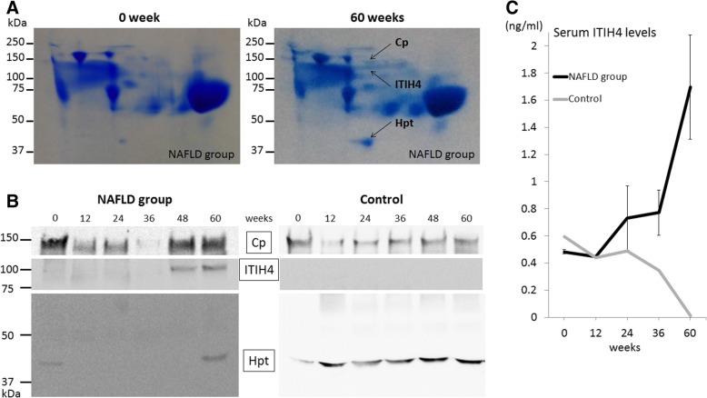
Methods: Microminipigs were fed a high-fat diet to induce NAFLD and a normal diet as the control. To induce HCC, diethylnitrosamine was intraperitoneally administered. Biopsied liver samples were histopathologically analyzed every 12 weeks. Serum proteins were separated by blue native two-dimensional gel electrophoresis and proteins of interest were subsequently identified by MALDI-TOF MS/MS. Human serum samples were analyzed to validate the candidate protein using antibody-mediated characterization.
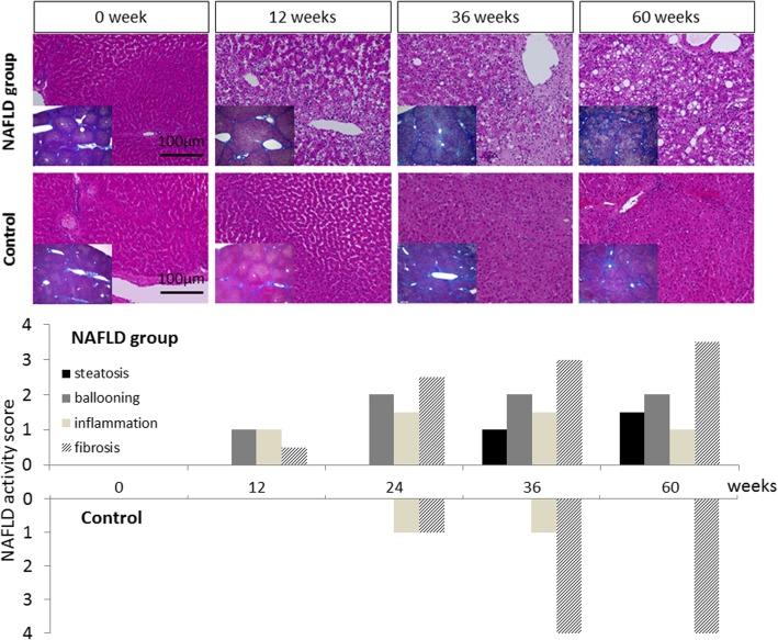
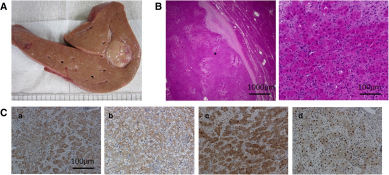
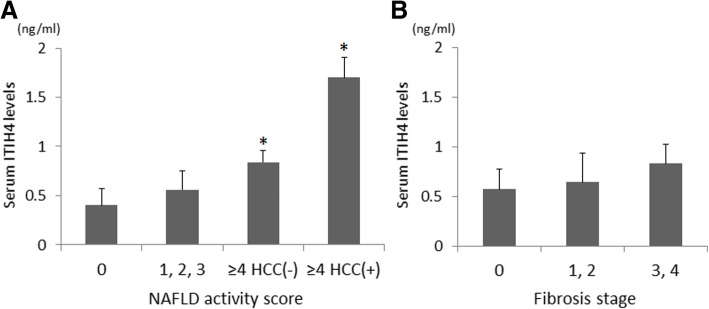
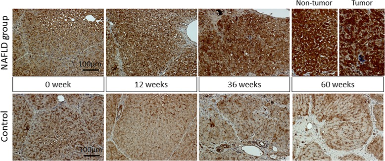
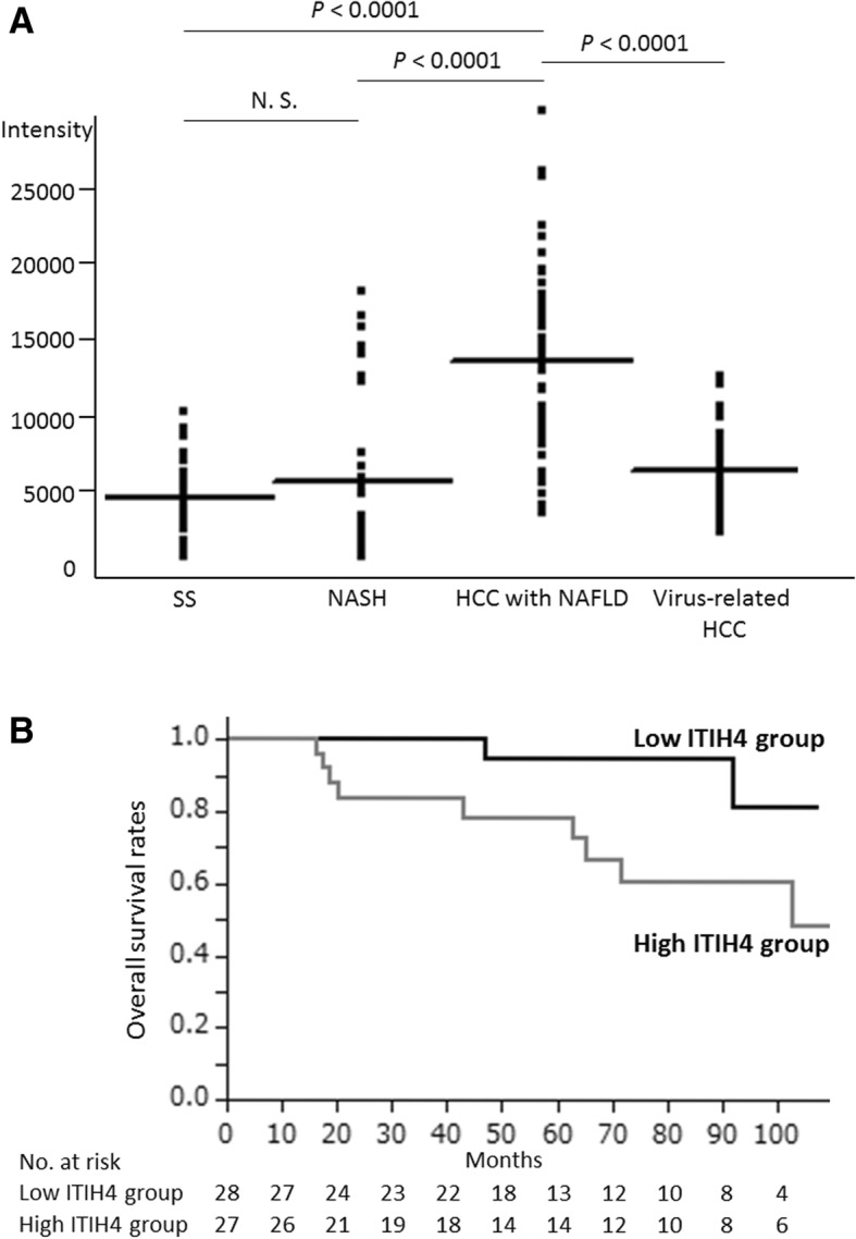
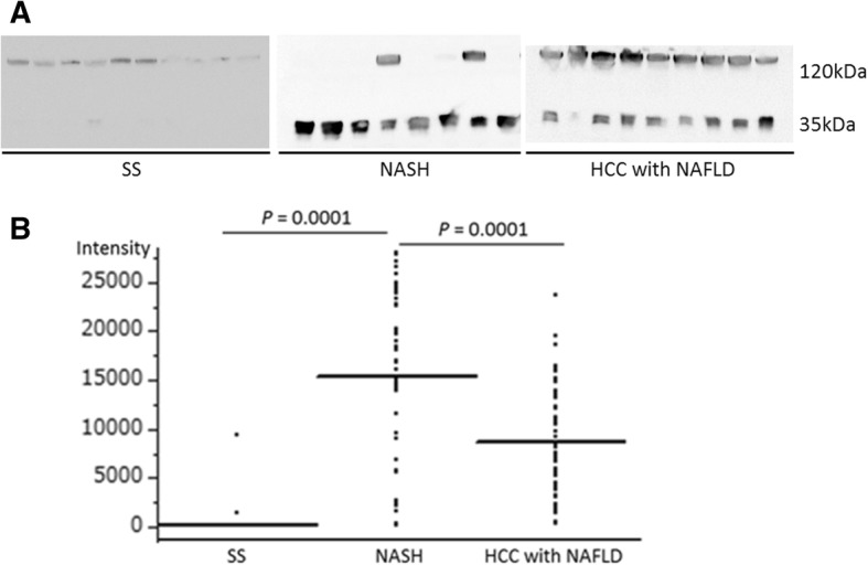
Results: In the NAFLD pigs, hepatic histology of nonalcoholic steatohepatitis (NASH) was observed at 36 weeks, and HCC developed at 60 weeks. Among serum proteins identified with MALDI-TOF MS/MS, serum inter-alpha-trypsin inhibitor heavy chain 4 (ITIH4), an acute response protein which is secreted primarily by liver, was identified as the most characteristic protein corresponding with NAFLD progression and HCC development in the NAFLD pigs. With immunoassay, serum ITIH4 levels in the NAFLD pigs were chronologically increased in comparison with those in control animal. Furthermore, immunohistochemistry showed ITIH4 expression in hepatocytes also increased in both the cancer lesions and parenchyma as NAFLD progressed. Human study is also consistent with this observation because serum ITIH4 levels were significantly higher in HCC-NAFLD patients than in the simple steatosis, NASH, and virus-related HCC patients. Of note, HCC-NAFLD patients who had higher serum ITIH4 levels exhibited poorer prognosis after hepatectomy.
Conclusions: We established an HCC pig model associated with NAFLD. Serum proteomics on the swine HCC with NAFLD model implicated ITIH4 as a non-invasive biomarker reflecting NAFLD progression as well as subsequent HCC development. Most importantly, the results in the swine study have been validated in human cohort studies. Dissecting speciation of serum ITIH4 promises to have clinical utility in monitoring the disease.
Keywords: Blue native/SDS gel electrophoresis; Hepatocellular carcinoma; Inter-alpha-trypsin inhibitor heavy chain 4; MALDI-TOF MS/MS; Nonalcoholic fatty liver disease; Pig model.
Conflict of interest statement
The authors declare that they have no competing interests.
Figures
Similar articles
-
ITIH4: Effective Serum Marker, Early Warning and Diagnosis, Hepatocellular Carcinoma.Pathol Oncol Res. 2018 Jul;24(3):663-670. doi: 10.1007/s12253-017-0285-4. Epub 2017 Aug 21. Pathol Oncol Res. 2018. PMID: 28828637
-
Differences in metabolic and liver pathobiology induced by two dietary mouse models of nonalcoholic fatty liver disease.Am J Physiol Endocrinol Metab. 2020 Nov 1;319(5):E863-E876. doi: 10.1152/ajpendo.00321.2020. Epub 2020 Sep 14. Am J Physiol Endocrinol Metab. 2020. PMID: 32924526
-
Inter-alpha-trypsin inhibitor heavy chain H4 as a diagnostic and prognostic indicator in patients with hepatitis B virus-associated hepatocellular carcinoma.Clin Biochem. 2014 Sep;47(13-14):1257-61. doi: 10.1016/j.clinbiochem.2014.05.002. Epub 2014 May 14. Clin Biochem. 2014. PMID: 24836184
-
Molecular Mechanisms: Connections between Nonalcoholic Fatty Liver Disease, Steatohepatitis and Hepatocellular Carcinoma.Int J Mol Sci. 2020 Feb 23;21(4):1525. doi: 10.3390/ijms21041525. Int J Mol Sci. 2020. PMID: 32102237 Free PMC article. Review.
-
Histopathology of nonalcoholic fatty liver disease/nonalcoholic steatohepatitis.World J Gastroenterol. 2014 Nov 14;20(42):15539-48. doi: 10.3748/wjg.v20.i42.15539. World J Gastroenterol. 2014. PMID: 25400438 Free PMC article. Review.
Cited by
-
Hepatocellular Carcinoma in Non-Alcoholic Fatty Liver Disease: From Epidemiology to Diagnostic Approach.Cancers (Basel). 2021 Nov 21;13(22):5844. doi: 10.3390/cancers13225844. Cancers (Basel). 2021. PMID: 34830997 Free PMC article. Review.
-
Systemic Evaluation of the Effect of Diabetes Mellitus on Breast Cancer in a Mouse Model.Front Oncol. 2022 Apr 29;12:829798. doi: 10.3389/fonc.2022.829798. eCollection 2022. Front Oncol. 2022. PMID: 35578660 Free PMC article.
-
Oxidised Apolipoprotein Peptidome Characterises Metabolic Dysfunction-Associated Steatotic Liver Disease.Liver Int. 2025 Feb;45(2):e16200. doi: 10.1111/liv.16200. Liver Int. 2025. PMID: 39822152 Free PMC article.
-
Translatome profiling reveals Itih4 as a novel smooth muscle cell-specific gene in atherosclerosis.Cardiovasc Res. 2024 Jul 2;120(8):869-882. doi: 10.1093/cvr/cvae028. Cardiovasc Res. 2024. PMID: 38289873 Free PMC article.
-
The function of the inter-alpha-trypsin inhibitors in the development of disease.Front Med (Lausanne). 2024 Aug 1;11:1432224. doi: 10.3389/fmed.2024.1432224. eCollection 2024. Front Med (Lausanne). 2024. PMID: 39149600 Free PMC article. Review.
References
-
- Williams CD, Stengel J, Asike MI, Torres DM, Shaw J, Contreras M, et al. Prevalence of nonalcoholic fatty liver disease and nonalcoholic steatohepatitis among a largely middle-aged population utilizing ultrasound and liver biopsy: a prospective study. Gastroenterology. 2011;140:124–131. doi: 10.1053/j.gastro.2010.09.038. - DOI - PubMed
MeSH terms
Substances
LinkOut - more resources
Full Text Sources
Medical
Miscellaneous
