

Anterior chamber depth - a predictor of refractive outcomes after age-related cataract surgery
- PMID: 31238910
- PMCID: PMC6591866
- DOI: 10.1186/s12886-019-1144-8
Anterior chamber depth - a predictor of refractive outcomes after age-related cataract surgery
Abstract
Background: Anterior chamber depth (ACD) is becoming a hot topic and plays an important role in correcting the refractive errors (REs) after cataract surgery. The aim of this study was to assess the ACD changes and their relationship with the REs after phacoemulsification and intraocular lens (IOL) implantation in patients with age-related cataracts.
Methods: One hundred forty-five eyes of 125 age-related cataract patients from the Department of Ophthalmology, Tangdu Hospital, China, were recruited. IOL Master was used for axial length (AL) and the IOL power calculation measurements, and the Pentacam HR device was used for the ACD and lens thickness (LT) measurements. Every patient underwent uncomplicated phacoemulsification by a single surgeon using a single technique. Postoperative refraction results were obtained at 1 month. The appropriate formula used for the IOL power calculation was chosen depending on the AL, specifically the Hoffer Q (AL < 22.0 mm), SRK/T (22.0 mm ≤ AL ≤ 30.0 mm), and Haigis (AL > 30.0 mm) formulas.
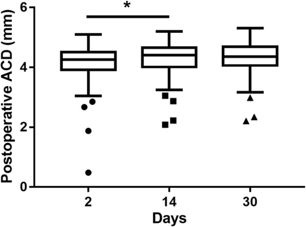
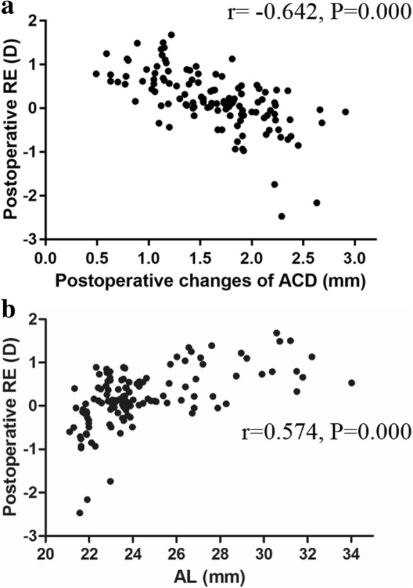
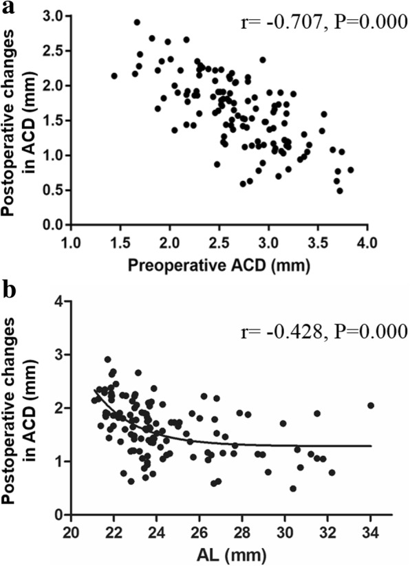
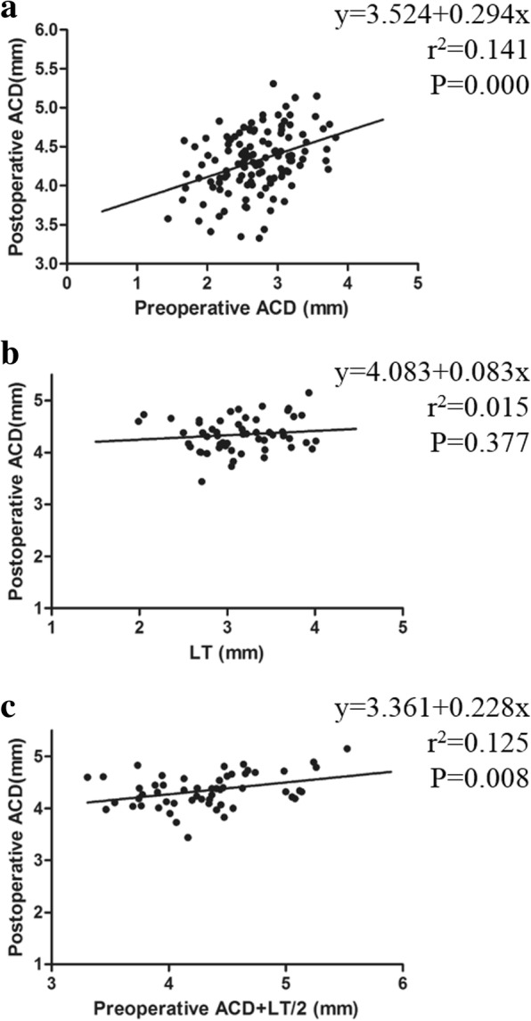
Results: The postoperative ACD was deepened and tended to stabilize gradually after 2 weeks. A concurrent hyperopic shift (0.57 ± 0.47 D) was observed when the change in the ACD was less than 1.65 mm, whereas a myopic shift (- 0.18 ± 0.62 D) occurred contrarily, and the difference between the two groups was statistically significant (P < 0.0001). The change in ACD was significantly larger in the shallow anterior chamber (1.92 ± 0.40 mm) than in the deep chamber (1.33 ± 0.42 mm) (P < 0.0001). Similarly, the change in ACD was larger in the short AL (2.12 ± 0.37 mm) than in the long AL (1.32 ± 0.49 mm). The postoperative ACD and refractive changes were correlated with the preoperative ACD and AL (P < 0.0001), respectively. Two regression formulas were proposed: postoperative ACD = 3.524 + 0.294 × preoperative ACD and postoperative ACD = 3.361 + 0.228× (preoperative ACD + 1/2 LT).
Conclusions: The results of this study showed that the ACD deepened and was associated with a concurrent RE after cataract surgery. Postoperative changes in the ACD were related to the preoperative ACD and AL, which determined the refraction status and visual quality. The regression formula of the postoperative ACD could provide a theoretical basis for predicting refractive errors in the clinic.
Keywords: Anterior chamber depth; Axial length; Cataract; Pentacam HR; Refractive errors.
Conflict of interest statement
The authors declare that they have no competing interests.
Figures
Similar articles
-
[The analysis of refractive error of long axial high myopic eyes after IOL implantation].Zhonghua Yan Ke Za Zhi. 2015 Apr;51(4):276-81. Zhonghua Yan Ke Za Zhi. 2015. PMID: 26081231 Chinese.
-
Effect of anterior chamber depth on the choice of intraocular lens calculation formula in patients with normal axial length.Middle East Afr J Ophthalmol. 2014 Oct-Dec;21(4):307-11. doi: 10.4103/0974-9233.142266. Middle East Afr J Ophthalmol. 2014. PMID: 25371635 Free PMC article.
-
The effect of ocular biometric factors on the accuracy of various IOL power calculation formulas.BMC Ophthalmol. 2017 May 2;17(1):62. doi: 10.1186/s12886-017-0454-y. BMC Ophthalmol. 2017. PMID: 28464806 Free PMC article.
-
Calculation of intraocular lens power: a review.Acta Ophthalmol Scand. 2007 Aug;85(5):472-85. doi: 10.1111/j.1600-0420.2007.00879.x. Epub 2007 Apr 2. Acta Ophthalmol Scand. 2007. PMID: 17403024 Review.
-
Refractive enhancements for residual refractive error after cataract surgery.Curr Opin Ophthalmol. 2021 Jan;32(1):54-61. doi: 10.1097/ICU.0000000000000717. Curr Opin Ophthalmol. 2021. PMID: 33122488 Review.
Cited by
-
Hyperopia shift in the long term after cataract surgery: a case report.BMC Ophthalmol. 2024 Dec 18;24(1):537. doi: 10.1186/s12886-024-03811-0. BMC Ophthalmol. 2024. PMID: 39696077 Free PMC article.
-
Measurements of white-to-white corneal diameter and anterior chamber parameters using the Pentacam AXL wave and their correlations in the adult Saudi population.PeerJ. 2025 Apr 1;13:e19227. doi: 10.7717/peerj.19227. eCollection 2025. PeerJ. 2025. PMID: 40191754 Free PMC article.
-
Mispositioned Hydrus Microstents: A Case Series Imaged with NIDEK GS-1 Gonioscope.J Ophthalmol. 2022 Sep 8;2022:1605195. doi: 10.1155/2022/1605195. eCollection 2022. J Ophthalmol. 2022. PMID: 36119138 Free PMC article.
-
Factors affecting the refractive error after cataract surgery.Int Ophthalmol. 2025 May 3;45(1):163. doi: 10.1007/s10792-025-03543-0. Int Ophthalmol. 2025. PMID: 40319199 Review.
-
The accuracy of intraocular lens power calculation formulas based on artificial intelligence in highly myopic eyes: a systematic review and network meta-analysis.Front Public Health. 2023 Nov 9;11:1279718. doi: 10.3389/fpubh.2023.1279718. eCollection 2023. Front Public Health. 2023. PMID: 38026369 Free PMC article.
References
MeSH terms
Grants and funding
LinkOut - more resources
Full Text Sources
Medical
Research Materials
