

Primary pseudomyogenic hemangioendothelioma of the vulva: a rare location for a rare entity
- PMID: 31238962
- PMCID: PMC6593540
- DOI: 10.1186/s13000-019-0846-9
Primary pseudomyogenic hemangioendothelioma of the vulva: a rare location for a rare entity
Abstract
Background: Pseudomyogenic hemangioendothelioma (PMHE) is a recently described vascular neoplasm which typically occurs in the lower extremities of young to middle-aged adults.
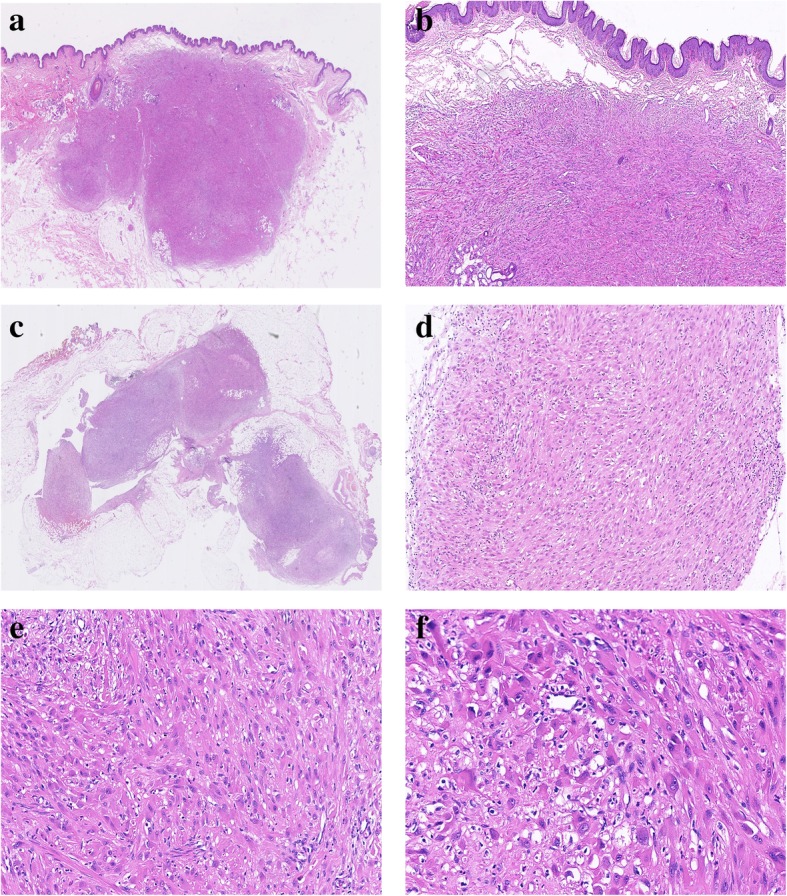
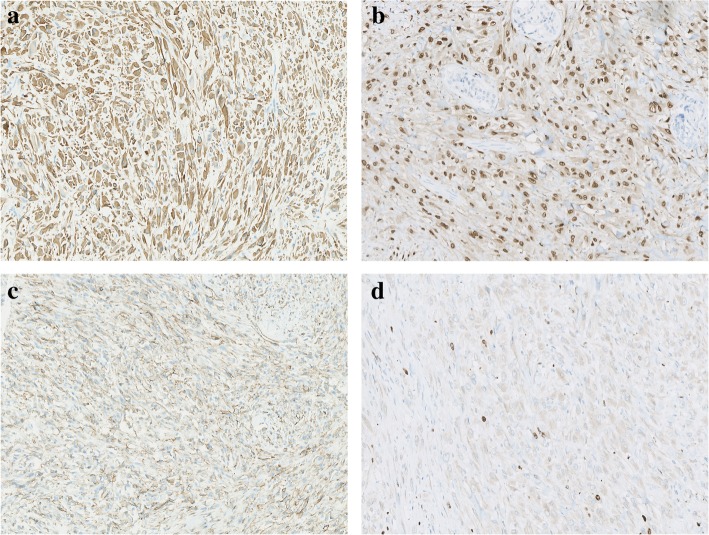
Case presentation: We present here a unique case of PMHE arising primarily in the vulva of a 51-year-old woman who presented with a painful vulvar nodule. Clinically, it was thought as Bartholin gland cyst, vulvar hematoma or papilloma. On surgery, two nodules were found with one located in the superficial dermis and the other in the deep subcutis. Histologically, these two nodules showed similar features, composed of fascicles or sheets of plump spindled to epithelioid cells with eosinophilic cytoplasm. Given the morphological resemblance to a myogenic tumor, the lesion was initially diagnosed as a rhabdomyosarcoma by the referring pathologist. However, a comprehensive reevaluation of the submitted slides made us reconsider a PMHE, which was subsequently confirmed by immunohistochemical study.
Conclusion: This case demonstrates that PMHE can also develop in the female external genitalia albeit extremely rare. This disease should be included in the differential diagnostic list of vulvar tumors with spindled to epithelioid morphology and cytokeratin-positive immunophenotypes.
Keywords: Immunohistochemistry; Pseudomyogenic hemangioendothelioma; Vulva.
Conflict of interest statement
The authors declare that they have no competing interests.
Figures
Similar articles
-
The clinicopathological spectrum of pseudomyogenic hemangioendothelioma: report of an additional series with review of the literature.Virchows Arch. 2020 Aug;477(2):231-240. doi: 10.1007/s00428-020-02753-4. Epub 2020 Jan 24. Virchows Arch. 2020. PMID: 31980959
-
FOSB is a Useful Diagnostic Marker for Pseudomyogenic Hemangioendothelioma.Am J Surg Pathol. 2017 May;41(5):596-606. doi: 10.1097/PAS.0000000000000795. Am J Surg Pathol. 2017. PMID: 28009608
-
A rare case of pseudomyogenic hemangioendothelioma (PHE)/epithelioid sarcoma-like hemangioendothelioma (ES-H) of the breast first misdiagnosed as metaplastic carcinoma by FNAB and review of the literature.Diagn Pathol. 2019 Jul 17;14(1):79. doi: 10.1186/s13000-019-0857-6. Diagn Pathol. 2019. PMID: 31311568 Free PMC article. Review.
-
[Pseudomyogenic hemangioendothelioma in the upper limb: A case report and literature review].Rev Esp Patol. 2017 Jan-Mar;50(1):49-53. doi: 10.1016/j.patol.2015.12.007. Epub 2016 Mar 11. Rev Esp Patol. 2017. PMID: 29179965 Review. Spanish.
-
Two Cases of Intraosseous Pseudomyogenic (Epithelioid Sarcoma-Like) Hemangioendothelioma With Unusual Features, Expanding the Clinicopathological Spectrum.Int J Surg Pathol. 2021 Jun;29(4):454-461. doi: 10.1177/1066896920951841. Epub 2020 Aug 27. Int J Surg Pathol. 2021. PMID: 32851904
Cited by
-
Clinicopathological study of pseudomyogenic hemangioendothelioma.Diagn Pathol. 2023 Feb 20;18(1):25. doi: 10.1186/s13000-023-01309-9. Diagn Pathol. 2023. PMID: 36803395 Free PMC article.
-
The clinicopathological spectrum of pseudomyogenic hemangioendothelioma: report of an additional series with review of the literature.Virchows Arch. 2020 Aug;477(2):231-240. doi: 10.1007/s00428-020-02753-4. Epub 2020 Jan 24. Virchows Arch. 2020. PMID: 31980959
References
-
- Fletcher CDM, Bridge JA, Hogendoorn PCW, et al. WHO classification of Tumours of soft tissue and bone. Lyon: IARC Press; 2013.
Publication types
MeSH terms
LinkOut - more resources
Full Text Sources
Medical
