. 2019 Jul 9;116(28):13937-13942.
doi: 10.1073/pnas.1906024116. Epub 2019 Jun 25.

Allosteric modulation of a human protein kinase with monobodies

Affiliations

Allosteric modulation of a human protein kinase with monobodies

Adelajda Zorba et al. Proc Natl Acad Sci U S A. .

Abstract

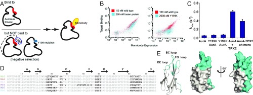
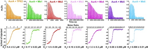
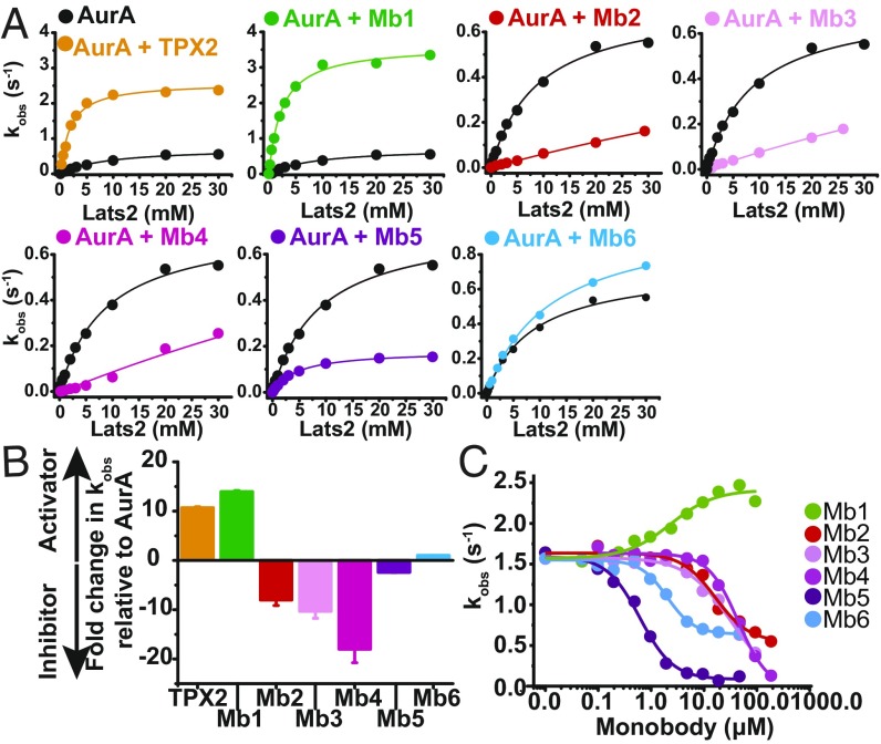
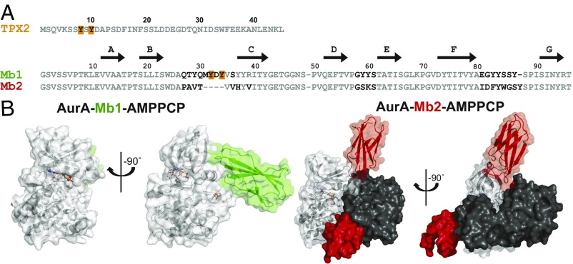
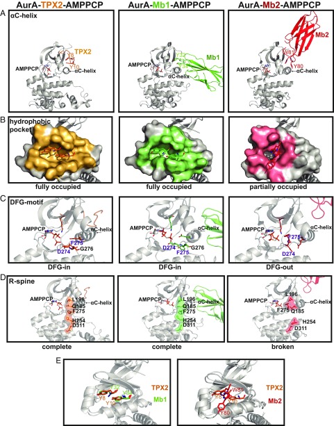
Despite being the subject of intense effort and scrutiny, kinases have proven to be consistently challenging targets in inhibitor drug design. A key obstacle has been promiscuity and consequent adverse effects of drugs targeting the ATP binding site. Here we introduce an approach to controlling kinase activity by using monobodies that bind to the highly specific regulatory allosteric pocket of the oncoprotein Aurora A (AurA) kinase, thereby offering the potential for more specific kinase modulators. Strikingly, we identify a series of highly specific monobodies acting either as strong kinase inhibitors or activators via differential recognition of structural motifs in the allosteric pocket. X-ray crystal structures comparing AurA bound to activating vs inhibiting monobodies reveal the atomistic mechanism underlying allosteric modulation. The results reveal 3 major advantages of targeting allosteric vs orthosteric sites: extreme selectivity, ability to inhibit as well as activate, and avoidance of competing with ATP that is present at high concentrations in the cells. We envision that exploiting allosteric networks for inhibition or activation will provide a general, powerful pathway toward rational drug design.

Keywords: Aurora A; allosteric drugs; allostery; kinase; monobody.


Conflict of interest statement

Conflict of interest statement: D.K. and A.Z. are the inventors on pending patents applied for by Brandeis University that describe compositions and methods for modulating kinase activity (US20180334510A1 and US20190038582A1). A.K. and S. Koide are listed as inventors on issued and pending patents on the monobody technology filed by The University of Chicago (US Patent 9512199 B2 and related pending applications).

Figures

Fig. 1.
Fig. 1.
Generation of monobodies that bind to the allosteric hydrophobic pocket of AurA. (A) Schematic of monobody selection design. Monobodies were chosen for their ability to bind to AurA but not Y199H or Y199K AurA mutants or AurA-TPX2 chimeric protein. (B) Representative monobody populations after negative selection with AurA-TPX2 chimera or Y199K AurA mutants, as tested using yeast display. (C) Mutation of AurA Y199 to either H or K does not change the activity of the mutant AurA proteins. Activities were measured by the ADP/NADH coupled assay at 25 °C with 1 μM AurA, 1 mM Lats2 peptide and saturating concentrations of ATP and MgCl2. AurA-TPX2 chimera closely mimics the activity of WT protein in the presence of 5 µM TPX2, ideally suiting this decoy protein to be used in monobody counter selection screens. (D) Amino acid sequences of monobody clones. Residues at diversified positions in the combinatorial libraries are shown in bold. Amino acid numbering is shown for Mb1. Although subsequent monobody sequences are shown with gaps for purposes of visual alignment among each other, we do not count gaps in sequence numbering of these monobodies. (E) Mapping of diversified positions (cyan) onto the structure of a prototypical monobody (PDB ID code 1FNF). Errors in C are SD from triplicates.
Fig. 2.
Fig. 2.
Monobodies bind to AurA with high affinity. Binding of TPX2 and Mb1-6 to AurA at 25 °C as measured by ITC. Fitted dissociation constants, Kd, are shown with the SE for the estimate of Kd, which is a measure of the goodness of fit of the data.
Fig. 3.
Fig. 3.
Monobodies as activators and inhibitors of AurA activity. (A) The ADP/NADH coupled assay was used to monitor Lats2 peptide phosphorylation by AurA in the absence (black) or presence of TPX2 or monobodies Mb1 to Mb6. Fits to Michaelis Menten kinetics, normalized by the enzyme concentration (kobs), show that the monobodies affect both the kcat and KM of AurA for Lats2. Assays were carried out at 25 °C in the presence of 1 or 0.25 μM AurA and saturating concentrations of monobody or TPX2, ATP, and MgCl2. (B) Comparison of kobs values of AurA at 1 mM Lats2 showing that Mb1 activates AurA 15-fold, a value comparable to allosteric activation by TPX2, whereas monobodies Mb2-Mb5 inhibit AurA between 2-fold and 20-fold, with Mb6 not significantly affecting AurA kinetics. (C) Monobodies can outcompete TPX2 from AurA in a manner consistent with their relative AurA affinities and reflective of their inhibitory/neutral (Mb2-Mb6) or activating (Mb1) propensity. Experiments were conducted in the presence of 1 μM AurA, 5 μM TPX2, 3 mM Lats2, using the same coupled assay as described earlier. Errors in B were determined from jackknifing of data in A.
Fig. 4.
Fig. 4.
Structures of AurA bound to activating or inhibiting monobody. (A) Sequences of AurA activators, TPX2 and Mb1, and inhibitor, Mb2. The 2 Tyr critical for TPX2-dependent AurA activation (Y8 and Y10; shown in bold, orange), and the structurally equivalent residues in Mb1 (also bold and orange), as well as sequence difference between Mb1 and Mb2 (black bold), are highlighted. Residue numbering is based on a previously published system (–19). (B) X-ray crystal structures of AurA (gray) bound to AMPPCP and either Mb1 (green, 2.06 Å, PDB ID code 5G15) or Mb2 (red, 2.55Å, PDB ID code 6C83), exposing extensive but different contacts between AurA and Mb1/Mb2.
Fig. 5.
Fig. 5.
Monobodies stabilize either an active or inactive conformation of AurA. (A) Structures of AurA bound to TPX2 (PDB ID code 4C3P), Mb1 (PDB ID code 5G15), or Mb2 (PDB ID code 6C83) highlighting (A) differential engagement of AurA’s αC-helix by 2 aromatic residues within TPX2 or Mb1/Mb2, (B) AurA residues interfacing with TPX2 (orange), Mb1 (green) or Mb2 (pink) were calculated using PISA (40), (C) Hallmarks of an active kinase: DFG-in motif, K162-E181 salt bridge, and (D) completion of the R-spine are present in the AurA-TPX2 or AurA-Mb1 complexes and absent in the AurA-Mb2 structure. (E) Y32 and Y34 of Mb1 are superimposable with Y8 and Y10 of TPX2 (Left), whereas Y80 and W81 of Mb2 interact very differently with AurA (Right); see also SI Appendix, Fig. S5.

Similar articles

Cited by

References

    1. Zhou H., et al. , Tumour amplified kinase STK15/BTAK induces centrosome amplification, aneuploidy and transformation. Nat. Genet. 20, 189–193 (1998). - PubMed
    1. Glover D. M., Leibowitz M. H., McLean D. A., Parry H., Mutations in aurora prevent centrosome separation leading to the formation of monopolar spindles. Cell 81, 95–105 (1995). - PubMed
    1. Asteriti I. A., Giubettini M., Lavia P., Guarguaglini G., Aurora-A inactivation causes mitotic spindle pole fragmentation by unbalancing microtubule-generated forces. Mol. Cancer 10, 131 (2011). - PMC - PubMed
    1. Kufer T. A., et al. , Human TPX2 is required for targeting Aurora-A kinase to the spindle. J. Cell Biol. 158, 617–623 (2002). - PMC - PubMed
    1. Zorba A., et al. , Molecular mechanism of Aurora A kinase autophosphorylation and its allosteric activation by TPX2. eLife 3, e02667 (2014). - PMC - PubMed

Publication types

MeSH terms

LinkOut - more resources