. 2019 Jul 9;116(28):14339-14348.
doi: 10.1073/pnas.1815425116. Epub 2019 Jun 25.

A Lotus japonicus cytoplasmic kinase connects Nod factor perception by the NFR5 LysM receptor to nodulation

Affiliations

A Lotus japonicus cytoplasmic kinase connects Nod factor perception by the NFR5 LysM receptor to nodulation

Jaslyn E M M Wong et al. Proc Natl Acad Sci U S A. .

Abstract

The establishment of nitrogen-fixing root nodules in legume-rhizobia symbiosis requires an intricate communication between the host plant and its symbiont. We are, however, limited in our understanding of the symbiosis signaling process. In particular, how membrane-localized receptors of legumes activate signal transduction following perception of rhizobial signaling molecules has mostly remained elusive. To address this, we performed a coimmunoprecipitation-based proteomics screen to identify proteins associated with Nod factor receptor 5 (NFR5) in Lotus japonicus. Out of 51 NFR5-associated proteins, we focused on a receptor-like cytoplasmic kinase (RLCK), which we named NFR5-interacting cytoplasmic kinase 4 (NiCK4). NiCK4 associates with heterologously expressed NFR5 in Nicotiana benthamiana, and directly binds and phosphorylates the cytoplasmic domains of NFR5 and NFR1 in vitro. At the cellular level, Nick4 is coexpressed with Nfr5 in root hairs and nodule cells, and the NiCK4 protein relocates to the nucleus in an NFR5/NFR1-dependent manner upon Nod factor treatment. Phenotyping of retrotransposon insertion mutants revealed that NiCK4 promotes nodule organogenesis. Together, these results suggest that the identified RLCK, NiCK4, acts as a component of the Nod factor signaling pathway downstream of NFR5.

Keywords: Lotus; NFR5; NiCK4; RLCK; nodulation.


Conflict of interest statement

The authors declare no conflict of interest.

Figures

Fig. 1.
Fig. 1.
Co-IP of NFR5-eYFP and MS discovery of NiCK4. (A) SimplyBlue SafeStained gel of proteins coimmunoprecipitated with GFP-trap beads. The molecular weights of the marker proteins (in kilodaltons) are indicated next to their corresponding protein bands. The corresponding Western blot of crude extract (input) and coimmunoprecipitated NFR5-eYFP proteins with anti–GFP-HRP antibodies used for visualization. (−) indicates the nontreated WT Gifu root sample, while (M) and (NF) indicate mock- and NF-treated NFR5-eYFP–expressing root samples, respectively. The asterisks indicate the positions of NFR5-eYFP. Three biological replicates were prepared for each condition. (B) Phylogenetic tree of L. japonicus, M. truncatula, and A. thaliana members in the CRPK1 family. NiCK4 is indicated with a green filled circle, and M. truncatula and A. thaliana RLCKs are indicated with filled yellow circles or red squares, respectively. The phylogenetic tree was constructed with the CLC main workbench using the neighbor joining method and Jukes–Cantor protein distance measurement with 10,000 bootstrap replicates (https://www.qiagenbioinformatics.com). (C and D) Two representative mass spectra of 2 unique peptides of NiCK4, ASNVLLDKDLQPK (C) and LPVEEQYLLTR (D), identified in mock-treated samples, with Mascot ion scores of 49.8 and 47.2, respectively.
Fig. 2.
Fig. 2.
Binding studies of NiCK4 and NFR5. (A and B) FRET measurements performed for the NFR5-eGFP/NiCK4-mCherry (A) or eGFP-LjLTI6b/NiCK4-mCherry (B) pairs in epidermal N. benthamiana leaf cells. The bleached regions are outlined by a white box in the corresponding confocal microscopy images. FRET was detected as an increase in GFP fluorescence (marked with vertical lines) after bleaching (marked with diagonal lines) for the NFR5-eGFP/NiCK4-mCherry pair (A). No increase in GFP fluorescence after bleaching was observed for the eGFP-LjLTI6b/NiCK4-mCherry pair (B). The extracted fluorescence values for donor and acceptor prebleaching/postbleaching and FRET efficiencies are presented in the corresponding tables. (Scale bars: 20 µm.) (C) Western blots from anti-FLAG pulldown experiment with p35S:NiCK4-FLAG, p35S:eGFP-LjLTI6b, p35S:NFR5-eGFP, p35S:NFR1-eGFP, and p35S:SymRK-eGFP expressed in N. benthamiana. The eGFP fusion proteins were expressed alone (odd lanes, excluding lane 1 with no transgenic protein) or coexpressed NiCK4-FLAG (even lanes, excluding lane 2). Synthetic FLAG peptide was added to samples containing no transgenic protein or eGFP fusion proteins expressed alone (odd lanes) to prevent unspecific binding of eGFP proteins to the beads and to check for any unspecific binding of the eGFP fusion proteins to the FLAG tag. Crude lysate (Input) and immunoprecipitated (IP) proteins were detected with anti-FLAG or anti-GFP antibodies. The results were reproducible in 3 biological replicates. (DF) MST binding curves obtained from binding experiments with labeled NiCK4 and nonlabeled NFR5-CD298-595, NFR1-CD, or SymRK-CD. NiCK4 directly binds NFR5-CD298-595 (D) and NFR1-CD (E) but not SymRK-CD (F). Binding curves and errors were obtained from 3 biological replicates.
Fig. 3.
Fig. 3.
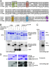
Kinase activity assessment of NiCK4. (A) Amino acid sequence alignment (118) of NiCK4 with the kinase domains of NFR1b, NFR5, and SymRK. NiCK4 possess all features of an active kinase; an intact glycine-rich loop (green) and AXK motif (red); the HRD motif in the catalytic loop (yellow); the DFG motif (blue); and the APE motif in the activation loop (magenta). The threonine residue corresponding to NFR1-CD-T483 is underlined. (BD) SDS/PAGE gels from in vitro kinase assays visualized by Coomassie staining or autoradiography. NiCK4 transphosphorylates myelin basic protein (MBP) and NFR5-CD276-595 more strongly than TRX-NFR1-CD or SymRK-CD, but none of the 3 active kinases phosphorylated BSA (B). The nonphosphorylation of BSA by TRX-NFR1-CD, which has a similar molecular weight to BSA, was confirmed by NFR-CD lacking the thioredoxin tag (C). NFR5-CD298-595 and NFR1-CD, but not SymRK-CD were strongly phosphorylated in the presence of NiCK4, while NiCK4 was strongly phosphorylated in the presence of both NFR1-CD and NFR5 (D). The phosphorylation results could be reproduced in 3 biological replicates.
Fig. 4.
Fig. 4.
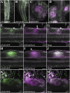
NiCK4 and Nfr5 display similar expression patterns. (AD) Confocal microscopy of L. japonicus roots individually expressing pNick4:tYFP-NLS (A and C) or pNfr5:tYFP-NLS (B and D). In noninoculated roots, pNick4:tYFP-NLS (A) and pNfr5:tYFP-NLS (B) are expressed in epidermal cells including root hair cells. In nodule primordia, (C) pNick4:tYFP-NLS expression is maintained in the nodule parenchyma (PA), cortex, and epidermis of mature nodules 14 d postinoculation (dpi) with M. loti strain MAFF303099 expressing dsRed. (D) The expression of pNfr5:tYFP-NLS is strongly down-regulated in mature nodules 14 dpi with M. loti strain MAFF303099. (EM) Confocal microscopy of L. japonicus roots coexpressing pNick4:tYFP-NLS and pNfr5:mCherry-NLS. pNick4:tYFP-NLS and pNfr5:mCherry-NLS are coexpressed in cortical cells (EG) and nodule primordia (HJ) 14 dpi with M. loti strain MAFF303099 expressing dsRed. pNick4:tYFP-NLS and pNfr5:mCherry-NLS are also coexpressed in root hair cells (KM) 11 dpi with M. loti strain MAFF303099. The asterisks (*) indicate root hairs that coexpress pNfr5:mCherry-NLS and pNick4:tYFP-NLS. Arrowheads depict infection threads. Autofluorescence, YFP, and mCherry/dsRed channels are represented in white, green, and magenta, respectively. White nuclei indicate merged green and magenta nuclei. (Scale bars: 50 µm.)
Fig. 5.
Fig. 5.
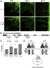
NiCK4 shuttles to the nucleus after Nod factor (NF) treatment and promotes nodulation. (A) In pLjUbi:NiCK4-eGFP transformed root systems, NiCK4-eGFP relocates to the nucleus (indicated with arrowheads) 90 min after NF treatment in roots of WT Lotus plants but not in transformed nfr1-1 or nfr5-2 mutant roots. Inset shows nuclear localization of NiCK4-eGFP at higher magnification. T = 0 and T = 90 represent images obtained from the same root hairs before and 90 min after NF treatment, respectively. The number of WT, nfr5-2, and nfr1-1 mutant plants imaged are 8, 11, and 9, respectively. (Scale bars: 50 µm.) (B) The predicted gene structure of Nick4 with six exons indicated. The untranslated regions and coding sequences are represented by filled dark gray and light gray boxes, respectively, and the intronic regions are represented a black line. LORE1 insertions for nick4-1, nick4-2, and nick4-3 alleles before the sequences encoding residues S14, F59, and R127 in exons 1, 2, and 4, respectively, are indicated below the exons. The direction of LORE1 insertion is indicated by the direction of the arrow. (C) Nodulation counts of nick4 mutants and WT plants grown on agar plates 21 d after inoculation with M. loti strain NZP2235. Reduced nodulation was observed in nick4 mutants compared with WT plants. *P < 0.05 and **P < 0.01 (t test). White and pink nodules are represented by filled dark gray and light gray bars, respectively. (D) Working model for NiCK4 involvement in symbiosis signaling. In the presence of NF, NiCK4 phosphorylates NFR5, possibly improving its own docking site(s) or sites for hitherto-unknown NFR5 interactors. NiCK4 then phosphorylates NFR1, which in turn phosphorylates NiCK4 and leads to NiCK4 dissociation and migration to the nucleus. This mechanism would relay the NF signal from the PM-localized receptors to the nuclear components involved in promoting nodule organogenesis. Given the nodulation phenotype of nick4 mutants, NICK4 is not solely responsible for this relay.

Similar articles

Cited by

References

    1. Fisher R. F., Long S. R., Rhizobium–plant signal exchange. Nature 357, 655–660 (1992). - PubMed
    1. Denarie J., et al. , “Rhizobium and legume nodulation—a molecular dialog” in New Horizons in Nitrogen Fixation. Current Plant Science and Biotechnology in Agriculture, R. Palacios, J. Mora, W. E. Newton, Eds. (Springer, Dordrecht, 1993), vol. 17, pp. 19–30.
    1. Mulligan J. T., Long S. R., Induction of Rhizobium meliloti nodC expression by plant exudate requires nodD. Proc. Natl. Acad. Sci. U.S.A. 82, 6609–6613 (1985). - PMC - PubMed
    1. Madsen L. H., et al. , The molecular network governing nodule organogenesis and infection in the model legume Lotus japonicus. Nat. Commun. 1, 10 (2010). - PMC - PubMed
    1. Madsen E. B., et al. , A receptor kinase gene of the LysM type is involved in legume perception of rhizobial signals. Nature 425, 637–640 (2003). - PubMed

Publication types

MeSH terms

Substances