

An Overview of the Thyroid Gland and Thyroid-Related Deaths for the Forensic Pathologist
- PMID: 31239894
- PMCID: PMC6507001
- DOI: 10.23907/2016.024
An Overview of the Thyroid Gland and Thyroid-Related Deaths for the Forensic Pathologist
Abstract
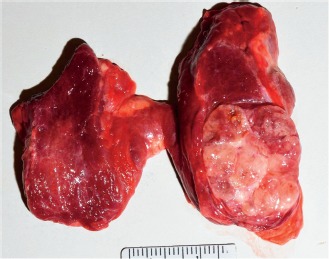
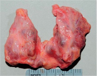
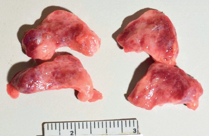
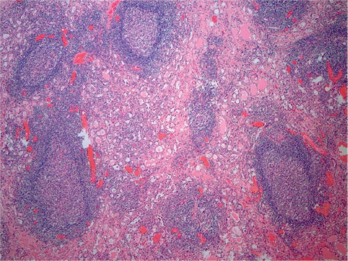
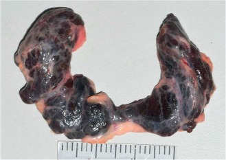
The thyroid gland is a butterfly-shaped organ situated in the anterior neck whose functions have system-wide effects. Thyroid diseases represent some of the most commonly encountered endocrine disorders and therefore are commonly encountered at the time of autopsy. Knowing how the gland functions and the effects it may have on vital organs is important when determining the cause of death and significant contributory conditions. Endocrine-related deaths may be anatomically subtle, therefore histologic examination, review of medical records, and selected postmortem testing must be performed to correctly identify and document their presence. For this reason, it is recommended that pathologists consider regularly examining the thyroid gland histologically, particularly on decedents where no apparent anatomic cause of death is identified after the autopsy. This article provides an in-depth review of the thyroid gland, thyroid hormones, and thyroid diseases, including potential thyroid-related deaths and incidental autopsy findings.
Keywords: Forensic pathology; Postmortem thyroid hormone; Thyroid cancer; Thyroid gland; Thyrotoxicosis.
Conflict of interest statement
Disclosures & Declaration of Conflicts of Interest: The authors, reviewers, editors, and publication staff do not report any relevant conflicts of interest
Figures
References
-
- Maitra A. Thyroid gland. In: chmidt W., Gruliow R., editors. Robbins and Cotran pathologic basis of disease. 8th ed. Philadelphia: Saunders Elsevier; 2010. p. 1107–30.
-
- Mescher A.L. Junqueira's basic histology text & atlas. 12th ed. New York: McGraw-Hill Medical; c2010. Chapter 20, Endocrine glands; p. 348–70.
-
- Rosai J., Tallini G. Rosai and Ackerman's surgical pathology. 10th ed. New York: Mosby Elsevier; c2011. Chapter 9, Thyroid gland; p. 487–565.
Publication types
LinkOut - more resources
Full Text Sources