

Superficial Myofibroblastoma in the Vulva Mimicking Aggressive Angiomyxoma: A Case Report and Review of the Literature
- PMID: 31240143
- PMCID: PMC6556266
- DOI: 10.1155/2019/1582714
Superficial Myofibroblastoma in the Vulva Mimicking Aggressive Angiomyxoma: A Case Report and Review of the Literature
Abstract
Background: Superficial myofibroblastoma (SMF) is a very rare benign mesenchymal tumor in the female lower genital tract. Only 46 cases have been reported in the English language literature, among which only 7 cases arose in the vulva. Sometimes SMF histologically mimics aggressive angiomyxoma (AA) in which massive myxoid change in stroma is characteristic. We herein report a case of vulvar SMF with prominent myxoid stroma and review the literature with the emphasis on the differential diagnosis of SMF and AA.
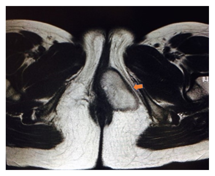
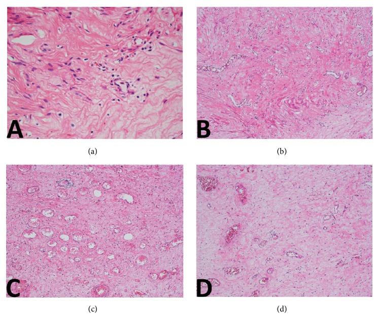
Case presentation: a 37-year-old woman presented with a painless mass in the vulva. Magnetic resonance imaging (MRI) showed a well-circumscribed 7 cm mass in the subcutis of the vulva. The tumor was resected. Histopathologically, the tumor was characterized by sparsely populated spindle-shaped cells in the fibromyxoid stroma. Thin-walled blood vessels were detected. Mitoses or pleomorphism was not found. Tumor cells were positive for vimentin, ER, PgR, and desmin. Some cells were positive for alpha-SMA and CD34. All cells were negative for S100 protein.
Conclusions: because SMF and AA show different clinical prognoses, distinguishing SMF from AA is important. However, SMF may share many common histological features with AA: superficial localization (above fascia), sharp borderline from adjacent tissue, expansive growth pattern; a specific vascular pattern will lead to an accurate diagnosis of SMF. Familiarization with the histological characteristics of the two entities will help to make a prognostic prediction.
Figures


References
-
- Laskin W. B., Fetsch J. F., Tavassoli F. A. Superficial cervicovaginal myofibroblastoma: Fourteen cases of a distinctive mesenchymal tumor arising from the specialized subepithelial stroma of the lower female genital tract. Human Pathology. 2001;32(7):715–725. doi: 10.1053/hupa.2001.25588. - DOI - PubMed
-
- Olinici C. D., Crisan D., Zolog A., Puscas M. Vaginal superficial myofibroblastoma. Case report and review of the literature. Romanian Journal of Morphology and Embryology. 2007;48:165–170. - PubMed
-
- Wang Q. F., Wu Y. Y., Wang J. Superficial cervicovaginal myofibroblastoma: report of four cases and literature review. Chinese Medical Journal. 2010;123:1093–1096. - PubMed
Publication types
LinkOut - more resources
Full Text Sources
Research Materials
