. 2019 Jun 20:2:227.
doi: 10.1038/s42003-019-0478-3. eCollection 2019.

MCRIP1 promotes the expression of lung-surfactant proteins in mice by disrupting CtBP-mediated epigenetic gene silencing

Affiliations

MCRIP1 promotes the expression of lung-surfactant proteins in mice by disrupting CtBP-mediated epigenetic gene silencing

Jane S Weng et al. Commun Biol. .

Abstract

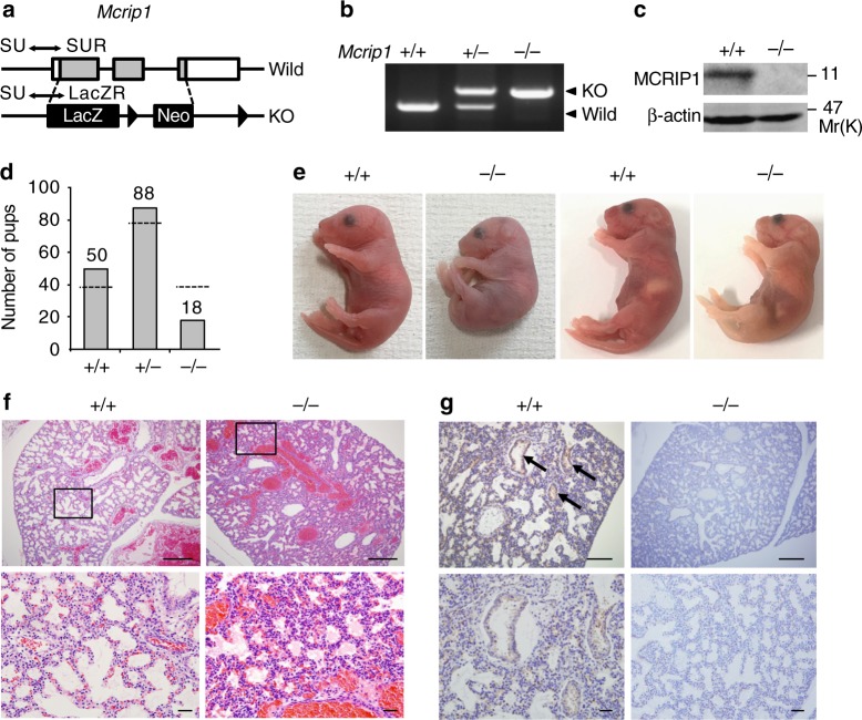
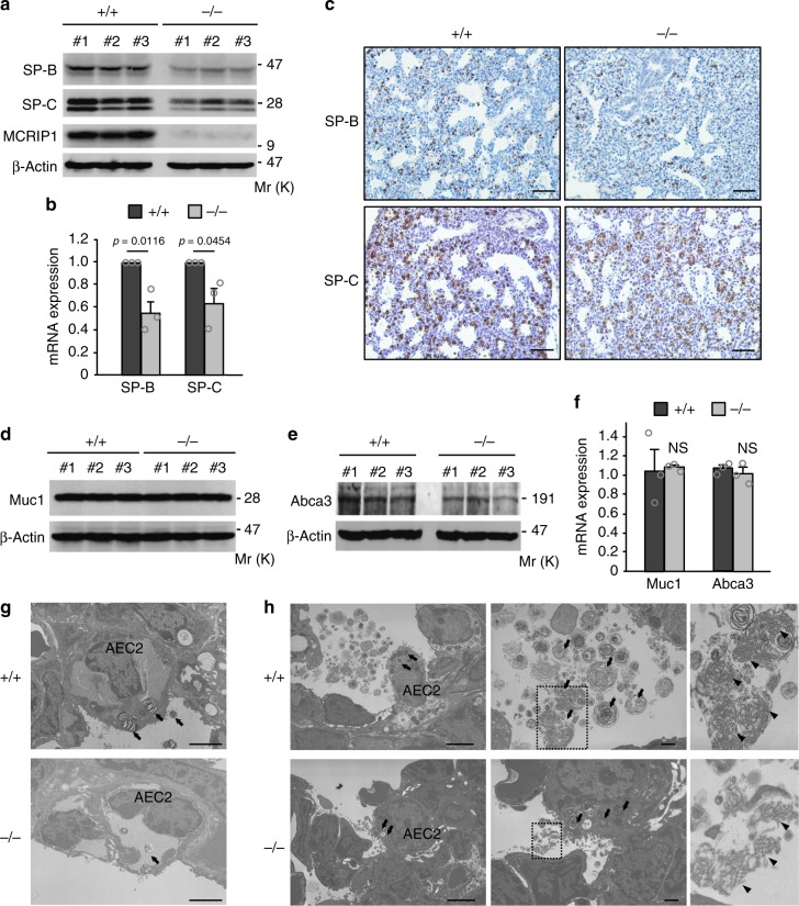
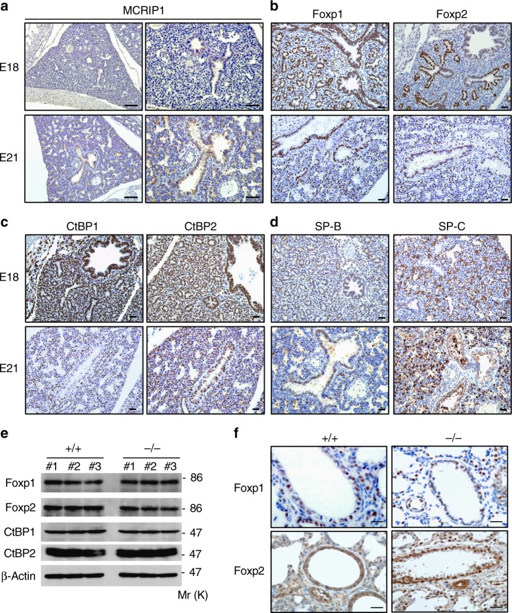
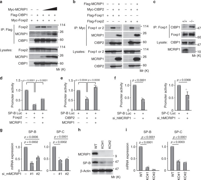
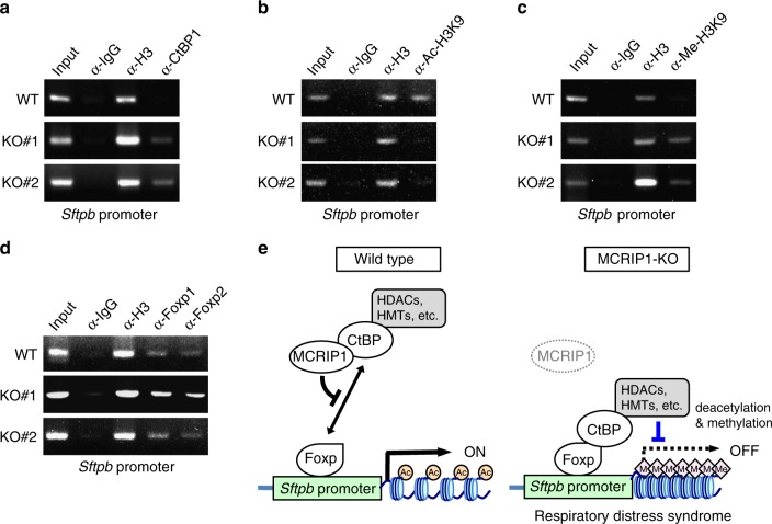
Proper regulation of epigenetic states of chromatin is crucial to achieve tissue-specific gene expression during embryogenesis. The lung-specific gene products, surfactant proteins B (SP-B) and C (SP-C), are synthesized in alveolar epithelial cells and prevent alveolar collapse. Epigenetic regulation of these surfactant proteins, however, remains unknown. Here we report that MCRIP1, a regulator of the CtBP transcriptional co-repressor, promotes the expression of SP-B and SP-C by preventing CtBP-mediated epigenetic gene silencing. Homozygous deficiency of Mcrip1 in mice causes fatal respiratory distress due to abnormal transcriptional repression of these surfactant proteins. We found that MCRIP1 interferes with interactions of CtBP with the lung-enriched transcriptional repressors, Foxp1 and Foxp2, thereby preventing the recruitment of the CtBP co-repressor complex to the SP-B and SP-C promoters and maintaining them in an active chromatin state. Our findings reveal a molecular mechanism by which cells prevent inadvertent gene silencing to ensure tissue-specific gene expression during organogenesis.

Keywords: Development; Differentiation; Gene silencing.


Conflict of interest statement

Competing interestsThe authors declare no competing financial or non-financial interests.

Figures

Fig. 1
Fig. 1
Mcrip1-KO mice die at the neonatal stage due to respiratory failure. a The targeting strategy for Mcrip1-KO mouse generation. Positions of the PCR primers (SU, SUR, and LacZR; for details see methods) that were used for genotyping are indicated. b PCR analysis of genomic DNA isolated from Mcrip1 wild-type (+/+), heterozygous (+/−), or homozygous (−/−) KO mice. c Immunoblot analysis of MCRIP1 expression in MEFs isolated from Mcrip1+/+ or Mcrip1−/− mice. β-actin, loading control. d Genotype distribution of mice from a pool of 156 recorded pups that were still alive at P7. The bar graph indicates the observed number of pups; the dashed line indicates the expected number of pups according to Mendel’s law. e Gross morphology of Mcrip1+/+ and Mcrip1−/− newborn mice. f Hematoxylin and eosin staining of lungs from Mcrip1+/+ or Mcrip1−/− mice at P0. The areas in the small squares in the upper panels were enlarged and are shown below. g Immunohistochemical analysis of MCRIP1 expression in Mcrip1+/+ and Mcrip1−/− lungs. Lung tissues isolated from the neonates were sectioned and stained with an anti-MCRIP1 antibody. Arrows indicate the MCRIP1-expressing epithelial layer in Mcrip1+/+ lungs. f, g Scale bars, 100 μm (upper), 50 μm (lower)
Fig. 2
Fig. 2
Surfactant proteins are down-regulated in Mcrip1-KO mouse lungs. a, b Immunoblotting a and qRT-PCR b analyses of SP-B and SP-C expression in whole-lung lysates isolated from Mcrip1+/+ and Mcrip1−/− mice. Data in b represent the mean ± SEM. obtained from three mice. c Immunohistochemical analyses of SP-B and SP-C expression in the lung tissues of Mcrip1+/+ and Mcrip1−/− neonates. Scale bars, 100 μm. df Immunoblot d, e and qRT-PCR f analyses of the expression of Muc1 and Abca3 in whole-lung lysates isolated from Mcrip1+/+ and Mcrip1−/− mice. Data in f represent the mean ± SEM obtained from three mice. NS, not significant. g, h Ultrastructure of AEC2, lamellar bodies, and tubular myelin in Mcrip1+/+ and Mcrip1−/− lungs at E21 g and at P0 h. h The areas in the small squares in the middle sets of panels were enlarged and are shown on the right. Arrows indicate lamellar bodies and arrowheads indicate tubular myelin. Scale bars, 2 μm in g. Scale bars, 2 and 1 μm in left and middle panels of h, respectively
Fig. 3
Fig. 3
MCRIP1 is expressed in the lung epithelium during lung development. ad Immunohistochemical analysis of the expression of MCRIP1 a, Foxp1 and Foxp2 b, CtBP1 and CtBP2 c, and SP-B and SP-C d in Mcrip1+/+ lungs at the embryonic stage of E18 and E21. Scale bars, 100 and 50 μm in left and right panels of a, respectively. Scale bar, 50 μm in bd. e Western blot analysis of whole-lung lysates isolated from Mcrip1+/+ and Mcrip1−/− mice. The levels of the indicated proteins were analyzed by immunoblotting with their specific antibodies. β-actin, loading control. f Immunohistochemical analysis of Foxp1 and Foxp2 in lung tissues isolated from Mcrip1+/+ and Mcrip1−/− mice. Scale bars, 20 μm
Fig. 4
Fig. 4
MCRIP1 promotes the expression of SP-B and SP-C by disrupting the CtBP-Foxp interactions. a, b MCRIP1 inhibits Foxp-CtBP interactions. U2OS cells were transiently transfected as indicated. a Flag-CtBP1 was immunoprecipitated, and co-precipitated Myc-MCRIP1 and Myc-Foxp2 were probed with an anti-Myc antibody. Increasing amounts of Myc-MCRIP1 expression vector were used. b Myc-CtBP2 was immunoprecipitated, and co-precipitated Flag-MCRIP1, Flag-Foxp1, or Flag-Foxp2 were probed with an anti-Flag antibody. c Co-precipitation of endogenous CtBP1 and Foxp1 in lung lysates from Mcrip1+/+ and Mcrip1−/− mice. Endogenous Foxp1 was immunoprecipitated and co-precipitated CtBP1 was detected by immunoblotting. ac The levels of protein expression are shown in the lower rows. d, e MCRIP1 inhibits the repression of the SP-B promoter induced by Foxp2 d or CtBP2. e A549 cells were transfected with the SP-B-luciferase reporter plasmid (SP-B Luc) and the pMCRIP1 plasmid, together with pFoxp2 d or pCtBP2 e as indicated, and SP-B promoter activity was analyzed (fold induction). f SP-B and SP-C promoter assays. A549 cells were transfected with siRNA targeting human MCRIP1, cultured for 48 h, and then re-transfected with SP-B-Luc or SP-C-luciferase reporter plasmid (SP-CLuc). After an additional 48-h incubation, SP-B, or SP-C promoter activities were analyzed. df Data are means ± SEM from three independent experiments performed in triplicate. g MCRIP1 siRNA-mediated depletion decreases the mRNA expression of SP-B and SP-C. MLE12 cells were transfected with siRNAs targeting mouse Mcrip1 (#1 or #2). Total RNA was then extracted and analyzed for endogenous SP-B (left) or SP-C (right) mRNA expression (fold change) using qRT-PCR. h, i Mcrip1-KO MLE12 cells were established using the CRISPR/Cas9 approach with two different guide RNAs (KO#1 or KO#2). h The expression levels of endogenous MCRIP1 and SP-B proteins were analyzed by immunoblotting. β-actin, loading control. i The expression levels of SP-B and SP-C mRNAs were analyzed using qRT-PCR. g, i The data represent the mean ± SEM from three independent experiments
Fig. 5
Fig. 5
MCRIP1 depletion allows the recruitment of a CtBP co-repressor complex to the SP-B promoter and modulates histone modifications. ad Chromatin immunoprecipitation (ChIP) assays were performed using parental (WT) and Mcrip1-KO (KO#1 and KO#2) MLE12 cells. Cross-linked chromatin was immunoprecipitated with antibodies specific for CtBP1 a, histone H3 acetylated at Lys9 (Ac-H3K9) b, histone H3 methylated at Lys9 (Me-H3K9) c, or Foxp1, or Foxp2 d. Immunoprecipitated DNAs were amplified with PCR primers specific for the SP-B promoter. Rabbit IgG and an anti-Histone H3 antibody were used as negative and positive controls, respectively. The input lanes represent 2% of the total chromatin used for each immunoprecipitation. e A schematic model of MCRIP1-dependent epigenetic regulation of SP-B and SP-C expression. In the lung epithelial cells of Mcrip1+/+ (wild-type) mice, MCRIP1 binds to CtBP and thereby disrupts the interaction of CtBP with DNA-binding transcriptional repressors (e.g., Foxp1/2). As a result, CtBP cannot be recruited to the promoters of target genes (e.g., SP-B and SP-C). In contrast, in Mcrip1−/− (MCRIP1-KO) lungs, the absence of MCRIP1 allows CtBP to interact with Foxp1/2 on the promoter regions of SP-B and SP-C. When bound to the promoters, the CtBP co-repressor complex, which includes histone-remodeling enzymes (e.g., HDACs and HMTs), modulates chromatin conformation via deacetylation and methylation of histones, resulting in transcriptional silencing of the surfactant proteins and the induction of respiratory failure that resembles human infant RDS

Similar articles

Cited by

References

    1. Reik W, Dean W, Walter J. Epigenetic reprogramming in mammalian development. Science. 2001;293:1089–1093. doi: 10.1126/science.1063443. - DOI - PubMed
    1. Zernicka-Goetz M, Morris SA, Bruce AW. Making a firm decision: multifaceted regulation of cell fate in the early mouse embryo. Nat. Rev. Genet. 2009;10:467–477. doi: 10.1038/nrg2564. - DOI - PubMed
    1. Rosenfeld MG, Lunyak VV, Glass CK. Sensors and signals: a coactivator/corepressor/epigenetic code for integrating signal-dependent programs of transcriptional response. Genes Dev. 2006;20:1405–1428. doi: 10.1101/gad.1424806. - DOI - PubMed
    1. Allis CD, Jenuwein T. The molecular hallmarks of epigenetic control. Nat. Rev. Genet. 2016;17:487–500. doi: 10.1038/nrg.2016.59. - DOI - PubMed
    1. Atlasi Y, Stunnenberg HG. The interplay of epigenetic marks during stem cell differentiation and development. Nat. Rev. Genet. 2017;18:643–658. doi: 10.1038/nrg.2017.57. - DOI - PubMed

Publication types

MeSH terms

Substances