. 2019 Jun:9:26-33.
doi: 10.1016/j.cophys.2019.04.022. Epub 2019 May 2.

Redox Signaling in Sickle Cell Disease

Affiliations

Redox Signaling in Sickle Cell Disease

Deirdre Nolfi-Donegan et al. Curr Opin Physiol. 2019 Jun.

Abstract

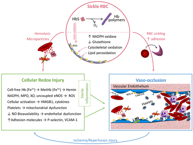
Sickle cell disease (SCD) is characterized by chronic hemolysis and repeated episodes of vascular occlusion leading to progressive organ injury. SCD is characterized by unbalanced, simultaneous pro-oxidant and anti-oxidant processes at the molecular, cellular and tissue levels, with the majority of reactions tipped in favor of pro-oxidant pathways. In this brief review we discuss new findings regarding how oxidized hemin, hemolysis, mitochondrial dysfunction and the innate immune system generate oxidative stress while hemopexin, haptoglobin, heme oxygenase-1 (HO-1) and nuclear factor erythroid 2-related factor 2 (Nrf2) may provide protection in human and murine SCD. We will also describe recent clinical trials showing beneficial effects of antioxidant therapy in SCD.

Keywords: hemoglobin; hemolysis; inflammation; oxidative stress; sickle cell disease.


Conflict of interest statement

Declaration of Interests: None

Figures

Figure 1:
Figure 1:. Redox Pathophysiology of Sickle Cell Disease (SCD).
Sickle RBC: A single point mutation in the beta globin (HBB) gene results in sickle hemoglobin (HbS), which reversibly polymerizes upon deoxygenation leading to red blood cell (RBC) sickling. Auto-oxidation of unstable HbS forms reactive oxygen species (ROS), upregulation of nicotinamide adenine dinucleotide phosphate (NADPH) oxidase and depletion of glutathione, further contributing to RBC oxidative stress, cytoskeletal oxidation and lipid membrane peroxidation. Fragile sickle RBCs are prone to lysis, releasing intracellular components and microparticles into the circulation. Cellular Redox Injury: Cell-free Hb (Fe2+) can be oxidized to MetHb (Fe3+) by scavenging nitric oxide (NO) or reacting with H2O2 (Fenton reaction); once oxidized, hemin is readily released from MetHb. Increased circulating or cellular NADPH oxidase, myeloperoxidase (MPO), xanthine oxidase (XO) and uncoupled endothelial nitric oxide synthase (eNOS) also generate ROS in SCD. Immune cell and platelet activation release high mobility group box protein 1 (HMGB1) and cytokines. Hemin, ROS, HMGB1 and cytokines all promote sterile inflammation, endothelial dysfunction and increased expression of adhesion molecules, such as P-selectin and vascular cell adhesion molecule-1 (VCAM-1). Vaso-occlusion: RBC sickling and increased adhesion between the neutrophils, platelets, sickle RBCs and the vascular endothelium lead to stasis of blood flow, known as vaso-occlusion in SCD. Repeated episodes of vaso-occlusion produce ischemia/reperfusion injury that further contributes to cellular redox injury and sterile inflammation. Used with permission from Cheryl A. Hillery.
Figure 2:
Figure 2:. Protective Mechanisms and Therapeutic Targets.
Endogenous molecules and exogenous drugs target different pathways to protect against redox injury in SCD. Sickle RBC: Fetal hemoglobin (HbF), glutathione, NADH, hydroxyurea, GBT440 (voxelotor), omega-3-fatty acids (SC411) and oral carbon monoxide help prevent RBC injury by reducing HbS polymerization or by stabilizing the RBC membrane. Cellular Redox Injury: Haptoglobin, hemopexin, heme oxygenase-1 (HO-1), Nuclear factor erythroid 2-related factor 2 (Nrf2), L-glutamine, coenzyme Q10, a wide variety of antioxidants and inhibition of cytokines or leukotrienes are both natural protective mechanisms against redox injury and potential therapeutic strategies to reduce redox stress in SCD. Vaso-occlusion: RBC transfusion, nitric oxide (NO), hydroxyurea, L-arginine, crizanlizumab (P-selectin inhibitor), rivipansel (pan-selection inhibitor), intravenous immune globulin (IVIG), CCP-224 (glycoprotein Ibα inhibitor), mitogen-activated protein kinase enzyme (MEK) inhibitors and anti-platelet agents are aimed at improving endothelial dysfunction and preventing cell-cell interactions, which play vital roles in vaso-occlusion. Only hydroxyurea and l-glutamine are FDA approved for the treatment of SCD. Used with permission from Cheryl A. Hillery.

References

    1. Kato GJ, Piel FB, Reid CD, Gaston MH, Ohene-Frempong K, Krishnamurti L, Smith WR, Panepinto JA, Weatherall DJ, Costa FF, Vichinsky EP. Sickle cell disease. Nat Rev Dis Primers. 2018;4:18010. doi: 10.1038/nrdp.2018.10. PubMed PMID: 29542687. - DOI - PubMed
    1. Sundd P, Gladwin MT, Novelli EM. Pathophysiology of Sickle Cell Disease. Annu Rev Pathol. 2019;14:263–92. doi: 10.1146/annurev-pathmechdis-012418-012838. PubMed PMID: 30332562. - DOI - PMC - PubMed
    1. Alayash AI. Oxidative pathways in the sickle cell and beyond. Blood Cells Mol Dis. 2018;70:78–86. doi: 10.1016/j.bcmd.2017.05.009. PubMed PMID: 28554826; PMCID: PMC5696113. - DOI - PMC - PubMed
    1. Romana M, Connes P, Key NS. Microparticles in sickle cell disease. Clin Hemorheol Microcirc. 2018;68(2–3):319–29. doi: 10.3233/CH-189014. PubMed PMID: 29614639. - DOI - PubMed
    1. Camus SM, Gausseres B, Bonnin P, Loufrani L, Grimaud L, Charue D, De Moraes JA, Renard JM, Tedgui A, Boulanger CM, Tharaux PL, Blanc-Brude OP. Erythrocyte microparticles can induce kidney vaso-occlusions in a murine model of sickle cell disease. Blood. 2012; 120(25): 5050–8. doi: 10.1182/blood-2012-02-413138. PubMed PMID: 22976952. - DOI - PubMed

LinkOut - more resources