Review
. 2020 Jun;35(6):937-957.
doi: 10.1007/s00467-019-04271-1. Epub 2019 Jun 25.

Assessing bone mineralisation in children with chronic kidney disease: what clinical and research tools are available?

Affiliations
Review

Assessing bone mineralisation in children with chronic kidney disease: what clinical and research tools are available?

A D Lalayiannis et al. Pediatr Nephrol. 2020 Jun.

Abstract

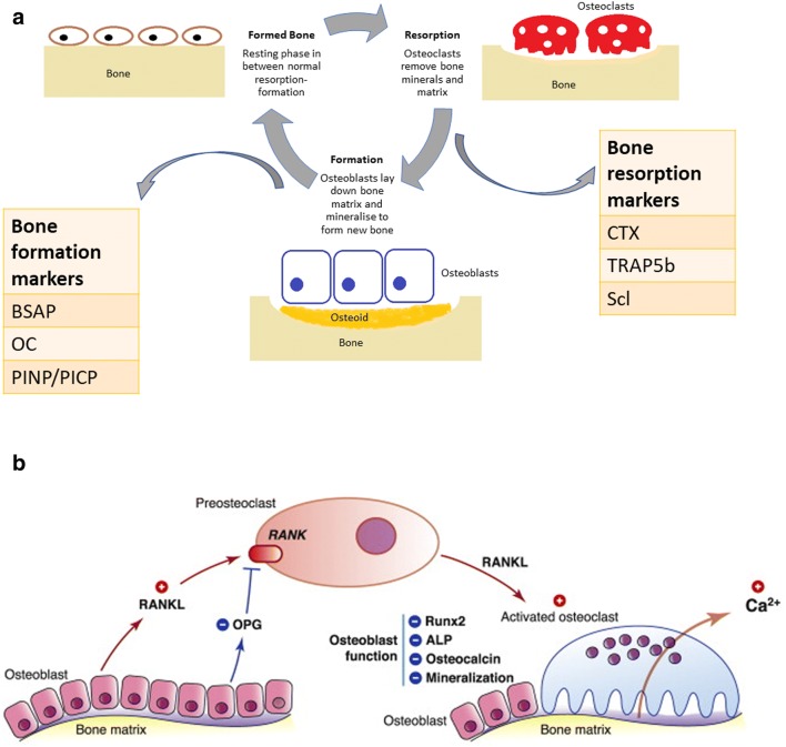
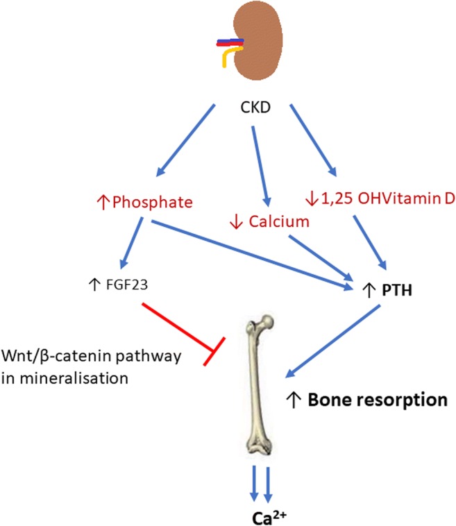
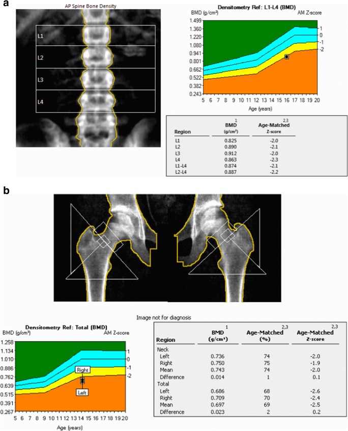
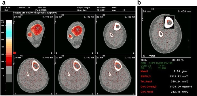
Mineral and bone disorder in chronic kidney disease (CKD-MBD) is a triad of biochemical imbalances of calcium, phosphate, parathyroid hormone and vitamin D, bone abnormalities and soft tissue calcification. Maintaining optimal bone health in children with CKD is important to prevent long-term complications, such as fractures, to optimise growth and possibly also to prevent extra-osseous calcification, especially vascular calcification. In this review, we discuss normal bone mineralisation, the pathophysiology of dysregulated homeostasis leading to mineralisation defects in CKD and its clinical consequences. Bone mineralisation is best assessed on bone histology and histomorphometry, but given the rarity with which this is performed, we present an overview of the tools available to clinicians to assess bone mineral density, including serum biomarkers and imaging such as dual-energy X-ray absorptiometry and peripheral quantitative computed tomography. We discuss key studies that have used these techniques, their advantages and disadvantages in childhood CKD and their relationship to biomarkers and bone histomorphometry. Finally, we present recommendations from relevant guidelines-Kidney Disease Improving Global Outcomes and the International Society of Clinical Densitometry-on the use of imaging, biomarkers and bone biopsy in assessing bone mineral density. Given low-level evidence from most paediatric studies, bone imaging and histology remain largely research tools, and current clinical management is guided by serum calcium, phosphate, PTH, vitamin D and alkaline phosphatase levels only.

Keywords: Bone biopsy; Bone mineralisation; Chronic kidney disease (CKD); Dual-energy X-ray absorptiometry (DXA); Peripheral quantitative CT (pQCT).


Conflict of interest statement

The authors declare that they have no conflict of interest.

Figures

Fig. 1
Fig. 1
a Remodelling of bone is controlled by osteoblasts and osteoclasts. Bone formation happens through organic matrix formation (osteoid), that gets mineralised to form bone, and finally undergoes remodelling by resorption and reformation. Calcium and phosphate form hydroxyapatite that deposits in the extracellular compartment, between collagen fibres. Osteoclasts are responsible for bone resorption, removing bone minerals and matrix. Certain biochemical markers reflect bone turnover and bone cell activity. Bone regulators can be grouped broadly into bone turnover factors (e.g. PTH, sclerostin) and bone cell activity indicators (bone formation, e.g. bone-specific alkaline phosphatase (BSAP), osteocalcin (OC), procollagen type I N propeptide (PINP), procollagen type I C propeptide (PICP); bone resorption, e.g. carboxyterminal cross-linking telopeptide of bone collagen (CTX), tartrate-resistant acid phosphatase (TRAP5b)). b Bone resorption is activated by the RANK-RANKL-OPG pathway, which regulates osteoclast differentiation and activation. Osteoclast precursors express RANK, which is activated by its ligand, RANKL, produced by osteoblasts and osteocytes. Osteoprotegerin (OPG), also a product of osteoblasts and osteocytes, is a decoy receptor for RANKL, neutralising the osteoclastic function activated by the RANKL-RANK complex. Thus, the RANKL/OPG ratio is an important determinant of bone mass as it affects mineralisation, alkaline phosphatase, Runx2 and osteocalcin which reflect osteoblast differentiation and bone formation rate. Figure adapted from Charoenphandhu et al. [117]
Fig. 2
Fig. 2
In chronic kidney disease (CKD), hypocalcaemia, low 1,25 OH vitamin D levels and hyperphosphataemia develop. In an attempt to increase phosphaturia, and thus decrease serum phosphate levels, FGF23 production increases. Raised FGF23 may directly inhibit Wnt signalling pathways which are needed in bone mineralisation. Low 1,25OHVitD and low serum calcium lead to increased PTH production. This in turn causes increased bone turnover with the aim of restoring normocalcaemia, by mobilising calcium out of bone. The reduced production of active vitamin D from the kidneys perpetuates hypocalcaemia further fuelling this cycle. This demineralisation affects bone quality as a whole leading to an increased risk of fractures and decreased bone strength
Fig. 3
Fig. 3
a DXA images. This is an example of DXA imaging of the L1-4 spine of a 16-year-old male with chronic kidney disease. His mean L1-4 age-matched Z-score is − 2.2. However, when adjusted for his shorter height and poor growth, his BMAD Z-score is − 0.8 (the BMAD value is obtained by adding the bone mineral content of the L1-L4 vertebrae and dividing by the total volume of the 4 vertebrae). b This is an example of a DXA image of both hips of a 14-year-old girl with chronic kidney disease on home nocturnal haemodialysis. Her mean age-matched Z-score for both hips is − 2.5
Fig. 4
Fig. 4
a pQCT images. This is an example of pQCT imaging of the left tibia of a 16-year-old male with chronic kidney disease. The images have been obtained at 4 different sites along the tibia. The software then proceeds to automatic analysis of the bone parameters. In this example, the images are from the 3%, 4%, 38% and 66% sites. b This is an example of the analysis of the 38% site of the left tibia of a 16-year-old male with chronic kidney disease. In this particular analysis, the total mass, total area, cortical area and cortical density have been given

References

    1. Moe S, Drueke T, Cunningham J, Goodman W, Martin K, Olgaard K, Ott S, Sprague S, Lameire N, Eknoyan G. Definition, evaluation, and classification of renal osteodystrophy: a position statement from kidney disease: improving global outcomes (KDIGO) Kidney Int. 2006;69:1945–1953. - PubMed
    1. Wesseling-Perry K, Salusky IB. Phosphate binders, vitamin D and calcimimetics in the management of chronic kidney disease-mineral bone disorders (CKD-MBD) in children. Pediatric nephrology (Berlin, Germany) 2013;28:617–625. - PMC - PubMed
    1. Ketteler M, Block GA, Evenepoel P, Fukagawa M, Herzog CA, McCann L, Moe SM, Shroff R, Tonelli MA, Toussaint ND, Vervloet MG, Leonard MB. Diagnosis, evaluation, prevention, and treatment of chronic kidney disease-mineral and bone disorder: synopsis of the kidney disease: improving global outcomes 2017 clinical practice guideline update. Ann Intern Med. 2018;168:422–430. - PubMed
    1. Fang Y, Ginsberg C, Sugatani T, Monier-Faugere M-C, Malluche H, Hruska KA. Early chronic kidney disease-mineral bone disorder stimulates vascular calcification. Kidney Int. 2014;85:142–150. - PMC - PubMed
    1. Ketteler M, Block GA, Evenepoel P, Fukagawa M, Herzog CA, McCann L, Moe SM, Shroff R, Tonelli MA, Toussaint ND, Vervloet MG, Leonard MB Executive summary of the 2017 KDIGO chronic kidney disease–mineral and bone disorder (CKD-MBD) guideline update: what’s changed and why it matters. Kidney Int 92:26–36 - PubMed

Publication types