

The role of receptor MAS in microglia-driven retinal vascular development
- PMID: 31240418
- PMCID: PMC6863789
- DOI: 10.1007/s10456-019-09671-3
The role of receptor MAS in microglia-driven retinal vascular development
Abstract
Objective: The receptor MAS, encoded by Mas1, is expressed in microglia and its activation has been linked to anti-inflammatory actions. However, microglia are involved in several different processes in the central nervous system, including the promotion of angiogenesis. We therefore hypothesized that the receptor MAS also plays a role in angiogenesis via microglia.
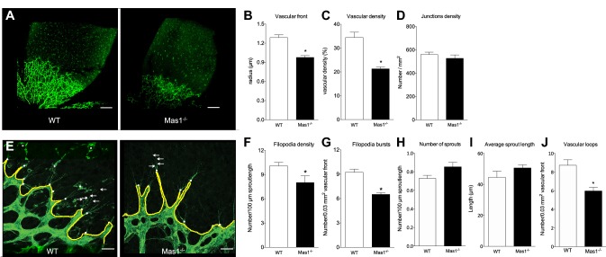
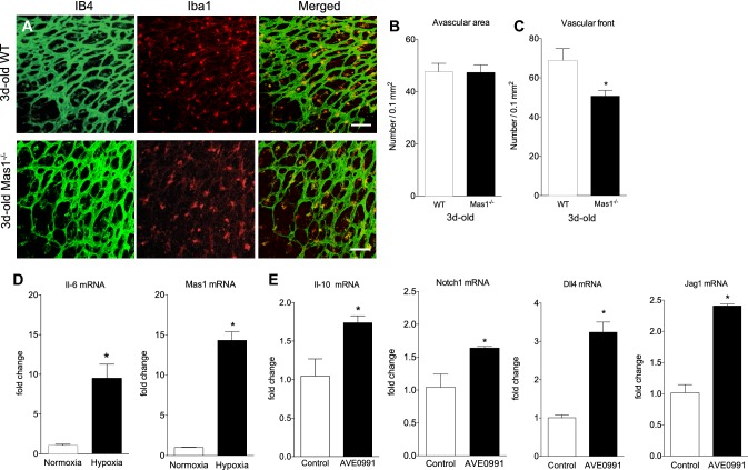
Approach and results: To assess the role of MAS on vascular network development, flat-mounted retinas from 3-day-old wild-type (WT) and Mas1-/- mice were subjected to Isolectin B4 staining. The progression of the vascular front was reduced (- 24%, p < 0.0001) and vascular density decreased (- 38%, p < 0.001) in Mas1-/- compared to WT mice with no change in the junction density. The number of filopodia and filopodia bursts were decreased in Mas1-/- mice at the vascular front (- 21%, p < 0.05; - 29%, p < 0.0001, respectively). This was associated with a decreased number of vascular loops and decreased microglial density at the vascular front in Mas1-/- mice (-32%, p < 0.001; - 26%, p < 0.05, respectively). As the front of the developing vasculature is characterized by reduced oxygen levels, we determined the expression of Mas1 following hypoxia in primary microglia from 3-day-old WT mice. Hypoxia induced a 14-fold increase of Mas1 mRNA expression (p < 0.01). Moreover, stimulation of primary microglia with a MAS agonist induced expression of Notch1 (+ 57%, p < 0.05), Dll4 (+ 220%, p < 0.001) and Jag1 (+ 137%, p < 0.001), genes previously described to mediate microglia/endothelial cell interaction during angiogenesis.
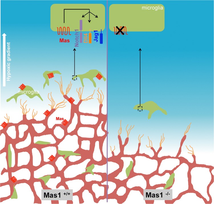
Conclusions: Our study demonstrates that the activation of MAS is important for microglia recruitment and vascular growth in the developing retina.
Keywords: Angiogenesis; Angiotensin receptors; CNS; Developmental biology; Endothelium; Macrophage; Renin angiotensin system; Vascular biology.
Figures
Similar articles
-
Microglia in retinal angiogenesis and diabetic retinopathy.Angiogenesis. 2024 Aug;27(3):311-331. doi: 10.1007/s10456-024-09911-1. Epub 2024 Apr 2. Angiogenesis. 2024. PMID: 38564108 Free PMC article. Review.
-
Single and Compound Knock-outs of MicroRNA (miRNA)-155 and Its Angiogenic Gene Target CCN1 in Mice Alter Vascular and Neovascular Growth in the Retina via Resident Microglia.J Biol Chem. 2015 Sep 18;290(38):23264-81. doi: 10.1074/jbc.M115.646950. Epub 2015 Aug 4. J Biol Chem. 2015. PMID: 26242736 Free PMC article.
-
Up-regulated basigin-2 in microglia induced by hypoxia promotes retinal angiogenesis.J Cell Mol Med. 2017 Dec;21(12):3467-3480. doi: 10.1111/jcmm.13256. Epub 2017 Jun 29. J Cell Mol Med. 2017. PMID: 28661035 Free PMC article.
-
Laminin-Dependent Interaction between Astrocytes and Microglia: A Role in Retinal Angiogenesis.Am J Pathol. 2017 Sep;187(9):2112-2127. doi: 10.1016/j.ajpath.2017.05.016. Epub 2017 Jul 8. Am J Pathol. 2017. PMID: 28697326 Free PMC article.
-
Asthma: role of the angiotensin-(1-7)/Mas (MAS1) pathway in pathophysiology and therapy.Br J Pharmacol. 2021 Nov;178(22):4428-4439. doi: 10.1111/bph.15619. Epub 2021 Aug 21. Br J Pharmacol. 2021. PMID: 34235725 Review.
Cited by
-
Microglia Contribution to the Regulation of the Retinal and Choroidal Vasculature in Age-Related Macular Degeneration.Cells. 2020 May 14;9(5):1217. doi: 10.3390/cells9051217. Cells. 2020. PMID: 32423062 Free PMC article. Review.
-
Microglia in retinal angiogenesis and diabetic retinopathy.Angiogenesis. 2024 Aug;27(3):311-331. doi: 10.1007/s10456-024-09911-1. Epub 2024 Apr 2. Angiogenesis. 2024. PMID: 38564108 Free PMC article. Review.
-
Vessel-Associated Immune Cells in Cerebrovascular Diseases: From Perivascular Macrophages to Vessel-Associated Microglia.Front Neurosci. 2019 Dec 4;13:1291. doi: 10.3389/fnins.2019.01291. eCollection 2019. Front Neurosci. 2019. PMID: 31866808 Free PMC article. Review.
-
Noteworthy perspectives on microglia in neuropsychiatric disorders.J Neuroinflammation. 2023 Oct 4;20(1):223. doi: 10.1186/s12974-023-02901-y. J Neuroinflammation. 2023. PMID: 37794488 Free PMC article. Review.
-
Microglia Colonization Associated with Angiogenesis and Neural Cell Development.Adv Neurobiol. 2024;37:163-178. doi: 10.1007/978-3-031-55529-9_10. Adv Neurobiol. 2024. PMID: 39207692 Review.
References
MeSH terms
Substances
LinkOut - more resources
Full Text Sources
Molecular Biology Databases
Research Materials
