. 2019 Dec;56(12):8345-8363.
doi: 10.1007/s12035-019-01659-w. Epub 2019 Jun 25.

NMDA Receptors Regulate Neuregulin 2 Binding to ER-PM Junctions and Ectodomain Release by ADAM10 [corrected]

Affiliations

NMDA Receptors Regulate Neuregulin 2 Binding to ER-PM Junctions and Ectodomain Release by ADAM10 [corrected]

Detlef Vullhorst et al. Mol Neurobiol. 2019 Dec.

Erratum in

Abstract

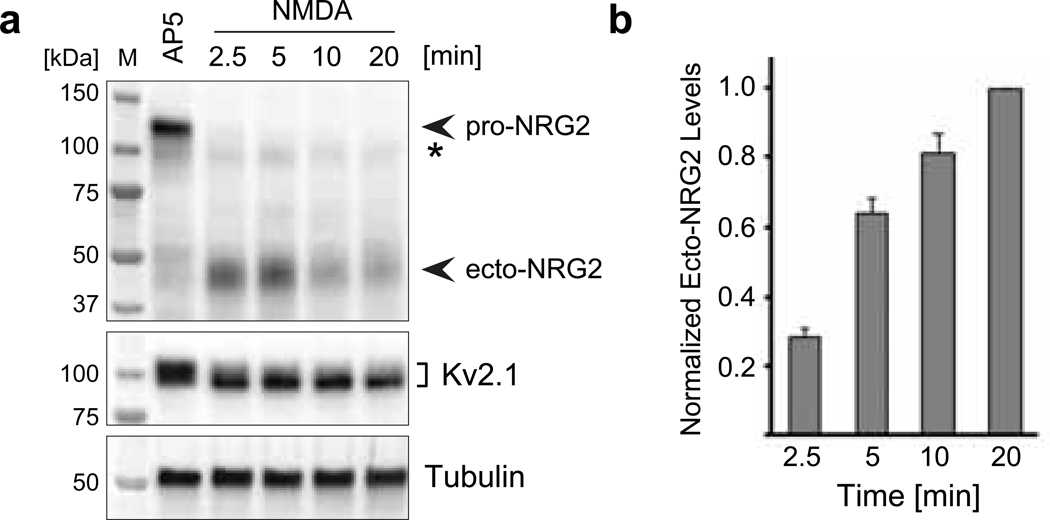
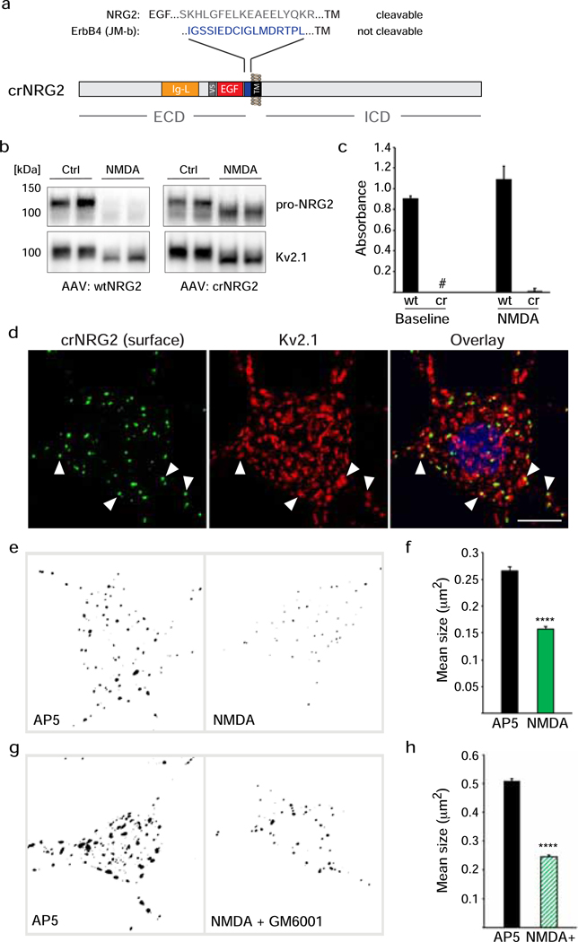
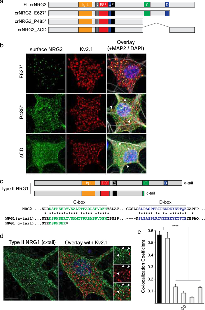
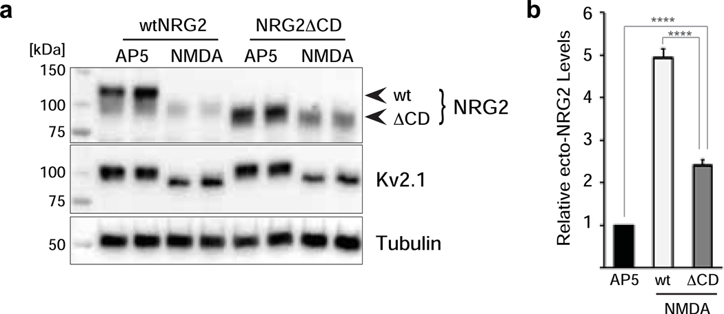
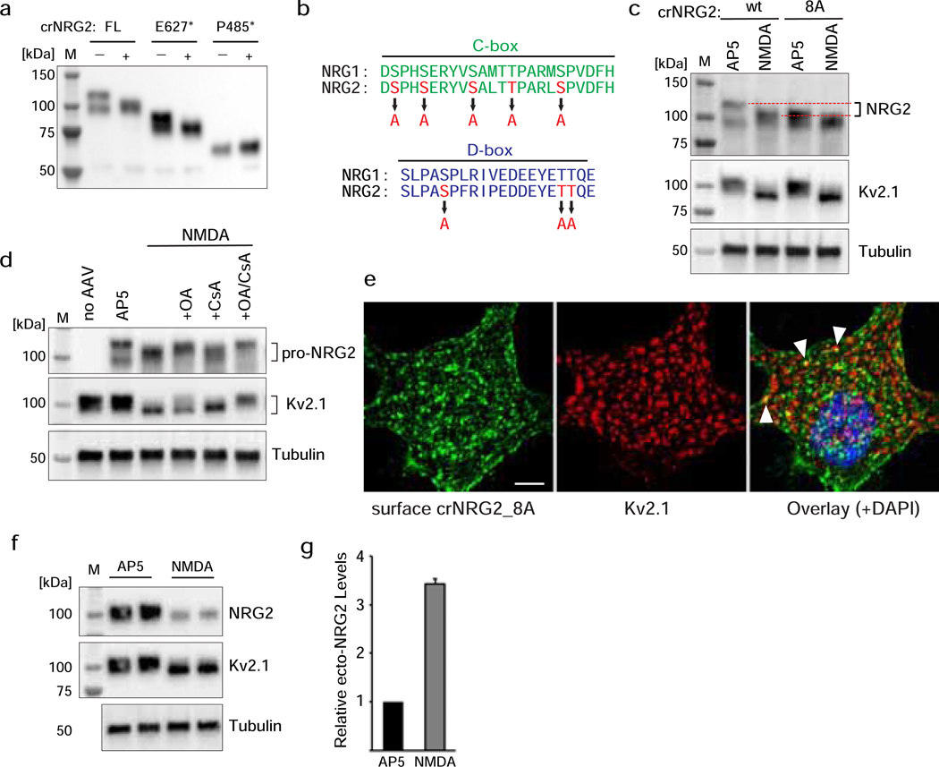
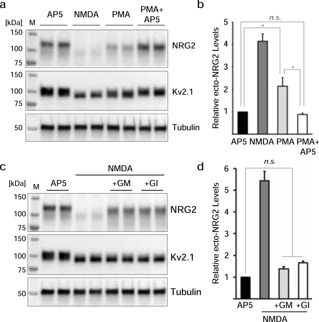
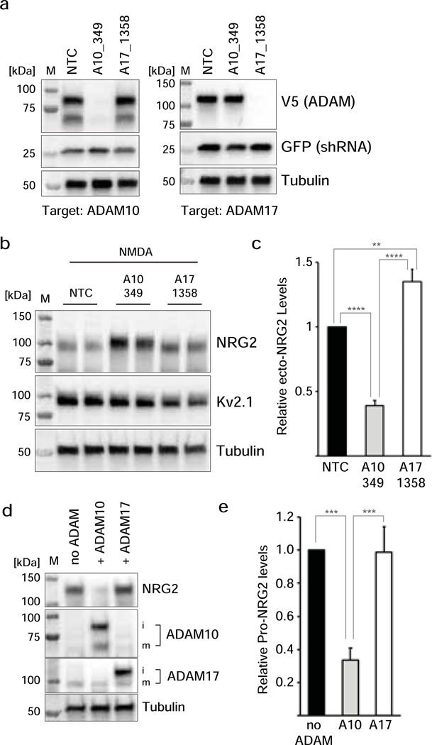
Unprocessed pro-neuregulin 2 (pro-NRG2) accumulates on neuronal cell bodies at junctions between the endoplasmic reticulum and plasma membrane (ER-PM junctions). NMDA receptors (NMDARs) trigger NRG2 ectodomain shedding from these sites followed by activation of ErbB4 receptor tyrosine kinases, and ErbB4 signaling cell-autonomously downregulates intrinsic excitability of GABAergic interneurons by reducing voltage-gated sodium channel currents. NMDARs also promote dispersal of Kv2.1 clusters from ER-PM junctions and cause a hyperpolarizing shift in its voltage-dependent channel activation, suggesting that NRG2/ErbB4 and Kv2.1 work together to regulate intrinsic interneuron excitability in an activity-dependent manner. Here we explored the cellular processes underlying NMDAR-dependent NRG2 shedding in cultured rat hippocampal neurons. We report that NMDARs control shedding by two separate but converging mechanisms. First, NMDA treatment disrupts binding of pro-NRG2 to ER-PM junctions by post-translationally modifying conserved Ser/Thr residues in its intracellular domain. Second, using a mutant NRG2 protein that cannot be modified at these residues and that fails to accumulate at ER-PM junctions, we demonstrate that NMDARs also directly promote NRG2 shedding by ADAM-type metalloproteinases. Using pharmacological and shRNA-mediated knockdown, and metalloproteinase overexpression, we unexpectedly find that ADAM10, but not ADAM17/TACE, is the major NRG2 sheddase acting downstream of NMDAR activation. Together, these findings reveal how NMDARs exert tight control over the NRG2/ErbB4 signaling pathway, and suggest that NRG2 and Kv2.1 are co-regulated components of a shared pathway that responds to elevated extracellular glutamate levels.

Keywords: ADAM10; Activity-dependent; ER-PM junction; Kv2.1; Neuregulin; Sheddase.


Figures

Fig. 1
Fig. 1. Rapid NRG2 ectodomain shedding after NMDAR stimulation in cultured hippocampal neurons.
(a) Representative Western blot of whole-cell lysates prepared from AAV-transduced hippocampal neurons expressing wt NRG2 and treated with 100 μM AP5 or with 50 μM NMDA for the indicated times. The blot was probed with anti-NRG2 (Ab 7215) raised against its extracellular domain (top). It illustrates the rapid decrease of pro-NRG2 signals at ~120 kDa apparent molecular mass in response to NMDAR activation, and a concomitant increase in ecto-NRG2 levels at ~45 kDa apparent molecular mass. The weak band marked with an asterisk (*) is only detected in transduced neurons and likely reflects immature NRG2 protein. The blot was re-probed with anti-Kv2.1 to show the characteristic downward electrophoretic mobility shift of Kv2.1 protein in response to NMDAR stimulation (middle). Tubulin signals served as reference (bottom). (b) Corresponding ELISA measurements of ecto-NRG2 accumulation in conditioned cell culture supernatants demonstrating the parallel increase in levels of soluble ecto-NRG2. Data are normalized to NMDA at 20 min (by which time pro-NRG2 protein signals were consistently undetectable in Western blots) and represent the mean ± SEM from 3 independent experiments.
Fig. 2
Fig. 2. Cleavage-resistant NRG2 reveals NMDAR-dependent downregulation of pro-NRG2 at ER-PM junctions in the absence of ectodomain shedding.
(a) Diagram of cleavage-resistant NRG2 (crNRG2), showing the replaced juxtamembrane extracellular sequence of wtNRG2 and the inserted non-cleavable juxtamembrane sequence of ErbB4 JM-b (see also [36]). Ig-like (Ig-L) and EGF-like domains are also indicated, as well as the location of a V5 epitope tag present in all NRG constructs described in this study that was used for ELISA and immunofluorescence cytochemistry. (b) Western blots of lysates prepared from hippocampal neurons expressing wtNRG2 (left) or crNRG2 (right), showing the effects of 10 min of 50 μM NMDA on pro-NRG2 protein levels. Note that NMDAR stimulation has little, if any, effect on crNRG2 protein levels (top) but causes a downward shift in its apparent electrophoretic mobility that parallels the shift in Kv2.1 mobility (below). (c) ELISA of ecto-NRG2 in the corresponding culture supernatants, demonstrating ectodomain shedding of wtNRG2 but not crNRG2. The (#) denotes that levels were below the limits of detection. Data represent the means of two biological replicates. (d) Representative confocal Z-projection of a hippocampal neuron expressing crNRG2 (surface-labeled with anti-V5), showing colocalization with endogenous Kv2.1 clusters. DAPI was included in the overlay image on the right. Arrowheads point to examples of closely associated signals for crNRG2 and Kv2.1. Scale bar = 10 μm (e) Representative binary images of crNRG2 puncta in hippocampal neurons following 10-min treatments with 100 μM AP5 (left) or 50 μM NMDA (right). (f) Quantitative analysis reveals reduction of mean crNRG2 puncta size after NMDAR stimulation (n=752 puncta from 11 neurons (AP5) and 397 puncta from 12 neurons (NMDA)). (g,h) Similar analysis of neurons expressing wtNRG2 and treated with AP5 or NMDA plus GM6001 (10 μM) (n=846 puncta from 12 neurons (AP5) and 566 puncta from 12 neurons (NMDA+GM6001)). ****, p<0.0001 (unpaired t-test).
Fig. 3
Fig. 3. NRG2 accumulates at ER-PM junctions via conserved sequence elements in its ICD.
(a) Schematic overview of NRG constructs used in this figure; motifs upstream of the TM are depicted as in Fig. 2a. Locations of conserved NRG2-ICD C/D-boxes are illustrated in green and blue, respectively. All deletions were introduced into crNRG2. (b) Representative Z-projected confocal images of neurons transduced with crNRG2_E627* (top), crNRG2_P485* (middle) or crNRG2_ΔCD (bottom). Cells were first surface-labeled with anti-V5 and subsequently labeled with anti-Kv2.1 and MAP-2 antibodies following permeabilization. Arrowheads in the crNRG2_E627* and crNRG2_P485* overlay images indicate puncta that overlap with Kv2.1 (see also Fig. S3). (c) Top, Schematic representation of a- and c-tail isoforms of type II NRG1. Note that c-tail NRG1 terminates upstream of the C-box. Bottom, Sequence comparison of the human NRG2 C- and D-boxes with the homologous sequences in the human NRG1 a-tail ICD. Conserved amino acids are marked by asterisks (*). The truncated c-tail is also shown. (d) Representative Z-projected confocal image of a transduced hippocampal neurons expressing type II NRG1 with the c-tail ICD and treated for 4 days with GM6001 (10 μM) and BACE-IV (1 μM) to block shedding. The image shows surface-labeled NRG1 using anti-V5 (left) and Kv2.1 labeled after permeabilization (right). The area marked by the bounding box in the overlay is magnified on the right and illustrates the lack of colocalization of most (white arrowheads) but not all (red arrowhead) NRG1 puncta with Kv2.1. Scale bar = 10 μM. (e) Quantitative analysis of colocalization of NRG2 variants and c-tail NRG1 with Kv2.1. Full-length crNRG2 (black) is included as reference. Data are plotted as weighted colocalization coefficients and represent the mean ± SEM (n=12 neurons per group). Note that the graph also includes data for the variant crNRG2_8A that is discussed in Fig. 5. ****, p<0.0001 (F (5,66) = 79.23; 1-way ANOVA with Tukey’s multiple comparison).
Fig. 4
Fig. 4. NMDAR activation promotes NRG2 shedding in the absence of ER-PM junction interactions.
(a) Stimulation with 50 μM NMDA for 10 min promotes processing of both wtNRG2 and, to a lesser extent, NRG2_ΔCD. Treatments were run in duplicates. Kv2.1 was included to show its electrophoretic mobility shift in response to NMDA treatment and tubulin signals served as an internal control. (b) ELISA measurements of ecto-NRG2 in culture supernatants from NMDA-treated neurons (50 μM) expressing wtNRG2 or NRG2_ΔCD, relative to their respective AP5-treated controls (100 μM). Values represent the mean ± SEM of 6 (wtNRG2) and 5 (NRG2_ΔCD) independent experiments. ****, p<0.0001 (F(2,16) = 242; 1-way ANOVA with Tukey’s multiple comparison).
Fig. 5
Fig. 5. Phosphorylation of conserved Ser/Thr residues in the NRG2 C/D-boxes regulate association with ER-PM junctions.
(a) Western blot of whole cell lysates from transduced hippocampal neurons illustrating increased electrophoretic mobility of full-length (FL) crNRG2 and crNRG2_E627*, but not crNRG2_P485*, after NMDAR stimulation with 20 μM glutamate for 20 min. (b) Conversion of Ser/Thr residues in the C- and D-boxes to Ala to generate constructs NRG2_8A and crNRG2_8A. (c) Western blot of crNRG2 vs. crNRG2_8A after 10-min treatment with AP5 or NMDAR. Note that the electrophoretic mobility of crNRG2_8A is increased compared to crNRG2 and similar to crNRG2 after NMDA treatment (red dotted lines). NMDA further increases the electrophoretic mobility of crNRG2_8A, albeit modestly. (d) Representative Western blot showing the effects of pre-treatment with Ser/Thr phosphatase inhibitors okadaic acid (500 nM; OA) or cyclosporin A (20 μM; CsA) on NMDA-mediated changes in crNRG2 (top) and Kv2.1 (middle) electrophoretic mobility. (e) Representative Z-projected confocal image of a cultured neuron expressing crNRG2_8A, surface-labeled with anti-V5 and then with anti-Kv2.1 following permeabilization. Arrowheads point to examples of some crNRG2_8A puncta that co-localize with Kv2.1, likely as a result of association with endogenous NRG2 puncta (see also Fig. S3). Scale bar = 5 μm. (f) Representative Western blot of whole cell lysates from transduced cultured neurons showing processing of pro-NRG2_8A in response to treatment with 50 μM NMDA (10 min). The panel includes Kv2.1 as positive control for the NMDA treatment and tubulin as loading control. (g) ELISA measurements of ecto-NRG2 in culture supernatants from NMDA-treated neurons expressing NRG2_8A. Data are normalized to AP5 controls and represent the mean ± SEM of 4 data points from 3 independent experiments.
Fig. 6
Fig. 6. NRG2 shedding by PMA requires NMDAR activity and the ADAM10 selective blocker GI254023X blocks NMDAR-mediated NRG2 shedding.
(a) Representative Western blot of whole cell lysates from transduced hippocampal neurons showing the effects of PMA (0.2 μM, 10 min), without or with 100 μM AP5 pre-treatment, on pro-NRG2 protein levels and electrophoretic mobility. AP5 and NMDA treatments are included as controls. (b) Corresponding ELISA measurements of ecto-NRG2 in culture supernatants treated as described in (a). Data are normalized to AP5 control and represent the mean ± SEM of 6 data points from 3 independent experiments. *, p<0.05; n.s., not significant (F(3,20) = 36.54; 1-way ANOVA with Tukey’s multiple comparison). (c) Representative Western blot of whole cell lysates from transduced hippocampal neurons showing inhibition of NMDAR-dependent pro-NRG2 processing in cultures that were pre-incubated with GM6001 (10 μM) or GI254023X (3 μM). (d) Corresponding ELISA measurements of ecto-NRG2 in culture supernatants treated as described in (d). Data are normalized to AP5 controls and represent the mean ± SEM of 6 data points from 3 independent experiments. n.s., not significant (F(3,20) = 85.27; 1-way ANOVA with Tukey’s multiple comparison).
Fig. 7
Fig. 7. ADAM10, but not ADAM17, mediates NRG2 shedding in response to NMDAR activation.
(a) Selective and potent shRNA-mediated knockdown of ADAM10 (shRNA A10_349) and of ADAM17 (shRNA A17_1358) in cultured hippocampal neurons. Neurons were transduced at DIV3 with AAVs driving a nontargeting control (NTC), shRNA A10_349 or shRNA A17_1358, and at DIV10 with AAVs expressing rat ADAM10 (left) or ADAM17 (right). (b,c) shRNA-mediated knockdown of ADAM10, but not of ADAM17, reduces NMDAR-mediated pro-NRG2 processing and ecto-NRG2 shedding. Values are normalized to the nontargeting control (NTC) and represent the mean ± SEM of 8 data points from 4 independent experiments. **, p<0.01; ****, p<0.0001 (F(2,21) = 67.14; 1-way ANOVA with Tukey’s multiple comparison). (d) AAV-driven cDNA expression of ADAM10, but not of ADAM17, in cultured hippocampal neurons decreases pro-NRG2 protein levels under baseline conditions (i.e., in the absence of exogenous NMDAR stimulation). Immature (i) and mature (m) forms of ADAM10 and ADAM17 are indicated. (e) Densitometric analysis of pro-NRG2 band intensities in control cultures and in cultures overexpressing ADAM10 or ADAM17. Data are normalized to controls and represent the mean ± SEM of 7 independent experiments. ***, p<0.001 (F(2,15) = 15.91; 1-way ANOVA with Tukey’s multiple comparison).

Similar articles

Cited by

References

    1. Gerecke KM, Wyss JM, Karavanova I, Buonanno A, Carroll SL (2001) ErbB transmembrane tyrosine kinase receptors are differentially expressed throughout the adult rat central nervous system. J Comp Neurol 433 (1):86–100 - PubMed
    1. Buonanno A, Fischbach GD (2001) Neuregulin and ErbB receptor signaling pathways in the nervous system. Curr Opin Neurobiol 11 (3):287–296. doi:S0959–4388(00)00210–5 [pii] - PubMed
    1. Birchmeier C, Nave KA (2008) Neuregulin-1, a key axonal signal that drives Schwann cell growth and differentiation. Glia 56 (14):1491–1497. doi:10.1002/glia.20753 - DOI - PubMed
    1. Buonanno A (2010) The neuregulin signaling pathway and schizophrenia: from genes to synapses and neuralcircuits. Brain Res Bull 83 (3–4):122–131. doi:S0361–9230(10)00169–3 [pii]10.1016/j.brainresbull.2010.07.012 - DOI - PMC - PubMed
    1. Mei L, Nave KA (2014) Neuregulin-ERBB Signaling in the Nervous System and Neuropsychiatric Diseases. Neuron 83 (1):27–49. doi:10.1016/j.neuron.2014.06.007 - DOI - PMC - PubMed

MeSH terms

LinkOut - more resources