. 2019 Aug 1;317(2):G98-G107.
doi: 10.1152/ajpgi.00081.2019. Epub 2019 Jun 26.

Temporal induction of intestinal epithelial hypoxia-inducible factor-2α is sufficient to drive colitis

Affiliations

Temporal induction of intestinal epithelial hypoxia-inducible factor-2α is sufficient to drive colitis

Sumeet Solanki et al. Am J Physiol Gastrointest Liver Physiol. .

Abstract

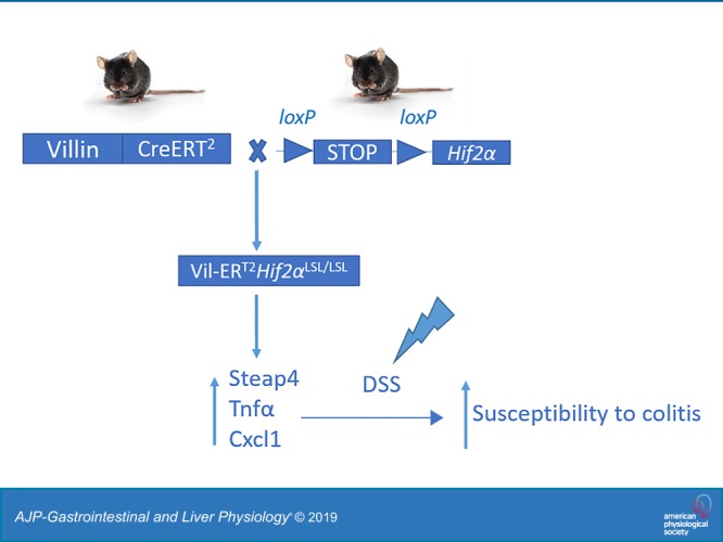
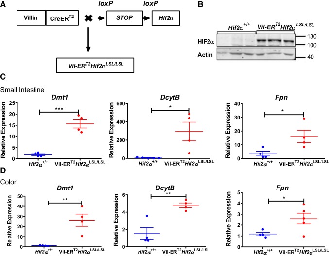
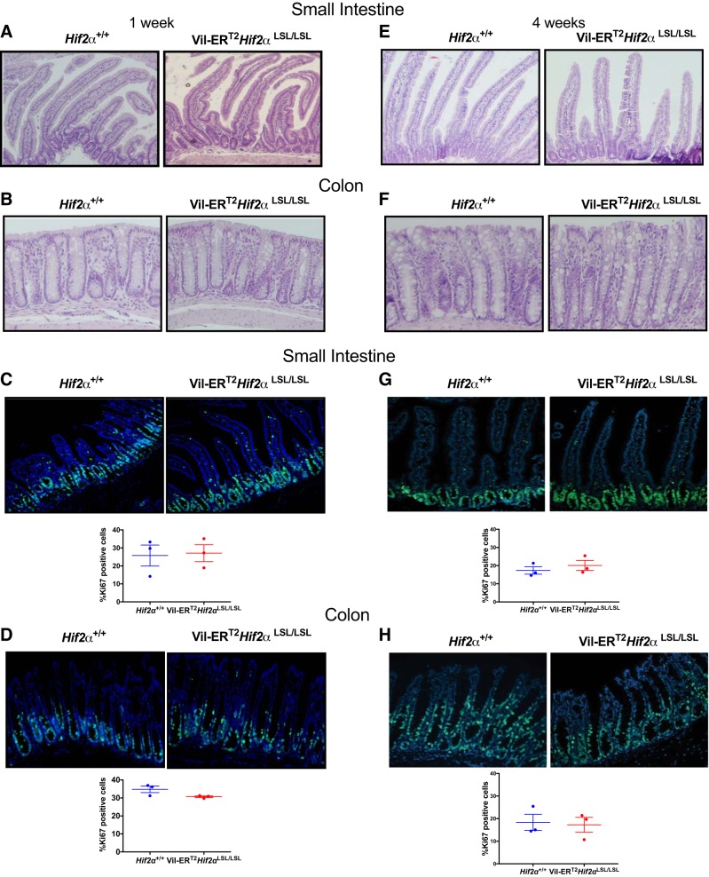
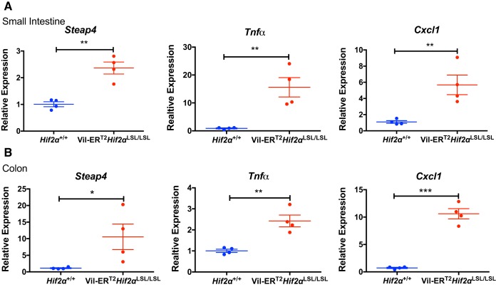
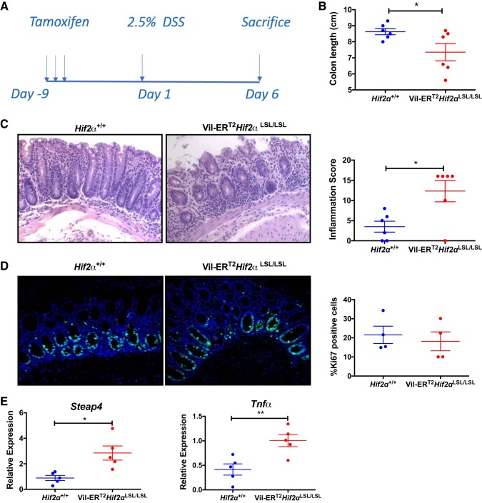
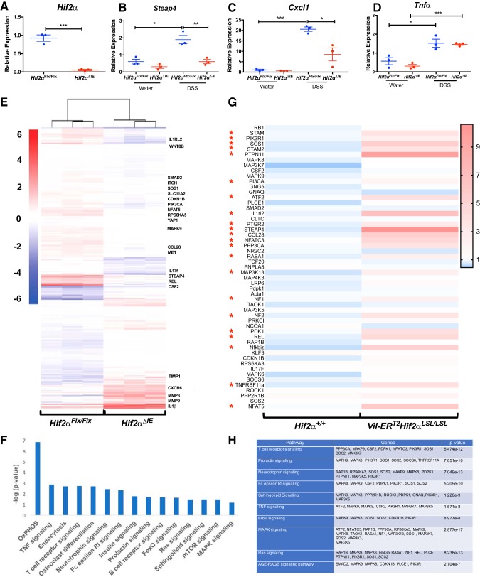
Hypoxia is a notable feature of inflammatory bowel disease and chronic induction of hypoxia-inducible factor (HIF)-1α and HIF-2α (endothelial PAS domain protein 1, EPAS1) play important, but opposing, roles in its pathogenesis. While activation of HIF-1α decreases intestinal inflammation and is beneficial in colitis, activation of HIF-2α exacerbates colitis and increases colon carcinogenesis in animal models, primarily due to the role of epithelial HIF-2α in mounting a potent inflammatory response. Previous work from our laboratory showed that mice overexpressing intestinal epithelial HIF-2α led to massive intestinal inflammation and decreased survival. As oxygen homeostasis and HIFs are critical in embryonic development, it is not clear whether the observed intestinal inflammatory response was secondary to developmental defects. To address this question, the present study used a mouse model to temporally modulate expression of intestinal epithelial HIF-2α to assess its role in mediating inflammatory response. Remarkably, activation of HIF-2α in intestinal epithelial cells in adult mice increased expression of proinflammatory mediators; however, no decrease in survival was observed. Furthermore, in an acute model of colitis, activation of HIF-2α was sufficient to exacerbate colitis. These data confirm our previous finding that epithelial HIF-2α mediates inflammatory response and demonstrates that activation of HIF-2α is sufficient to exacerbate colitis.NEW & NOTEWORTHY Inflammatory bowel disease (IBD) is a chronic relapsing inflammatory disease of the intestinal tract. Hypoxia and activation of its downstream transcription factors hypoxia-inducible factor (HIF)-1α and HIF-2α are notable features of IBD. HIF-1α has well-characterized protective roles in IBD; however, the role of HIF-2α has been less studied. Using novel HIF-2α mouse models, we show that activation of HIF-2α in intestinal epithelial cells is sufficient to exacerbate colitis.

Keywords: colitis; hypoxia; hypoxia-inducible factor-2α.


Conflict of interest statement

No conflicts of interest, financial or otherwise, are declared by the authors.

Figures

None
Graphical abstract
Fig. 1.
Fig. 1.
Temporal model of intestinal hypoxia-inducible factor-2α (HIF-2α) activation. A: schematic of a temporal HIF-2α-overexpressing mouse model (Vil-ERT2Hif2αLSL/LSL). B: Western blot analysis of HIF-2α from scraped intestinal mucosal cells of Vil-ERT2Hif2αLSL/LSL or littermate control Hif-2α+/+ mice. Quantitative PCR analysis for the expression of divalent metal transporter 1 (Dmt1), duodenal cytochrome b (DcytB), ferroportin (Fpn) in scraped mucosal cells from the small intestine (C) and colon (D) of Vil-ERT2Hif2αLSL/LSL or littermate control, Hif-2α+/+ mice. *P < 0.05, **P < 0.01, ***P < 0.001 for the difference between Vil-ERT2Hif2αLSL/LSL compared with littermate Hif-2α+/+ mice. Four mice were assessed from each group.
Fig. 2.
Fig. 2.
Temporal activation of intestinal hypoxia-inducible factor-2α (HIF-2α) does not lead to any basal histological changes. Representative hematoxylin-and-eosin staining of small intestine (duodenum) (A, E) and colon (B, F) from littermate control Hif2α+/+ and Vil-ERT2Hif2αLSL/LSL mice at 1 wk and 4 wk, respectively. Representative Ki67 staining and quantitation of small intestine (C, G) and colon (D, H) from Hif2α+/+ and Vil-ERT2Hif2αLSL/LSL mice at 1 wk and 4 wk, respectively. Four mice were assessed from each group.
Fig. 3.
Fig. 3.
Temporal activation of intestinal hypoxia-inducible factor-2α (HIF-2α) activates proinflammatory targets in the intestine. Quantitative PCR analysis of Steap4, Tnf-α, and Cxcl1 in the small intestine (A) and colon (B) from Vil-ERT2Hif2αLSL/LSL or Hif2α+/+ mice. *P < 0.05, **P < 0.01, ***P < 0.001 for the difference between Vil-ERT2Hif2αLSL/LSL and littermate control Hif2α+/+ mice. Four mice were assessed from each group.
Fig. 4.
Fig. 4.
Temporal activation of intestinal hypoxia-inducible factor-2α (HIF-2α) exacerbates colitis. A: schematic of DSS-induced colitis model. Colon length (n = 6; B), representative hematoxylin-and-eosin staining of colons and inflammation score from Hif2α+/+ and Vil-ERT2Hif2αLSL/LSL mice (n = 6; C), Ki67 staining and quantitation of colon from Hif2α+/+ and Vil-ERT2Hif2αLSL/LSL mice (n = 4; D), and qPCR analysis for the expression of Steap4 and Tnf-α in scraped colonic mucosal cells from Vil-ERT2Hif2αLSL/LSL or Hif2α+/+ mice (n = 5; E). *P < 0.05, **P < 0.01 for the differences between Vil-ERT2Hif2αLSL/LSL compared with littermate control Hif2α+/+ mice.
Fig. 5.
Fig. 5.
RNA-Seq analysis in the colons from Hif2αFlx/Flx and Hif2αΔIE mice. A: expression of HIF-2α in Hif2αFlx/Flx and Hif2αΔIE mice. Quantitative PCR analysis for the expression of Steap4 (B), Cxcl1 (C), and Tnf-α (D) in scraped colonic mucosal cells from Hif2αFlx/Flx and Hif2αΔIE mice. E: heat map generated from RNA-seq data on scrapped colonic mucosal cells isolated from Hif2αFlx/Flx and Hif2αΔIE mice. F: list of top 15 KEGG pathways altered in Hif2αFlx/Flx and Hif2αΔIE mice. G: qPCR array analysis from scraped colonic mucosal cells from Vil-ERT2Hif2αLSL/LSL and Hif2α+/+. H: list of top 10 KEGG pathways with respective altered genes in Hif2α+/+ and Vil-ERT2Hif2αLSL/LSL mice. Three mice were assessed for each group. *P < 0.05, **P < 0.01, ***P < 0.001 for the differences between Hif2αΔIE or Vil-ERT2Hif2αLSL/LSL mice compared with littermate control mice.

Similar articles

Cited by

References

    1. Campbell EL, Bruyninckx WJ, Kelly CJ, Glover LE, McNamee EN, Bowers BE, Bayless AJ, Scully M, Saeedi BJ, Golden-Mason L, Ehrentraut SF, Curtis VF, Burgess A, Garvey JF, Sorensen A, Nemenoff R, Jedlicka P, Taylor CT, Kominsky DJ, Colgan SP. Transmigrating neutrophils shape the mucosal microenvironment through localized oxygen depletion to influence resolution of inflammation. Immunity 40: 66–77, 2014. doi:10.1016/j.immuni.2013.11.020. - DOI - PMC - PubMed
    1. Cathomas G. PIK3CA in colorectal cancer. Front Oncol 4: 35, 2014. doi:10.3389/fonc.2014.00035. - DOI - PMC - PubMed
    1. Chassaing B, Aitken JD, Malleshappa M, Vijay-Kumar M. Dextran sulfate sodium (DSS)-induced colitis in mice. Curr Protoc Immunol 104: 25, 2014. - PMC - PubMed
    1. Chen W, Hill H, Christie A, Kim MS, Holloman E, Pavia-Jimenez A, Homayoun F, Ma Y, Patel N, Yell P, Hao G, Yousuf Q, Joyce A, Pedrosa I, Geiger H, Zhang H, Chang J, Gardner KH, Bruick RK, Reeves C, Hwang TH, Courtney K, Frenkel E, Sun X, Zojwalla N, Wong T, Rizzi JP, Wallace EM, Josey JA, Xie Y, Xie XJ, Kapur P, McKay RM, Brugarolas J. Targeting renal cell carcinoma with a HIF-2 antagonist. Nature 539: 112–117, 2016. doi:10.1038/nature19796. - DOI - PMC - PubMed
    1. Colombel JF, Mahadevan U. Inflammatory Bowel Disease 2017: innovations and changing paradigms. Gastroenterology 152: 309–312, 2017. doi:10.1053/j.gastro.2016.12.004. - DOI - PubMed

Publication types

Substances

LinkOut - more resources