. 2019 Jun 26;15(6):e1007850.
doi: 10.1371/journal.ppat.1007850. eCollection 2019 Jun.

The protective effect of inflammatory monocytes during systemic C. albicans infection is dependent on collaboration between C-type lectin-like receptors

Affiliations

The protective effect of inflammatory monocytes during systemic C. albicans infection is dependent on collaboration between C-type lectin-like receptors

Aiysha Thompson et al. PLoS Pathog. .

Abstract

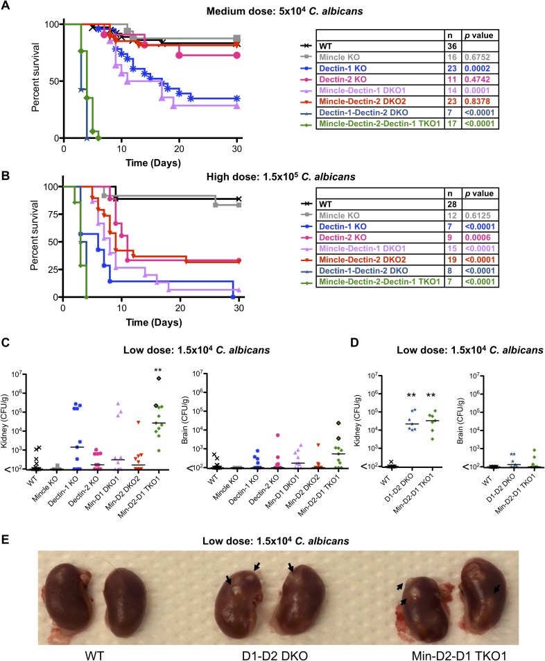
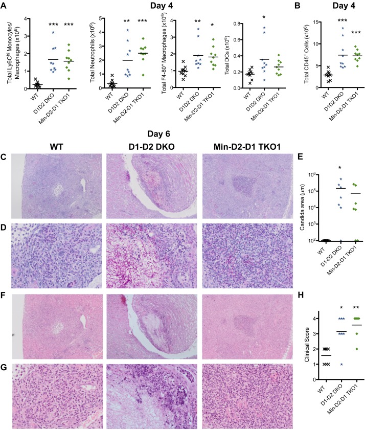
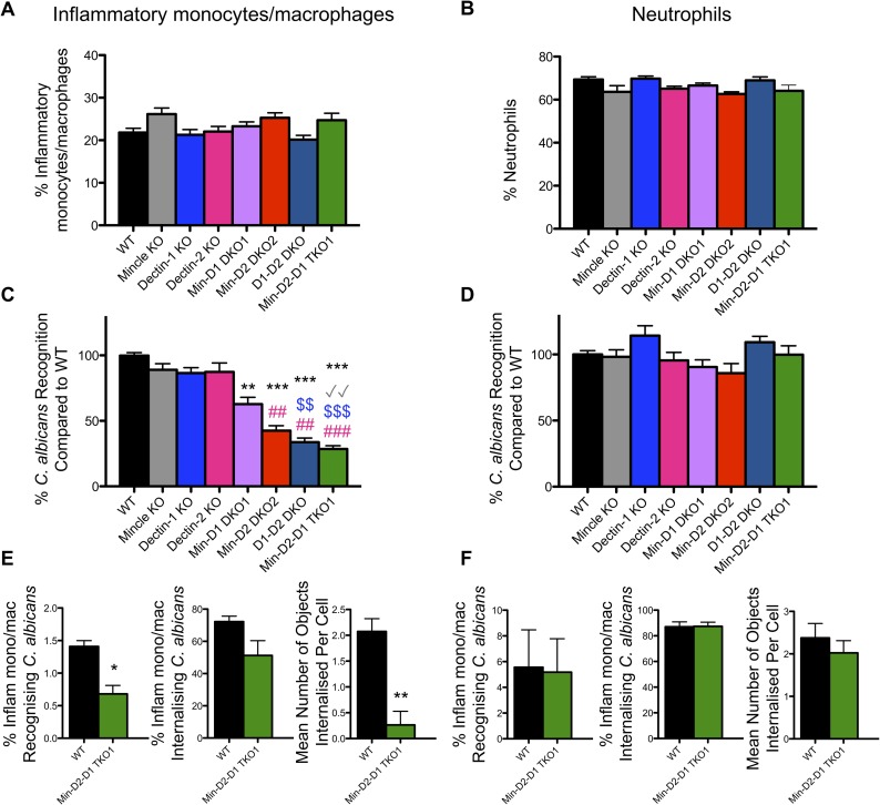
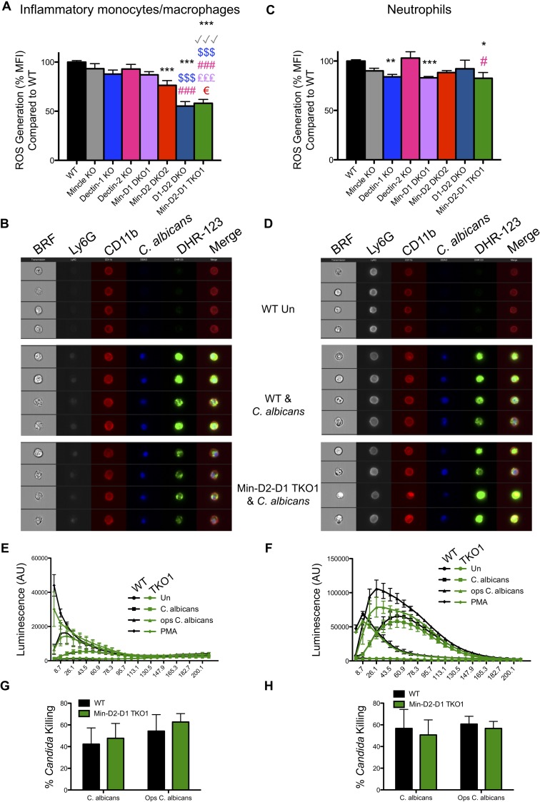
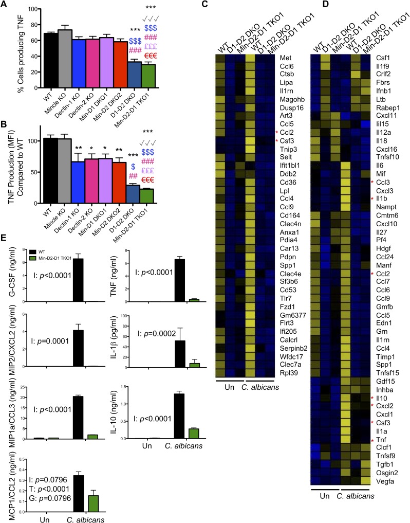
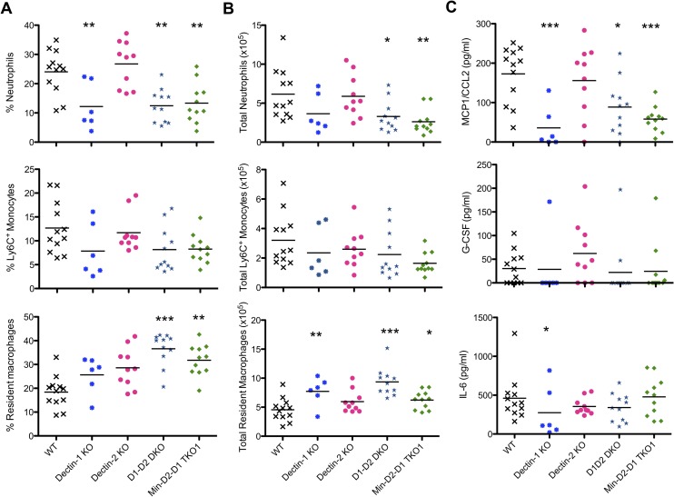
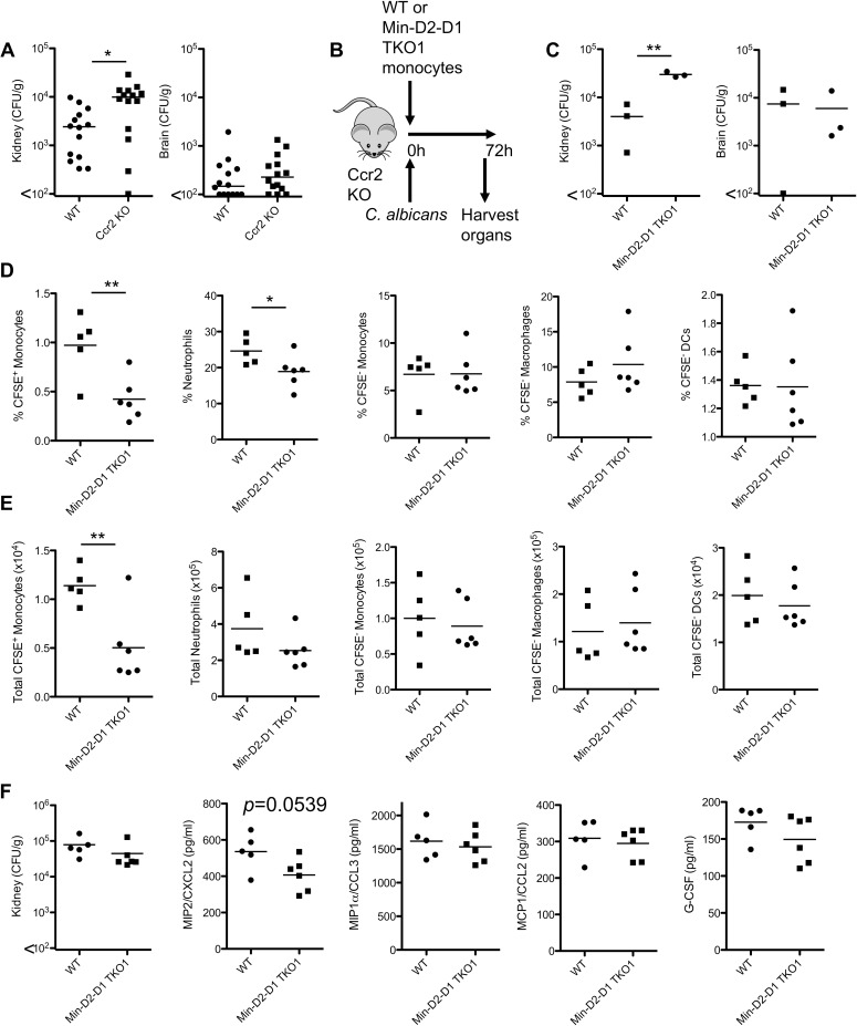
Invasive candidiasis, mainly caused by Candida albicans, is a serious healthcare problem with high mortality rates, particularly in immunocompromised patients. Innate immune cells express pathogen recognition receptors (PRRs) including C-type lectin-like receptors (CLRs) that bind C. albicans to initiate an immune response. Multiple CLRs including Dectin-1, Dectin-2 and Mincle have been proposed individually to contribute to the immune response to C. albicans. However how these receptors collaborate to clear a fungal infection is unknown. Herein, we used novel multi-CLR knockout (KO) mice to decipher the individual, collaborative and collective roles of Dectin-1, Dectin-2 and Mincle during systemic C. albicans infection. These studies revealed an unappreciated and profound role for CLR co-operation in anti-fungal immunity. The protective effect of multiple CLRs was markedly greater than any single receptor, and was mediated through inflammatory monocytes via recognition and phagocytosis of C. albicans, and production of C. albicans-induced cytokines and chemokines. These CLRs were dispensable for mediating similar responses from neutrophils, likely due to lower expression of these CLRs on neutrophils compared to inflammatory monocytes. Concurrent deletion of Dectin-1 and Dectin-2, or all three CLRs, resulted in dramatically increased susceptibility to systemic C. albicans infection compared to mice lacking a single CLR. Multi-CLR KO mice were unable to control fungal growth due to an inadequate early inflammatory monocyte-mediated response. In response to excessive fungal growth, the multi-CLR KO mice mounted a hyper-inflammatory response, likely leading to multiple organ failure. Thus, these data reveal a critical role for CLR co-operation in the effective control of C. albicans and maintenance of organ function during infection.


Conflict of interest statement

The authors have declared that no competing interests exist.

Figures

Fig 1
Fig 1. Multi-CLR KO mice are dramatically susceptible to C. albicans infection.
(A-B) Survival curves based on humane end point of WT and multi-CLR KO mice infected intravenously with 5x10⁴ CFU (A) or 1.5x10⁵ CFU (B) C. albicans SC5314. Graphs show cumulative data from 7 (A) or 6 (B) independent experiments. The total number of mice and p values for survival curves of CLR KO mice compared to WT mice by log-rank test are shown in the tables opposite each graph. (C-D) Quantification of fungal burden in the kidneys (left) or brains (right) of WT and multi-CLR KO mice 7 days (C) or 6 days (D) after intravenous infection with 1.5x10⁴ CFU C. albicans. Graphs display cumulative data from 3 (C) or 2 (D) independent experiments. Each symbol represents one mouse. Two Mincle-Dectin-2-Dectin-1 TKO1 mice had to be taken on day 6 post-infection (C). The symbols for these two mice have a black outline. (Kruskal-Wallis test with Dunn’s post-test on transformed data). (E) Photograph of representative kidneys from WT and multi-CLR KO mice 6 days after intravenous infection with 1.5x10⁴ CFU C. albicans.
Fig 2
Fig 2. C. albicans-infected multi-CLR KO mice display enhanced inflammation in association with uncontrolled fungal burden.
(A) Total myeloid cell numbers in the CD45+ cells and (B) total CD45+ cells from kidneys of WT and multi-CLR KO mice 4 days post-infection with 1.5x10⁴ CFU C. albicans were quantified by flow cytometry. Graphs display the cumulative data from 2 independent experiments. Each symbol represents an individual mouse. (1-way ANOVA with Bonferroni’s post-test, Total neutrophils: Kruskal-Wallis test with Dunn’s post-test). (C-D, E-F) Representative WT (left panels), D1-D2 DKO (middle panels) and Min-D2-D1 TKO1 (right panels) kidneys, 6 days after i.v. infection with 1.5x10⁴ CFU C. albicans. (C-D) Kidney sections were stained with Periodic acid Schiff reagent or (E-F) hematoxylin and eosin. Images shown are 4x magnification (C & F) and 20x magnification (D & G). Graphs display Candida area (μm) (E) and clinical score (H). Graphs are the cumulative result of 2 independent experiments. Each symbol represents an individual mouse. (1-way ANOVA with Bonferroni’s post-test: Performed on transformed data for Candida area).
Fig 3
Fig 3. CLR collaboration mediates C. albicans recognition by inflammatory monocytes/macrophages but not neutrophils.
(A-F) WT and CLR KO mice were injected with BIOgel i.p. and inflammatory cells were recovered by peritoneal lavage after 16–18 h. (A-D) Cells were stimulated for 15 min at 37°C with cell trace far red-labelled C. albicans at a 1:1 ratio. Cells were stained with anti-Ly6G and anti-CD11b. (A-B) Graphs display % recruited Ly6G-CD11b+ inflammatory monocytes/macrophages (A) and % Ly6G+CD11b+ neutrophils (B) in the peritoneal cavity 16–18 h after BIOgel injection. (1-way ANOVA with Bonferroni’s post-test: Performed on transformed data for Inflammatory monocytes/macrophages) (C) Ly6G-CD11b+ inflammatory monocytes/macrophages and (D) Ly6G+CD11b+ neutrophils that interacted with C. albicans were measured by flow cytometry. Percentage WT cells that recognised C. albicans was set at 100%. n = 7–16 mice. Graphs show cumulative data from 8 independent experiments. (Inflammatory monocytes/macrophages: Kruskal-Wallis test with Dunn’s post-test; Neutrophils: 1-way ANOVA with Bonferroni’s post-test). * compared to WT, √ compared to Mincle KO, $ compared to Dectin-1 KO, # compared to Dectin-2 KO (E-F) WT and Min-D2-D1 TKO1 cells were stained with anti-Ly6G and anti-CD11b. Cells were stimulated with cell trace far red-labelled C. albicans at a 1:1 ratio for 1.5 h and analysed by image flow cytometry (Amnis Imagestreamx MkII) for the course of the 1.5 h stimulation at room temperature. Inflammatory monocytes/macrophages (E) and neutrophils (F) that interacted with labelled C. albicans, internalised C. albicans and the mean number of objects internalised per cell that recognised C. albicans were quantified. n = 3 mice. (Student’s paired t test: % Inflam mono/mac recognising C. albicans on normalised data). (E-F) Graphs show the cumulative results from 3 independent experiments.
Fig 4
Fig 4. Monocytes/macrophages and neutrophils display limited dependence on CLRs for ROS production.
(A-D) WT and CLR KO mice were injected with BIOgel i.p. and inflammatory cells were recovered by peritoneal lavage after 16–18 h. Cells were stimulated for 15 min with cell trace far red-labelled C. albicans at a 1:1 ratio. Cells were loaded with the ROS indicator Dihydrorhodamine 123 (DHR-123) and stained with anti-Ly6G and anti-CD11b. (A & C) ROS production (DHR-123) was measured by flow cytometry in inflammatory monocytes/macrophages (A) and neutrophils (C) that interacted with labelled C. albicans. DHR-123 MFI for WT cells interacting with C. albicans was set at 100%. n = 7–16 mice. Graphs show cumulative data from 8 independent experiments. (Inflammatory monocytes/macrophages: 1-way ANOVA with Bonferroni’s post-test; Neutrophils: Kruskal-Wallis test with Dunn’s post-test). * compared to WT, √ compared to Mincle KO, $ compared to Dectin-1 KO, # compared to Dectin-2 KO, € compared to MincleDectin-2 DKO2 (B & D) ROS (DHR-123) was measured by image flow cytometry (Amnis Imagestreamx MkII) in inflammatory monocytes/macrophages (B) and neutrophils (D) that interacted with labelled C. albicans. Representative images from 3 independent experiments are shown. BRF = Brightfield. Merged lane consists of CD11b, C. albicans and DHR-123. Ly6G is not included in the merged lane. (E-H) WT and CLR KO mice were injected with BIOgel i.p. and inflammatory cells were recovered by peritoneal lavage after 16–18 h and Ly6G-CD11b+ inflammatory monocytes/macrophages and Ly6G+CD11b+ neutrophils were purified. (E-F) Purified cell populations were incubated with luminol for 30 min and stimulated with C. albicans, opsonised C. albicans or PMA immediately prior to measurement of luminol luminescence for the shown timecourse. Graphs show cumulative data from 3 independent experiments. (1-way ANOVA with Bonferroni’s post-test on area under the curve). (G-H) Purified Ly6G-CD11b+ inflammatory monocytes/macrophages and Ly6G+CD11b+ neutrophils were incubated with C. albicans. Cells were lysed after 3 h and candidacidal activity was analysed by measuring CFU in cell lysates. Graphs display cumulative data from 3 independent experiments. (2-way ANOVA with Bonferroni’s post-test).
Fig 5
Fig 5. CLR collaboration mediates C. albicans-induced cytokine/chemokine production from inflammatory monocytes/macrophages.
(A-D) WT and CLR KO mice were injected with BIOgel i.p. and inflammatory cells were recovered by peritoneal lavage after 16–18 h. (A-B) Cells were stimulated for 3 h with cell trace far red-labelled C. albicans at a 1:1 ratio in the presence of Brefeldin A. TNF levels were analysed by flow cytometry. Percentage Ly6G-CD11b+ inflammatory monocytes/macrophages that produced TNF—isotype (A) and MFI TNF production—isotype (B) was measured by flow cytometry in inflammatory monocytes/macrophages that interacted with labelled C. albicans. TNF MFI for WT cells interacting with C. albicans was set at 100%. (A-B) n = 2–7 mice. Graphs show cumulative data from 5 independent experiments. (1-way ANOVA with Bonferroni’s post-test). (C-D) Ly6Chi inflammatory monocytes were purified by cell sorting. Cells were stimulated with C. albicans at a ratio of 3:1 (Cells:Candida) for 3 h. RNA was extracted and RNAseq analysis was performed. Data shows the mean from two replicates each of WT and Min-D2-D1 and one replicate of D1D2 DKO cells. 177 protein coding transcripts that showed 10 or more reads and an adjusted p value of <0.05 (Benjamin-Hochberg correction for multiple testing) were selected from the RNAseq data. Following Z transformation across genes, genes were clustered by K means of FPKM values into 3 clusters using Genesis software (S8 Fig). (C) Genes from Cluster 1 are displayed. (D) Heatmap displays genes with the GO terms chemokine activity (GO:0008009), cytokine activity (GO:0005125) and growth factor activity (GO:0008083) for transcripts that showed 10 or more reads. (C-D) Data range -2.5 (blue) to +2.5 (yellow). Asterisks highlight the genes chosen for validation. (E) Bone marrow monocytes were bead purified from WT and Min-D2-D1 TKO1 mice and stimulated with C. albicans at a ratio of 1:1 for 24 h in the presence of Amphotericin B. Cytokines and chemokines in the supernatants were measured by ELISA. Graphs display cumulative data from 4 independent experiments. I = Interaction, T = Treatment, G = Genotype (matched 2-way ANOVA on transformed data).
Fig 6
Fig 6. Dectin-1 is important for early neutrophil recruitment.
(A-C) WT and CLR KO mice were injected with 1x10⁵ CFU C. albicans i.p. and inflammatory cells were recovered by peritoneal lavage after 4 h. (A-B) Percentage (A) and total number (B) of recruited neutrophils, Ly6+ monocytes and resident macrophages in the peritoneal lavages were measured by flow cytometry. (C) Chemokine and cytokine levels in the lavage fluid were measured by ELISA. (A-C) n = 6–12 mice. Graphs show cumulative data from 3 independent experiments. (1-way ANOVA with Bonferroni’s post-test–Performed on transformed data for total resident macrophages, G-CSF and IL-6; Kruskal-Wallis test with Dunn’s post-test for % Resident macrophages).
Fig 7
Fig 7. CLR collaboration is responsible for the protective effect of inflammatory monocytes during infection with C. albicans.
(A) Quantification of fungal burden in the kidneys (left) or brains (right) of WT and Ccr2 KO mice 2 days after intravenous infection with 1x10⁴ CFU C. albicans. Graphs display cumulative data from 2 independent experiments. Each symbol represents one mouse. (Student’s t test on transformed data). (B) Protocol of WT or Min-D2-D1 TKO1 monocyte transfer and C. albicans infection in Ccr2 KO mice. (C) 3-4x10⁶ purified Ly6Chi monocytes from WT or Min-D2-D1 TKO1 mice were adoptively transferred to Ccr2 KO mice followed by intravenous injection with 1.5x10⁴ CFU C. albicans. Organs were harvested after 72 h and fungal burden was determined. Graphs display cumulative data from 3 independent experiments. Each symbol represents one mouse. (Student’s paired t test). (D-F) 2.5x10⁶ purified monocytes from WT or Min-D2-D1 TKO1 mice were adoptively transferred to Ccr2 KO mice followed by intravenous injection with 1.5x10⁴ CFU C. albicans. (D-E) Kidneys were harvested after 24 h and myeloid cell recruitment to the kidneys was determined by flow cytometry. (F) Fungal burden and chemokine/cytokine production in the kidneys was determined. Graphs display cumulative data from 3 independent experiments. Each symbol represents one mouse. (Student’s t test).

References

    1. Brown GD, Denning DW, Gow NA, Levitz SM, Netea MG, White TC. Hidden killers: human fungal infections. Science translational medicine. 2012;4(165):165rv13 Epub 2012/12/21. 10.1126/scitranslmed.3004404 4/165/165rv13 [pii]. . - DOI - PubMed
    1. Alangaden GJ. Nosocomial fungal infections: epidemiology, infection control, and prevention. Infect Dis Clin North Am. 2011;25(1):201–25. Epub 2011/02/15. S0891-5520(10)00090-5 [pii] 10.1016/j.idc.2010.11.003 . - DOI - PubMed
    1. Duggan S, Leonhardt I, Hunniger K, Kurzai O. Host response to Candida albicans bloodstream infection and sepsis. Virulence. 2015;6(4):316–26. Epub 2015/03/19. 10.4161/21505594.2014.988096 - DOI - PMC - PubMed
    1. Patin EC, Thompson A, Orr SJ. Pattern recognition receptors in fungal immunity. Seminars in cell & developmental biology. 2018. Epub 2018/03/10. 10.1016/j.semcdb.2018.03.003 . - DOI - PMC - PubMed
    1. Strasser D, Neumann K, Bergmann H, Marakalala MJ, Guler R, Rojowska A, et al. Syk kinase-coupled C-type lectin receptors engage protein kinase C-sigma to elicit Card9 adaptor-mediated innate immunity. Immunity. 2012;36(1):32–42. Epub 2012/01/24. S1074-7613(12)00002-7 [pii] 10.1016/j.immuni.2011.11.015 . - DOI - PMC - PubMed

Publication types