Review
. 2019 Sep:72:100767.
doi: 10.1016/j.preteyeres.2019.06.003. Epub 2019 Jun 23.

Cholinergic nervous system and glaucoma: From basic science to clinical applications

Affiliations
Review

Cholinergic nervous system and glaucoma: From basic science to clinical applications

Muneeb A Faiq et al. Prog Retin Eye Res. 2019 Sep.

Abstract

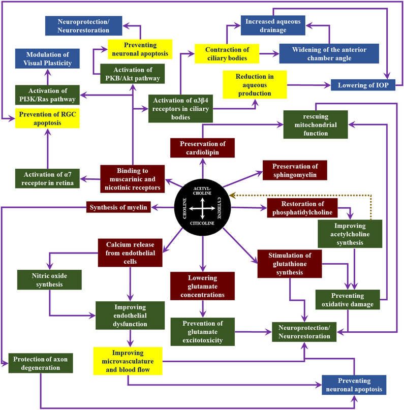
The cholinergic system has a crucial role to play in visual function. Although cholinergic drugs have been a focus of attention as glaucoma medications for reducing eye pressure, little is known about the potential modality for neuronal survival and/or enhancement in visual impairments. Citicoline, a naturally occurring compound and FDA approved dietary supplement, is a nootropic agent that is recently demonstrated to be effective in ameliorating ischemic stroke, traumatic brain injury, Parkinson's disease, Alzheimer's disease, cerebrovascular diseases, memory disorders and attention-deficit/hyperactivity disorder in both humans and animal models. The mechanisms of its action appear to be multifarious including (i) preservation of cardiolipin, sphingomyelin, and arachidonic acid contents of phosphatidylcholine and phosphatidylethanolamine, (ii) restoration of phosphatidylcholine, (iii) stimulation of glutathione synthesis, (iv) lowering glutamate concentrations and preventing glutamate excitotoxicity, (v) rescuing mitochondrial function thereby preventing oxidative damage and onset of neuronal apoptosis, (vi) synthesis of myelin leading to improvement in neuronal membrane integrity, (vii) improving acetylcholine synthesis and thereby reducing the effects of mental stress and (viii) preventing endothelial dysfunction. Such effects have vouched for citicoline as a neuroprotective, neurorestorative and neuroregenerative agent. Retinal ganglion cells are neurons with long myelinated axons which provide a strong rationale for citicoline use in visual pathway disorders. Since glaucoma is a form of neurodegeneration involving retinal ganglion cells, citicoline may help ameliorate glaucomatous damages in multiple facets. Additionally, trans-synaptic degeneration has been identified in humans and experimental models of glaucoma suggesting the cholinergic system as a new brain target for glaucoma management and therapy.

Keywords: Acetylcholine; Citicoline; Glaucoma; Neurodegeneration; Neuroprotection; Retinal ganglion cell.


Conflict of interest statement

Conflicts of interest: The author(s) have made the following disclosure(s): J.S.S.: Royalties e Zeiss, Dublin, CA (for intellectual property licensed by the Massachusetts Institute of Technology and Massachusetts Eye and Ear Infirmary)

Figures

Figure 1:
Figure 1:. Conceptual guide of the cholinergic system in vision.
This figure highlights the key messages of this paper that are important to the understanding of the cholinergic system and the therapeutic effects of cholinergic drugs including citicoline on the visual system.
Figure 2:
Figure 2:. Representative portrayal of the micro-anatomy and molecular biology of the cholinergic synapse.
This illustration gives an overview of the molecular processes, proteins, receptors and pathways of the cholinergic synapses and their locale in and outside of the neurons. The timeline of the synaptic function runs from left to right. In the presynaptic neuron, ACh is synthesized from the building blocks in the mitochondria, and is transported by the vesicles and released in the synapse. ACh signaling occurs through binding with the muscarinic and nicotinic receptors on the postsynaptic membrane. Such signaling leads to important molecular processes including neuronal plasticity, regulation of apoptosis and other cellular functions. Visual plasticity and perception are relevant to cholinergic signaling in the visual cortex, whereas RGC survival/apoptosis is imperative in glaucoma. This figure also depicts the subtle differences between central cholinergic synapse and the cholinergic synapse in the RGCs. Specifically, the ChAT in RGCs is in an alternative spliced form called pChAT. In case of insufficient production of acetylcholine in the presynaptic neuron, choline is taken back from the synapse in an autoregulatory attempt to the presynaptic membrane thereby rescuing the cellular reservoir of choline for other functions like vesicle formation and membrane component synthesis. ACh also interacts between neurons (including RGCs) and glia, which helps maintain their proper functioning and calcium uptake for the prevention and mediation of hyperactivation.
Figure 3:
Figure 3:. The cholinergic system in the eyes and brains of humans and rodents.
This figure illustrates the cholinergic mapping of the human and mouse brains. Note that the labels for the human brain also apply to the mouse brain. Humans and rodents share several similarities in the central cholinergic system. For example, the cholinergic neurons in the visual pathway mainly originate from the basal forebrain which may play a role in glaucoma in terms of visual plasticity, visual perception and regulation of intracranial pressure. The pedunculopontine-lateral dorsal tegmental projections have also been depicted in both human and mouse brains. Apart from the cholinergic innervations within the brain, the eye is sensitive to cholinergic function. Cholinergic modulation of ocular structures can help regulate the intraocular pressure (the only modifiable risk factor in glaucoma). Cholinomimetics cause contraction of ciliary bodies and widening of anterior chamber angle leading to higher rate of aqueous clearance. The muscarinic cholinergic activation decreases the aqueous production thereby leading to lowering intraocular pressure. In addition to aqueous humor dynamics, activation of α−7 nicotinic ACh receptors in the eye induces neuroprotection of retinal ganglion cells (Linn, 2016).
Figure 4:
Figure 4:. Chemical structure of citicoline.
The chemical name of citicoline is 5’-O-[hydroxy({hydroxy[2-(trimethylammonio)ethoxy]phosphoryl}Moxy)phosphoryl]cytidine. It contains two major structural components, choline and cytidine. Choline is bound to the ribose ring through a pyrophosphate bond. This chemical bridge between ribose and choline gives citicoline the chemical property to be easily broken down and readily resynthesized given the favorable conditions or the presence of relevant enzymes. This property is important to the delivery of citicoline to the CNS, as citicoline cannot cross the blood-brain barrier while choline and cytidine can, hence citicoline has to be hydrolyzed to cytidine and choline in the liver and resynthesized in the brain via the pyrophosphate bridge. Ribose and cytosine make citicoline a component important for RNA biology, though the exact role of which has not yet been deciphered. It is speculated that nucleic acid synthesis may be one of the roles of citicoline in the light of its chemical composition.
Figure 5:
Figure 5:. Citicoline synthesis and metabolism.
Citicoline and choline are closely related metabolically and are involved in the synthesis of a variety of active biochemical moieties that have widespread roles to play in membrane biology, neurotransmission, apoptosis and bioenergetics in the visual system. When being acted upon by the enzyme CDP-choline 1,2,-diacylglycerol cholinephosohotransferase, citicoline leads to the formation of phosphatidylcholine, which is an important component of neuronal membranes and is imperative to the membrane integrity of the retinal ganglion cells. Phosphatidylcholine can be converted to sphingomyelin and subsequently to myelin, a major white matter component in the brain. Phosphatidylcholine can also be converted to choline, which forms betaine upon catalytic reaction by choline oxidase, and subsequently to serine, which modulates the non-NMDA ionotropic glutamate receptors expressed by inner retinal neurons. By a variety of enzymes including choline acetyltransferase, choline is converted into ACh, which acts as a neurotransmitter and modulates aqueous humor production through parasympathetic activity. ACh can act as a substrate for the synthesis of choline, a process mediated by acetylcholinesterate.
Figure 6:
Figure 6:. Citicoline bioavailability and pharmacokinetics in different body compartments.
This schematic diagram outlines how citicoline behaves as an exogenous agent (a drug or a supplement) in the mammalian biological system and how this external agent enters the brain. After being administered via oral, intramuscular, ocular, intraperitoneal or intravenous route, citicoline enters the organ of first pass, followed by the systemic circulation and the liver. Since citicoline cannot cross the blood-brain barrier, it needs to be hydrolyzed into choline and cytidine in the liver, which readily cross the blood-brain barrier. Once choline and cytidine enter the brain via the systemic circulation, they recombine to form citicoline which can be used up for various cholinergic functions including neurotransmission, myelin regulation, neuronal membrane rescue and regeneration.
Figure 7:
Figure 7:. In vivo metabolic assessments of the brain in glaucoma.
Using magnetic resonance spectroscopy, the neurochemistry of the visual cortex in glaucoma can be evaluated non-invasively and can be compared across species spanning from the conventional experimental rat model of unilateral chronic ocular hypertension (A) to glaucoma patients (B). It is important to note that both humans and rodents show a lower choline (Cho) level in the glaucomatous visual cortex relative to the control visual cortex, whereas the creatine (Cr) level appears relatively comparable between glaucoma and control visual cortices. This suggests the reduction of choline-containing compounds in the glaucomatous visual cortex during trans-synaptic degeneration. The volumes of interests sampled are shown in the purple (A) and white boxes (B) in the multiplanar magnetic resonance brain images on the left for references. (Reproduced with permission from (Chan et al., 2009) and (Murphy et al., 2016))
Figure 8:
Figure 8:. The classical triad of citicoline actions on neurodegeneration.
This figure summarizes the biochemical and biological activities of citicoline into the triad of pharmacodynamics for treating neurodegeneration. Citicoline protects undamaged axons and hence is neuroprotective (Adibhatla et al., 2002; Bogdanov et al., 2018; Grieb, 2014; Hurtado et al., 2005; Parisi et al., 2018). It rescues the partially damaged neurons presumably through membrane re-integration and therefore is neurorestorative (Saver, 2008). The regenerative function of citicoline arises from the initial in vitro evidence for the drug to regenerate neuronal cells (Ozay et al., 2007; Skripuletz et al., 2015).
Figure 9:
Figure 9:. Representation of various severity of ocular and central vision loss in glaucoma and the candidate plan for neurotherapeutic intervention.
This figure explicates the idea of glaucoma being a neurodegenerative disorder with definite ocular and brain manifestations. (A) portrays a schematic representation of healthy (1st column), partially lost (2nd column) and completely lost visual function (3rd column). The corresponding clinical manifestations are illustrated in terms of peripapillary retinal nerve fiber layer (RNFL) thickness by optical coherence tomography (B) and visual field perimetry (C). The concomitant structural and functional brain changes in diffusion tensor MRI (D) and functional MRI (E) across increasing extents of vision loss bolsters the notion of glaucoma being a neurodegenerative disease of the visual system. This figure also considers the candidature of each condition for neurotherapeutic intervention. Since citicoline is neuroprotective, a healthy visual field (in high risk individuals) is a candidate for neuroprotection. The neurorestorative and neuroregenerative properties of citicoline make it a candidate for partially damaged and completely damaged visual field (A). Color representations for the principal diffusion directions in (D): blue, caudal-rostral; red, left-right; green, dorsal-ventral. (RNFL: retinal nerve fiber layer; FA: fractional anisotropy; OT: optic tract; VC: visual cortex; BOLD fMRI: blood-oxygenation-level-dependent functional MRI)
Figure 10:
Figure 10:. Overview of the involvements of cholinergic metabolism in neuroprotection, neurorestoration and vision rehabilitation in both basic and clinical domains.
ACh and citicoline are reciprocal precursors and are interconvertible through various enzymatic systems. Choline and cytidine are also the metabolites within this network. The reported effects of this pool of moieties (citicoline, ACh, choline and cytidine) include: (1) Preservation of cardiolipin for rescuing mitochondrial function and consequently aiding in neuroprotection and neurorestoration; (2) Preservation of sphingomyelin for myelin formation and thereby protection of neurons and assurance of proper membrane function; (3) Restoration of phosphatidylcholine for improved ACh synthesis. This may help prevent oxidative stress and is important in neuroprotective and neurorestorative processes by maintaining mitochondrial function and viability and preventing mitochondrial genome instability; (4) Stimulation of glutathione synthesis works through two main mechanisms including prevention of oxidative stress and direct neuroprotection. Prevention of oxidative stress, in particular, ensures better bioenergetics and prevents neuronal apoptosis; (5) Lowering of glutamate concentration. This primarily prevents glutamate excitotoxicity thereby promotes neuroprotection and neurorestoration; (6) Releasing calcium from endothelial cells for modulating nitric oxide and improving endothelial function. Proper functioning of the microvasculature and proper tissue perfusion is important for neuronal viability. Ameliorating endothelial dysfunction leads to improved microvasculature and better blood flow, which, in turn, prevents neuronal apoptosis; (7) Myelin synthesis for axon protection and prevention of the axonal degeneration; and (8) Binding to muscarinic and nicotinic receptors for activating molecular pathways that are involved in the prevention of RGC death and neuronal apoptosis, neurorestoration, modulation of neuroplasticity, contraction of ciliary bodies, and widening of the anterior chamber angle with increased aqueous outflow and consequent drop in IOP.

References

    1. Abudukeyoumu N, Hernandez-Flores T, Garcia-Munoz M, Arbuthnott GW, 2018. Cholinergic modulation of striatal microcircuits. Eur J Neurosci. - PMC - PubMed
    1. Acevedo-Rodriguez A, Zhang L, Zhou F, Gong S, Gu H, De Biasi M, Zhou FM, Dani JA, 2014. Cocaine inhibition of nicotinic acetylcholine receptors influences dopamine release. Front Synaptic Neurosci 6, 19. - PMC - PubMed
    1. Adhi M, Duker JS, 2013. Optical coherence tomography--current and future applications. Curr Opin Ophthalmol 24, 213–221. - PMC - PubMed
    1. Adibhatla RM, Hatcher JF, 2002. Citicoline mechanisms and clinical efficacy in cerebral ischemia. J Neurosci Res 70, 133–139. - PubMed
    1. Adibhatla RM, Hatcher JF, Dempsey RJ, 2001. Effects of citicoline on phospholipid and glutathione levels in transient cerebral ischemia. Stroke 32, 2376–2381. - PubMed

Publication types

MeSH terms