. 2019 Jun 25;24(12):2331.
doi: 10.3390/molecules24122331.

Design, Synthesis and Biological Evaluation of 7-Chloro-9 H-pyrimido[4,5- b]indole-based Glycogen Synthase Kinase-3β Inhibitors

Affiliations

Design, Synthesis and Biological Evaluation of 7-Chloro-9 H-pyrimido[4,5- b]indole-based Glycogen Synthase Kinase-3β Inhibitors

Stanislav Andreev et al. Molecules. .

Abstract

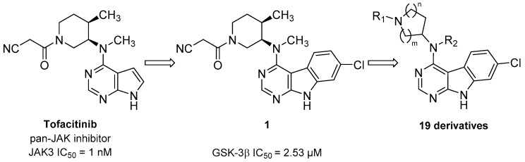
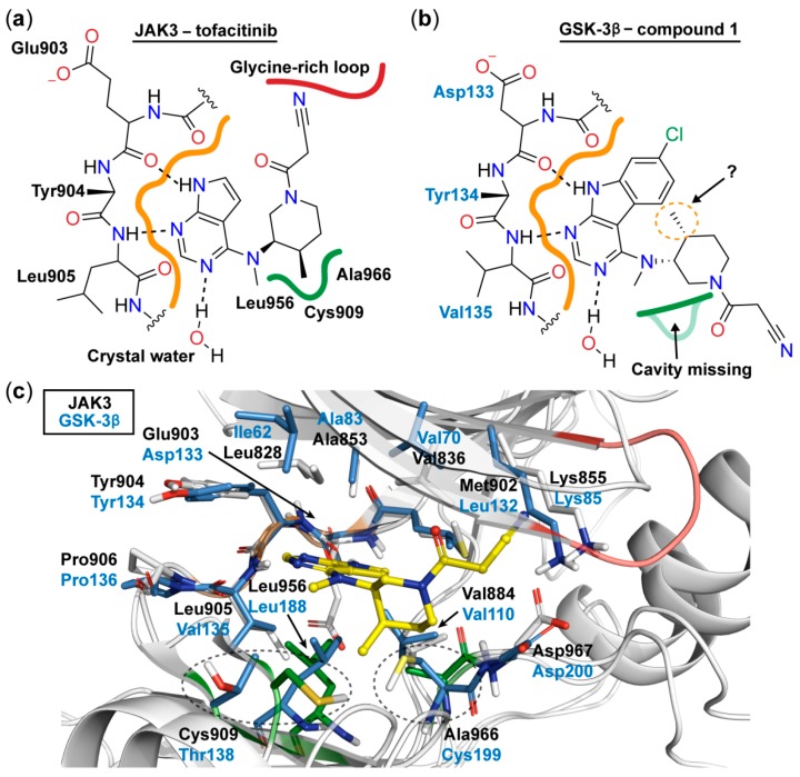
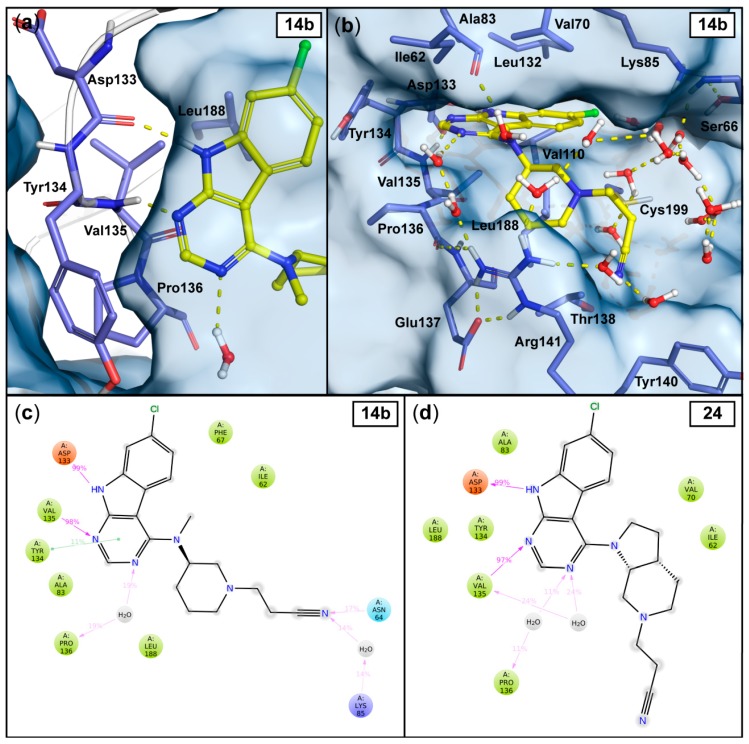
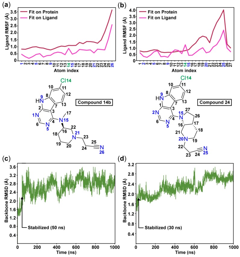
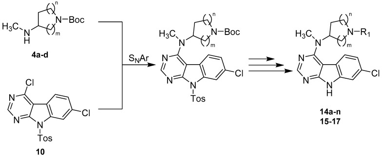
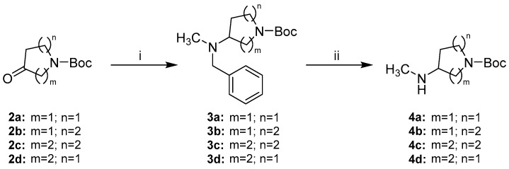
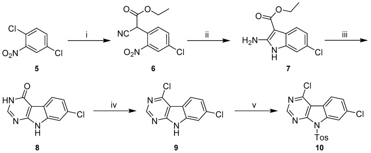
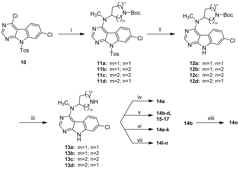
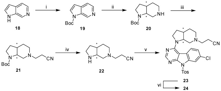
Glycogen synthase kinase-3β (GSK-3β) represents a relevant drug target for the treatment of neurodegenerative pathologies including Alzheimer's disease. We herein report on the optimization of a novel class of GSK-3β inhibitors based on the tofacitinib-derived screen hit 3-((3R,4R)-3-((7-chloro-9H-pyrimido[4,5-b]indol-4-yl)(methyl)amino)-4-methylpiperidin-1-yl)-3-oxopropanenitrile (1). We synthesized a series of 19 novel 7-chloro-9H-pyrimido[4,5-b]indole-based derivatives and studied their structure-activity relationships with focus on the cyanoacetyl piperidine moiety. We unveiled the crucial role of the nitrile group and its importance for the activity of this compound series. A successful rigidization approach afforded 3-(3aRS,7aSR)-(1-(7-chloro-9H-pyrimido[4,5-b]indol-4-yl)octahydro-6H-pyrrolo[2,3-c]pyridin-6-yl)-propanenitrile (24), which displayed an IC50 value of 130 nM on GSK-3β and was further characterized by its metabolic stability. Finally, we disclosed the putative binding modes of the most potent inhibitors within the ATP binding site of GSK-3β by 1 µs molecular dynamics simulations.

Keywords: 7-chloro-9H-pyrimido[4,5-b]indole; Glycogen synthase kinase-3β; kinase inhibitor; protein kinase; tofacitinib.


Conflict of interest statement

The authors declare no conflict of interest.

Figures

Scheme 1
Scheme 1
Development of glycogen synthase kinase-3β (GSK-3β) inhibitors derived from the pan-Janus kinase (JAK) inhibitor tofacitinib.
Figure 1
Figure 1
Schematic 2D representation of the binding mode of tofacitinib to JAK3 derived from its co-crystal structure (PDB ID: 3LXK) [16] (a) and the expected binding mode of 1 to GSK-3β (b). The lipophilic cavity is highlighted with green and the hinge region with orange. Hydrogen bonds are depicted as dashed lines. (c) 3D superposition based on the hinge regions of GSK-3β (PDB ID: 4PTC) and JAK3 (PDB ID: 3LXK) shows a high conservation of the residues in the ATP binding site [17]. The lipophilic cavity is missing in GSK-3β, as the spatial orientation of the Thr138 is totally different compared to the Cys909 in JAK3 and the residue Cys199 is bulkier than the Ala966 found in JAK3. Moreover, this Cys199 would clash with tofacitinib (yellow) in its JAK3 binding conformation. Residues near to tofacitinib are depicted as sticks. The stick coloring is the following: GSK-3β, blue; JAK3, grey; JAK3 residues forming the lipophilic cavity, green. The cartoon coloring is the following: hinge regions, orange; lipophilic cavity residues, green; JAK3 glycine-rich loop, red.
Figure 2
Figure 2
Observed interactions in the 1 μs MD simulations with the most potent compounds 14b and 24. The output conformation of compound 14b (at 1000 ns) illustrates the stable hinge interactions with the Asp133 and Val135 and to the solvent (similar in 24) (a), as well as the solvent exposure of the cyanoethyl moiety (b). Hydrogen bonds are depicted as yellow dashed lines and the water molecules within 4Å from the ligand are shown. The simulation interaction frequencies of compound 14b (c) and compound 24 (d) were analyzed using the stabilized part of the simulations: 50–1000 ns and 30–1000 ns for 14b and 24, respectively (see Figure 3c–d). Interactions that appeared with more than 10% frequency are shown; hydrogen bonds are depicted as purple arrows, π–π interactions are depicted as green lines.
Figure 3
Figure 3
The root-mean-square fluctuations (RMSF) of the ligands 14b (a) and 24 (b) illustrate the high flexibility of the cyanoethyl moieties throughout the simulations. The root-mean-square deviation (RMSD) of the protein shows that the simulations stabilize after 50 ns with compound 14b (c) and after 30 ns with compound 24 (d).
Scheme 2
Scheme 2
Synthetic strategy towards 7-chloro-9H-pyrimido[4,5-b]indole-based GSK-3β inhibitors.
Scheme 3
Scheme 3
Synthetic route to alicyclic secondary amine side chains 4a–d. Reagents and conditions: (i) N,N-benzylmethylamine, Na(OAc)3BH, AcOH, DCM, rt, (69–86%); (ii) H2 (5 bar), Pd/C, MeOH or EtOAc/MeOH 3:2, rt in case of 4a–4c or Pd/C and Pd(OH)2/C, MeOH, rt in case of 4d, (96–98%).
Scheme 4
Scheme 4
Synthetic route to 4,7-dichloro-9-tosyl-9H-pyrimido[4,5-b]indole (10). Reagents and conditions: (i) ethyl-2-cyanoacetate, NaH, DMF, 0 °C to 80 °C, (quant.); (ii) Zn, AcOH, 90 °C, (93%); (iii) NH4HCO2, formamide, 160 °C, (86%); (iv) POCl3, chlorobenzene, rt to 80 °C, (51%); (v) p-toluenesulfonyl chloride, NaH, THF, rt, (99%).
Scheme 5
Scheme 5
Synthetic route to final compounds 14a–o listed with their structures in Table 1 and 15–17 listed with their structures in Table 2. Reagents and conditions: (i) 4a–d, DIPEA, DMF, 80 °C, (93% to quant.); (ii) KtBuO, THF, rt, (65–78%); (iii) TFA, DCM, rt, (74–99%); (iv) 13b, cyanoacetic acid, PyBOP, DIPEA, DCM, rt, (59%); (v) 13a–d, acrylonitrile, MeOH, rt in case of 14b and 15–17 or 13b, corresponding acrylic acid derivative, MeOH, rt in case of 14c–d, (63–89%); (vi) 13b, corresponding aldehyde or ketone, AcOH, Na(OAc)3BH, DCM, rt, (43–74%); (vii) 13b, bromoacetonitrile, Et3N, DMF, rt in case of 14l or 13b, (bromomethyl)cyclopropane, Et3N, MeCN, 60 °C in case of 14m or 13b, 3-chloro-N,N-dimethylpropan-1-amine ·HCl, Et3N, MeCN, 90 °C in case of 14n, (39–91%); (viii) methyl iodide, NaH, THF, rt, (60%).
Scheme 6
Scheme 6
Synthetic route to final compound 24. Reagents and conditions: (i) Boc2O, THF, 0 °C to rt, (92%); (ii) H2 (5 bar), PtO2, AcOH, rt, (88%); (iii) acrylonitrile, MeOH, rt, (83%); (iv) 4N HCl in dioxane, DCM, rt, (83%); (v) 10, DIPEA, DMF, 80 °C, (64%); (vi) KtBuO, THF, rt, (55%).
Figure 4
Figure 4
X-ray crystal structure of compound 24.

Similar articles

Cited by

References

    1. Manning G., Whyte D.B., Martinez R., Hunter T., Sudarsanam S. The protein kinase complement of the human genome. Science. 2002;298:1912–1934. doi: 10.1126/science.1075762. - DOI - PubMed
    1. Manning G., Genomic Overview of Protein Kinases. WormBook The C. Elegans Research Community, WormBook. [(accessed on 13 December 2005)]; doi: 10.1895/wormbook.1.60.1. Available online: http://www.wormbook.org. - DOI - PMC - PubMed
    1. Sutherland C. What Are the Bona fide GSK3 Substrates? Int. J. Alzheimer’s Dis. 2011;2011:23. - PMC - PubMed
    1. Embi N., Rylatt D.B., Cohen P. Glycogen synthase kinase-3 from rabbit skeletal muscle. Eur. J. Biochem. 1980;107:519–527. doi: 10.1111/j.1432-1033.1980.tb06059.x. - DOI - PubMed
    1. Wang Y., Roach P.J. Inactivation of rabbit muscle glycogen synthase by glycogen synthase kinase-3. Dominant role of the phosphorylation of Ser-640 (site-3a) J. Biol. Chem. 1993;268:23876–23880. - PubMed

MeSH terms

Substances

LinkOut - more resources