. 2019 Jun 27;21(1):34.
doi: 10.1186/s12968-019-0546-3.

A simple measure of the extent of Ebstein valve rotation with cardiovascular magnetic resonance gives a practical guide to feasibility of surgical cone reconstruction

Affiliations

A simple measure of the extent of Ebstein valve rotation with cardiovascular magnetic resonance gives a practical guide to feasibility of surgical cone reconstruction

Marina L Hughes et al. J Cardiovasc Magn Reson. .

Abstract

Background: Once surgical management is indicated, variation of Ebstein valve morphology affects surgical strategy. This study explored practical, easily measureable, cardiovascular magnetic resonance (CMR)-derived attributes that may contribute to the complexity and risk of cone reconstruction.

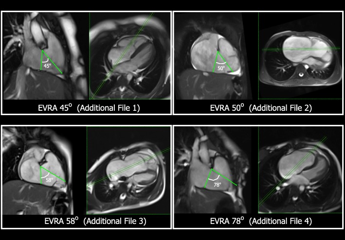
Methods: A retrospective assessment was performed of Ebstein anomaly patients older than 12 years age, with pre-operative CMR, undergoing cone surgical reconstruction by one surgeon. In addition to clinical data, the CMR-derived Ebstein valve rotation angle (EVRA), area ratios of chamber size, indexed functional RV (RVEDVi) and left ventricular (LV) volumes, tricuspid valve regurgitant fraction (TR%) and other valve attributes were related to early surgical outcome; including death, significant residual TR% or breakdown of repair.

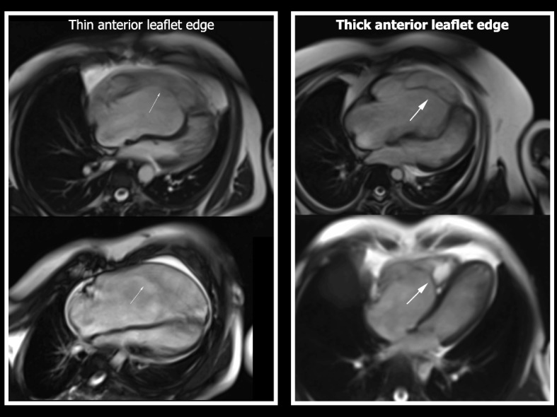
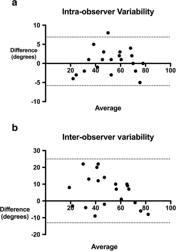
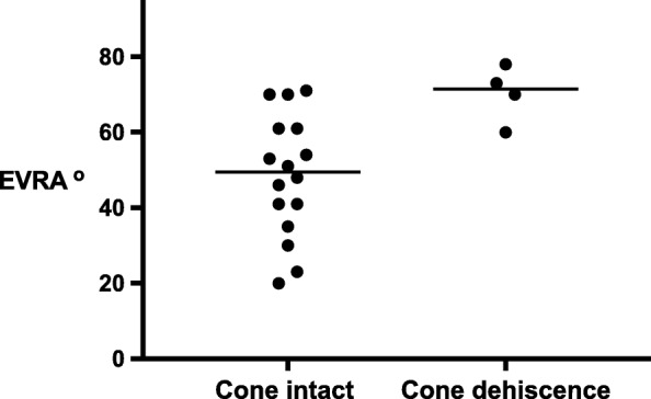
Results: Of 26 operated patients older than 12 years age, since program start, 20 had pre-op CMR and underwent surgery at median (range) age 20 (14-57) years. TR% was improved in all patients. Four of the 20 CMR patients (20%) experienced early surgical dehiscence of the paravalve tissue, with cone-shaped tricuspid valve intact; one of whom died. A larger EVRA correlated with Carpentier category and was significantly related to dehiscence. If EVRA >60o, relative risk of dehiscence was 3.2 (CI 1.3-4.9, p = 0.03). Those with dehiscence had thickened, more tethered anterior leaflet edges (RR 17, CI 3-100, p < 0.01), smaller pre-operative functional RVEDVi; (132 vs 177 mL/m2, p = 0.04), and were older (median 38 vs 19 years, p = 0.01). TR %, chamber area ratios and LV parameters were not different.

Conclusions: Comprehensive CMR assessment characterizes patients prior to cone surgical reconstruction of Ebstein anomaly. Pragmatic observation of larger EVRA, smaller RVEDVi and leaflet thickening, suggests risk of repair tension and dehiscence, and may require specific modification of cone surgical technique, such as leaflet augmentation.

Keywords: Cardiovascular magnetic resonance imaging; Cone reconstruction; Congenital heart disease; Ebstein anomaly; Tricuspid valve.


Conflict of interest statement

The authors declare that they have no competing interests.

Figures

Fig. 1
Fig. 1
These panels illustrate the method of Ebstein valve rotation angle (EVRA) measurement in 4 different patients, using the end-systolic frame from cardiovascular magnetic resonance (CMR) balanced steady state free precession (bSSFP) cine images. In each panel, the left-hand image is the right ventricular (RV) long axis cine view, from which the EVRA is calculated. The right-hand image in each panel is the 4-chamber cine view from the same patient. The green triple-line demonstrates the position of the RV long axis view relative to the 4-chamber view, which is a perpendicular plane through the centre of the right atrio-ventricular (AV) junction, parallel to the interventricular septum. The EVRA for each of these patients is shown in green, drawn with apex at the superior basal hingepoint of the TV anterior leaflet in the RV long axis cine view. Each of these image panels corresponds to Additional movie files 1–4 in the supplementary data
Fig. 2
Fig. 2
This is an end-diastolic frame from a 4-chamber view, using the CMR bSSFP sequence. The left-hand panel (a) shows the segmentation and tracing method for area ratios of the right and left heart, superimposed upon the image shown on the right, without segmentation (b). fRV = functional RV, RA = right atrium, LA + LV = left atrium + left ventricle
Fig. 3
Fig. 3
This figure shows end-diastolic frames from a 4-chamber view, using bSSFP images, demonstrating the morphologic characteristic of thickened edges of the anterior leaflet. Two patients with thickened anterior leaflet edges are shown on the right-sided panels (thick white arrows), compared to two patients in this surgical cohort with similar EVRA and RV size, with valves that did not exhibit thickened edges (thin white arrows) on cine images
Fig. 4
Fig. 4
Scattergram of the measured Ebstein valve rotation angle (EVRA) for the whole cohort, n = 20, stratified by Carpentier classification
Fig. 5
Fig. 5
a and b Bland Altman plots showing intra and inter-observer variability measuring Ebstein valve rotation angle (EVRA). The dotted lines show the 95% Limits of Agreement
Fig. 6
Fig. 6
Scattergram of the measured Ebstein valve rotation angle (EVRA), comparing patients with early surgical dehiscence of the cone reconstruction (n = 4), compared with patients with an intact repair. (p = .0.01). This indicates that the effective tricuspid valve orifice and plane of systolic closure was more rotated towards the RV outflow tract in the patients suffering early surgical dehiscence

Similar articles

Cited by

References

    1. Dearani JA, Danielson GK. Congenital heart surgery nomenclature and database project: Ebstein's anomaly and tricuspid valve disease. Ann Thorac Surg. 2000;69(4):S106–S117. doi: 10.1016/S0003-4975(99)01265-5. - DOI - PubMed
    1. Attenhofer Jost CH, Connolly HM, Edwards WD, Hayes D, Warnes CA, Danielson GK. Ebstein's anomaly - review of a multifaceted congenital cardiac condition. Swiss Med Wkly. 2005;135(19–20):269–281. - PubMed
    1. da Silva JP, Baumgratz JF, da Fonseca L, Franchi SM, Lopes LM, Tavares GM, et al. The cone reconstruction of the tricuspid valve in Ebstein's anomaly. The operation: early and midterm results. J Thorac Cardiovasc Surg. 2007;133(1):215–223. doi: 10.1016/j.jtcvs.2006.09.018. - DOI - PubMed
    1. Silva JP, Silva Lda F, Moreira LF, Lopez LM, Franchi SM, Lianza AC, et al. Cone reconstruction in Ebstein's anomaly repair: early and long-term results. Arq Bras Cardiol. 2011;97(3):199–208. doi: 10.1590/S0066-782X2011005000084. - DOI - PubMed
    1. Lange R, Burri M, Eschenbach LK, Badiu CC, da Silva JP, Nagdyman N, et al. Da Silva's cone repair for Ebstein's anomaly: effect on right ventricular size and function. Eur J Cardiothorac Surg. 2015;48(2):316–320. doi: 10.1093/ejcts/ezu472. - DOI - PubMed

Publication types

MeSH terms