. 2019 Aug 28;93(18):e00292-19.
doi: 10.1128/JVI.00292-19. Print 2019 Sep 15.

Model System for the Formation of Tick-Borne Encephalitis Virus Replication Compartments without Viral RNA Replication

Affiliations

Model System for the Formation of Tick-Borne Encephalitis Virus Replication Compartments without Viral RNA Replication

Wai-Lok Yau et al. J Virol. .

Abstract

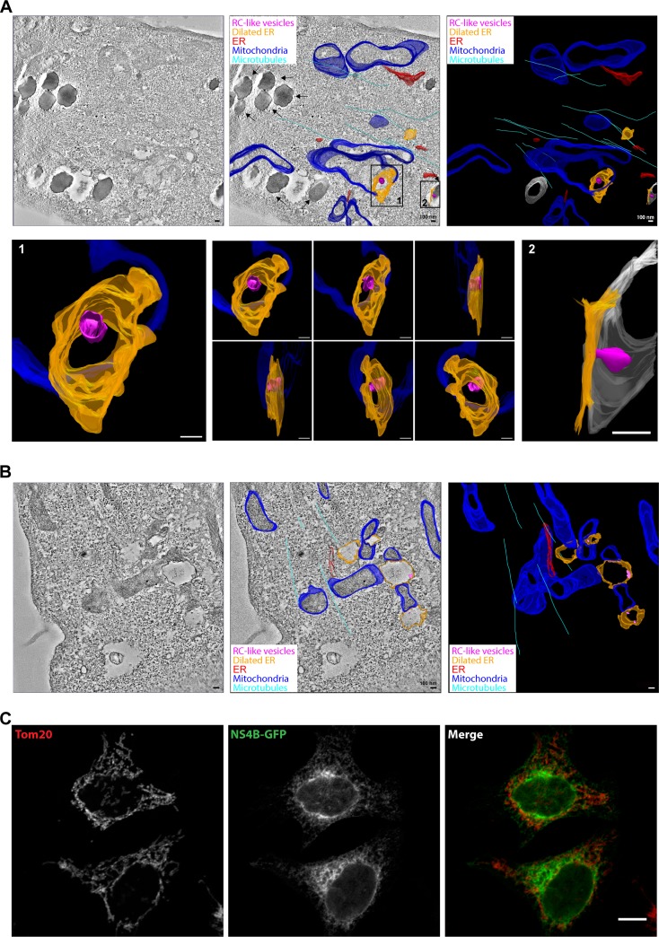
Flavivirus is a positive-sense, single-stranded RNA viral genus, with members causing severe diseases in humans such as tick-borne encephalitis, yellow fever, and dengue fever. Flaviviruses are known to cause remodeling of intracellular membranes into small cavities, where replication of the viral RNA takes place. Nonstructural (NS) proteins are not part of the virus coat and are thought to participate in the formation of these viral replication compartments (RCs). Here, we used tick-borne encephalitis virus (TBEV) as a model for the flaviviruses and developed a stable human cell line in which the expression of NS proteins can be induced without viral RNA replication. The model system described provides a novel and benign tool for studies of the viral components under controlled expression levels. We show that the expression of six NS proteins is sufficient to induce infection-like dilation of the endoplasmic reticulum (ER) and the formation of RC-like membrane invaginations. The NS proteins form a membrane-associated complex in the ER, and electron tomography reveals that the dilated areas of the ER are closely associated with lipid droplets and mitochondria. We propose that the NS proteins drive the remodeling of ER membranes and that viral RNA, RNA replication, viral polymerase, and TBEV structural proteins are not required.IMPORTANCE TBEV infection causes a broad spectrum of symptoms, ranging from mild fever to severe encephalitis. Similar to other flaviviruses, TBEV exploits intracellular membranes to build RCs for viral replication. The viral NS proteins have been suggested to be involved in this process; however, the mechanism of RC formation and the roles of individual NS proteins remain unclear. To study how TBEV induces membrane remodeling, we developed an inducible stable cell system expressing the TBEV NS polyprotein in the absence of viral RNA replication. Using this system, we were able to reproduce RC-like vesicles that resembled the RCs formed in flavivirus-infected cells, in terms of morphology and size. This cell system is a robust tool to facilitate studies of flavivirus RC formation and is an ideal model for the screening of antiviral agents at a lower biosafety level.

Keywords: Flaviviridae; Flp-In cell line; NS4B; flavivirus; replication compartment; replication vesicles; tick-borne encephalitis virus.


Figures

FIG 1
FIG 1
Dose- and time-dependent expression of NS1-NS4B-GFP in Flp-In T-REx HeLa cells. (A) Schematic illustration of the proposed membrane topology of the TBEV polyprotein, with color-coded representation of the individual structural (C, prM, and E) and nonstructural (NS1 to NS5) proteins. The GFP-tagged polyprotein (NS1 to NS4B) expressed following dox addition in the constructed NSP-GFP-Flp-In cells is shown below. (B) Immunoblotting expression analysis of the individual NS proteins, as indicated, following induction of NSP-GFP cells with different concentrations (0 to 10 ng/ml) of dox. Clathrin served as a loading control. (C) Immunoblotting analysis of the time-dependent expression of NS proteins, as indicated, following induction with 2 ng/ml dox in NSP-GFP cells (left), transfection with the TBEV DNA replicon (1 μg DNA per 5 × 106 cells) (middle), or infection with LGTV (MOI of 1) or TBEV Torö strain (MOI of 1) (right). Actin and clathrin served as loading controls. (D) Representative fluorescence micrographs of the time-dependent expression of NS4B-GFP in NSP-GFP cells induced by 2 ng/ml dox, as recorded by live cell imaging. Scale bars = 10 μm. (E) Schematic illustration of the organization of the TBEV DNA replicon. (F) Representative images from immunofluorescence analysis of Flp-In HeLa cells transfected with the TBEV DNA replicon. Nuclei were stained with 4′,6-diamidino-2-phenylindole (DAPI), and the ER marker calnexin was labeled with specific antibody, as indicated. dsRNA was labeled with the mouse anti-dsRNA monoclonal antibody J2. The GFP signal served as the reporter for replicon transfection. Scale bar = 10 μm. (G) Quantification of dsRNA in TBEV-replicon-transfected cells (n = 25) and untransfected cells (n = 25) from the immunofluorescence analysis in panel F. The dsRNA quantification was performed with Imaris v7.5 software (Bitplane). The statistical analysis was performed with GraphPad Prism software (GraphPad Software) (n = 25). ***, P ≤ 0.01 (t test).
FIG 2
FIG 2
NS proteins form a protein complex in the ER of NSP-GFP cells. (A) Representative fluorescence micrographs of dox-induced NSP-GFP cells expressing NS4B-GFP and immunostained against calreticulin or Sec61A, as indicated. Scale bars = 10 μm. (B) FRAP analysis of the dynamics of NS4B-GFP in dox-induced NSP-GFP cells. ER networks were stained with ER-tracker. Regions enriched in both NS4B-GFP and ER-tracker were photobleached, and then the fluorescence signal of the regions was traced for 300 s. Means ± standard errors of the means (SEMs) are shown. (C) Immunoblot analysis of the separation of NS proteins in the supernatant (S) and pellet (P), as indicated, following lysis and differential centrifugation of induced NS1-NS4B cells at the indicated speed (1,000 to 100,000 × g). Antibodies to Sec61A, calnexin, and calreticulin were used as markers of the ER. (D) OptiPrep density flotation analysis of the 20,000 × g pellet fraction in panel C, with or without pretreatment with1% Triton-X to dissolve membranes. Flotation of calnexin and individual NS proteins, as indicated, was analyzed by immunoblotting. (E) Immunoprecipitation of GFP and NS4B-GFP from dox-induced GFP (Ctl) or NS4B-GFP (4B) Flp-In cells coexpressing FLAG-tagged NS1, NS2A, NS2B, NS3, NS4A, NS4B, or NS5, as indicated. Input and immunoprecipitated (IP) material was analyzed by immunoblotting as indicated.
FIG 3
FIG 3
Replication-independent expression of NS1 to NS4B in HeLa cells results in dilation of the ER membrane and generation of RC-like structures. (A) Representative TEM images of control HeLa cells, cells infected with LGTV (MOI of 1), cells transfected with the TBEV DNA replicon, and uninduced and induced NSP-GFP cells. White arrowheads show dilated ER areas; black arrowheads denote replication-vesicle-like structures inside the dilated ER areas. Insets show magnifications of the indicated areas. Scale bars = 1 μm (except in insets [scale bars = 0.1 μm]). (B) Quantification of the number of dilated ER areas in replicon-transfected cells, LGTV-infected cells, and dox-induced NSP-GFP cells, compared with control HeLa cells (n = 5). Quantification was performed using ImageJ. **, P ≤ 0.05 (t test). (C) Membrane profiles and sizes of replication-vesicle-like structures in replicon-transfected cells, LGTV-infected cells, and dox-induced NSP-GFP cells. The membrane profiles were outlined using Adobe Photoshop. The diameters of at least 30 randomly chosen vesicles from each group were measured using ImageJ.
FIG 4
FIG 4
Localization of NS proteins in RC-like structures in NSP-GFP cells. (A) CLEM images of a NSP-GFP cell induced with dox. (Top) The left fluorescence micrograph shows NS4B-GFP (green) and ER-tracker (red), and the right panel shows the correlated electron micrograph, with arrows highlighting dilated ER membranes. (Bottom) The left panel shows an electron micrograph of the area indicated by the white square in higher magnification, and the right panel shows further magnification of the indicated area; the ER and dilated ER (dER) are indicated, and black arrowheads denote vesicle-like structures. Scale bars = 10 μm (top) or as indicated below the bars (bottom). (B) Immunofluorescence micrographs of the single and merged channels of a dox-induced NSP-GFP cell expressing NS4B-GFP and stained against NS1. Yellow and white arrowheads exemplify the punctuate NS1 localization on NS4B-GFP-positive ER tubules. Scale bar = 10 μm. (C) Immuno-EM images of NSP-GFP cells induced with dox. The cells were stained with anti-NS1 antibodies conjugated with gold particles. Scale bars = 200 nm.
FIG 5
FIG 5
ER dilation caused by NS proteins is linked to mitochondria. (A and B) Electron tomography analysis of induced NSP-GFP cells, showing close contacts of lipid droplets, mitochondria, and dilated ER. Tomograms were recorded and the membranes of mitochondria, ER, lipid droplets, and microtubules were manually assigned, yielding a schematic, color-coded, 3D membrane model overlaid on representative tomograms. A tomographic slice (left), overlaid 3D model (middle), and 3D model (right) are presented to show dilated ER (orange) and RC-like invaginations (magenta). ER tubules are depicted in red, mitochondria in blue, microtubules in cyan, and lipid droplets in gray. Scale bars = 100 nm. (A) In the overlaid 3D model, black arrows indicate lipid droplets. Insets show magnification and different angles of the indicated areas (also see movie S2 in the supplemental material). (B) Representative electron tomograms of dox-induced NSP-GFP cells show close contact of mitochondria and dilated ER. (C) Fluorescence micrographs of NS4B-GFP in NSP-GFP cells costained for mitochondria using antibodies to the mitochondrial protein Tom20. Scale bar, 10 μm.

Similar articles

Cited by

References

    1. Lindquist L. 2014. Tick-borne encephalitis. Handb Clin Neurol 123:531–559. doi:10.1016/B978-0-444-53488-0.00025-0. - DOI - PubMed
    1. Hrnjaković Cvjetković I, Cvjetković D, Patić A, Radovanov J, Kovacević G, Milosević V. 2016. Tick-borne encephalitis virus infection in humans. Med Pregl 69:93–98. doi:10.2298/MPNS1604093H. - DOI - PubMed
    1. Bogovic P, Strle F. 2015. Tick-borne encephalitis: a review of epidemiology, clinical characteristics, and management. World J Clin Cases 3:430–441. doi:10.12998/wjcc.v3.i5.430. - DOI - PMC - PubMed
    1. Gould EA, Solomon T. 2008. Pathogenic flaviviruses. Lancet 371:500–509. doi:10.1016/S0140-6736(08)60238-X. - DOI - PubMed
    1. Yun SI, Lee YM. 2017. Zika virus: an emerging flavivirus. J Microbiol 55:204–219. doi:10.1007/s12275-017-7063-6. - DOI - PubMed

Publication types

MeSH terms