Gliosarcoma in a Young Filipino Woman: A Case Report and Review of the Literature
- PMID: 31243260
- PMCID: PMC6613492
- DOI: 10.12659/AJCR.916020
Gliosarcoma in a Young Filipino Woman: A Case Report and Review of the Literature
Abstract
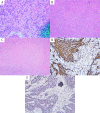
BACKGROUND Gliosarcoma (GS) is a rare variant of glioblastoma (GBM), which is typically seen in patients age 40-60 years and located in the supratentorial region. We present an unusual case of GS in a young patient with an unusual presentation, which eventually led to the finding of this neoplasm. CASE REPORT Our patient was a 38-year-old woman originally from the Philippines who was transferred to our institution with an isolated left foot drop that developed over the course of several months. Subsequent neuroimaging revealed an extensive mixed cystic and solid mass in the posterior mesial right frontal lobe. Subtotal surgical resection revealed a multi-lobed tumor with a malignant glioma-like surface component overlying a smooth, well-encapsulated, avascular, sarcoma-like component. Neuropathologic examination of the resected tumor revealed a biphasic histologic pattern of predominantly sarcomatous components with fewer adjacent-area glial components. Post-operatively, the patient was left with a mild worsening of left leg segmental strength. She was referred to our neurooncologist colleagues for adjuvant treatment options. CONCLUSIONS Our case is unique in that it represents a rare neoplasm in a patient whose demographics are atypical for this type of tumor, as well as the unusual presentation of isolated foot drop.
Conflict of interest statement
None.
Figures


References
-
- Morantz RA, Feigen I, Ransohoff J. Clinical and pathological study of 24 cases of gliosarcoma. J Neurosurg. 1976;45:398–408. - PubMed
-
- Meis JM, Martz Kl, Nelson JS. Mixed glioblastoma multiforme and sarcoma: A clinicopathologic study of 26 Radiation Therapy Oncology Group cases. Cancer. 1991;67:2342–49. - PubMed
-
- Zhang BY, Chen H, Geng DY, et al. Computed tomography and magnetic resonance features of gliosarcoma: A study of 54 cases. J Comput Assist Tomogr. 2011;35(6):667–73. - PubMed
Publication types
MeSH terms
LinkOut - more resources
Full Text Sources
Medical

