. 2019 Jun 26;10(7):504.
doi: 10.1038/s41419-019-1687-x.

The interaction between SPARC and GRP78 interferes with ER stress signaling and potentiates apoptosis via PERK/eIF2α and IRE1α/XBP-1 in colorectal cancer

Affiliations

The interaction between SPARC and GRP78 interferes with ER stress signaling and potentiates apoptosis via PERK/eIF2α and IRE1α/XBP-1 in colorectal cancer

Yi-Jye Chern et al. Cell Death Dis. .

Abstract

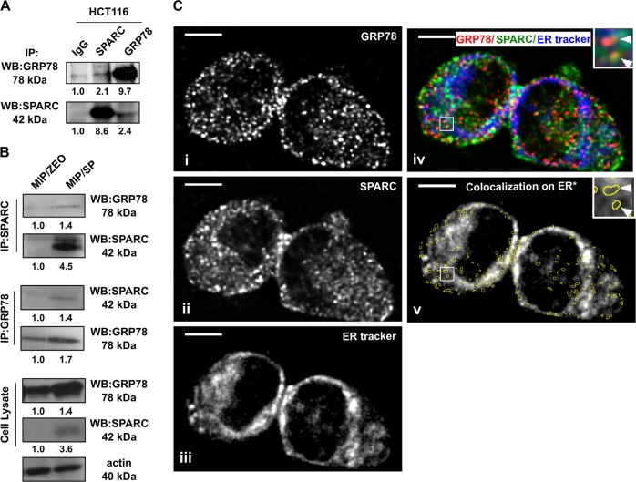
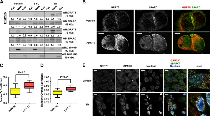
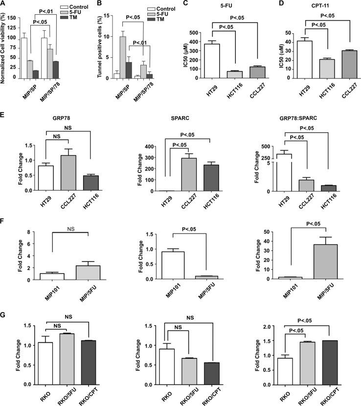
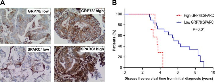
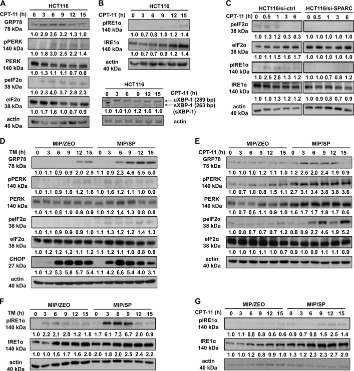
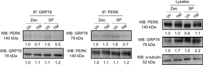
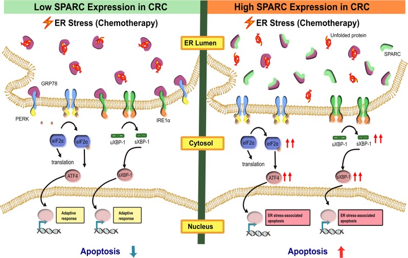
Therapy-refractory disease is one of the main contributors of treatment failure in cancer. In colorectal cancer (CRC), SPARC can function as a sensitizer to conventional chemotherapy by enhancing apoptosis by interfering with the activity of Bcl-2. Here, we examine a novel mechanism by which SPARC further potentiates apoptosis via its modulation of the unfolded protein response (UPR). Using mass spectrometry to identify SPARC-associated proteins, GRP78 was identified as a protein partner for SPARC in CRC. In vitro studies conducted to assess the signaling events resulting from this interaction, included induction of ER stress with tunicamycin, 5-fluorouracil (5-FU), and irinotecan (CPT-11). We found that the interaction between GRP78 and SPARC increased during exposure to 5-FU, CPT-11, and tunicamycin, resulting in an attenuation of GRP78's inhibition of apoptosis. In addition, we also show that SPARC can sensitize CRC cells to PERK/eIF2α and IRE1α/XBP-1 UPR signaling by interfering with ER stress following binding to GRP78, which leads to ER stress-associated cell death in CRC cells. In line with these findings, a lower expression of GRP78 relative to SPARC in CRC is associated with a lower IC50 for 5-FU in either sensitive or therapy-refractory CRC cells. Interestingly, this observation correlates with tissue microarray analysis of 143 human CRC, where low GRP78 to SPARC expression level was prognostic of higher survival rate (P = 0.01) in individuals with CRC. This study demonstrates that modulation of UPR signaling by SPARC promotes ER stress-associated death and potentiates apoptosis. This may be an effective strategy that can be combined with current treatment options to improve therapeutic efficacy in CRC.


Conflict of interest statement

The authors declare that they have no conflict of interest.

Figures

Fig. 1
Fig. 1. Interaction between GRP78 and SPARC in CRC.
Co-immunoprecipitation of SPARC with GRP78 in (a) HCT116 and confirmed in (b) MIP/Zeo and MIP/SP cells. (c) Colocalization of (i) GRP78 and (ii) SPARC in HCT116 by confocal microscopy and immunofluorescent analysis. (iii) The endoplasmic reticulum was stained with ER tracker blue-white DPX dye. (iv) Images were overlapped, and (v) the GRP78:SPARC colocalization pixels were identified by Colocalization Finder plugin in ImageJ and highlighted with yellow circles. The average ER tracker staining intensity in the highlighted areas was calculated to determine the percentage of GRP78:SPARC colocalization in the ER. Scale bar = 5 μm
Fig. 2
Fig. 2. Dynamic interaction between SPARC and GRP78 in the ER occurs with chemotherapy-induced ER stress.
(a) HCT116 were treated with 5-FU (25 μM, 12 h) and TM (1 μg/ml, 6 h) followed by cell fractionation, co-IP and Western blot analysis of cytoplasmic (Cyt), membranous (Mem), and nuclear (Nuc) fractions. Calnexin and DNA-PK served as quality control of cell fractionation. Densitometry quantification was performed, and the densitometry values were indicated below the blots. (b) Immunofluorescent analysis of HCT116 cells treated with CPT-11 (50 μM, 12 h) or vehicle by confocal microscopy. Cells were labeled with primary antibodies against SPARC (green) and GRP78 (red). Quantitative colocalization analysis was performed followed by deconvolution of the images. Scale bar = 5 μm. (c) The Pearson’s coefficient and (d) the overlap coefficient was calculated from 12 images (N = 12) for each group. (e) Immunofluorescent analysis of MIP/SP cells treated with TM (1 μg/ml, 6 h) or vehicle. Colocalization of GRP78 and SPARC was evident in TM-treated cells based on the analysis using the ImageJ Colocalization Finder plugin (white). Fluorescence intensities were gamma-corrected (gamma = 2.0) to reveal the colocalization pixels in the perinuclear region. Scale bar = 10 μm
Fig. 3
Fig. 3. SPARC attenuates GRP78’s pro-survival effects under ER stress.
The effects of SPARC and GRP78 expression on cell viability under ER stress were determined by using MIP/SP and GRP78-overexpressing MIP/SP cells (MIP/SP/78) following exposure to 5-FU (5 μM) and TM (1 μg/ml) for 48 h. Cell viability and apoptosis were analyzed by (a) MTS and (b) TUNEL assays, respectively. Cell viability and IC50 of CRC cell lines HT29, CCL227, and HCT116 following exposure to (c) 5-FU and (d) CPT-11 were determined by MTS assay. The mRNA expression levels of GRP78 (left), SPARC (middle), and the relative expression level (GRP78:SPARC) (right) in (e) CRC cell lines (HT29, CCL227, HCT116), (f) MIP101, and (g) RKO 5-FU- and CPT-11-resistant cell lines were determined by q-RT-PCR. (The data represent mean ± SEM, N = 3)
Fig. 4
Fig. 4. CRC patients with a low ratio of GRP78:SPARC protein expression have improved prognosis.
(a) Representative images of GRP78 and SPARC expression in CRC scored as high expression and low expression; x20, magnification. (b) Kaplan–Meier survival curves of patients with CRC. Individuals with tumor expressing low GRP78:SPARC ratio have significantly higher median disease-free survival
Fig. 5
Fig. 5. SPARC promotes early activation of PERK-eIF2α and IRE1α-sXBP ER stress signaling in endogenous HCT116 and MIP/SP cells.
HCT116 treated with CPT-11 (50 μM) for various time intervals were analyzed for the presence of the activation of (a) PERK/eIF2α and (b) IRE1α/XBP-1. The spliced form of XBP-1 was detected by RT-PCR analysis. (c) Western blot analysis of ER stress signaling in HCT116 cells following SPARC siRNA knockdown and exposure to CPT-11 (50 μM). Activation of (d, e) PERK/eIF2α and (f, g) IRE1α/XBP-1 signaling in MIP/Zeo and MIP/SP following treatment with TM (1 μg/ml) or CPT-11 (50 μM) were examined by Western blot analysis
Fig. 6
Fig. 6. SPARC interferes with the binding between GRP78 and PERK under ER stress.
MIP/Zeo and MIP/SP were treated with TM (1 μg/ml, 6 h) and the cell lysates were immunoprecipitated with anti-GRP78 and anti-PERK antibodies, respectively, followed by Western blot analysis
Fig. 7
Fig. 7. SPARC reduces the tolerance to ER stress in CRC cells.
SPARC interferes with the binding between GRP78 and ER stress sensors (such as PERK and IRE1α) via its interaction with GRP78. Under ER stress, the stress stimulus is amplified in high SPARC-expressing cells, as SPARC facilitates the dissociation of GRP78 from the ER stress sensors, thereby lowering the threshold of ER stress signaling and subsequent activation of the apoptotic cascade

References

    1. Torre LA, et al. Global cancer statistics, 2012. CA Cancer J. Clin. 2015;65:87–108. doi: 10.3322/caac.21262. - DOI - PubMed
    1. Luo B, Lee AS. The critical roles of endoplasmic reticulum chaperones and unfolded protein response in tumorigenesis and anticancer therapies. Oncogene. 2013;32:805–818. doi: 10.1038/onc.2012.130. - DOI - PMC - PubMed
    1. Wu J, Kaufman RJ. From acute ER stress to physiological roles of the unfolded protein response. Cell Death Differ. 2006;13:374–384. doi: 10.1038/sj.cdd.4401840. - DOI - PubMed
    1. Hetz C. The unfolded protein response: controlling cell fate decisions under ER stress and beyond. Nat. Rev. Mol. Cell Biol. 2012;13:89–102. doi: 10.1038/nrm3270. - DOI - PubMed
    1. Pootrakul L, et al. Expression of stress response protein Grp78 is associated with the development of castration-resistant prostate cancer. Clin Cancer Res. 2006;12:5987–5993. doi: 10.1158/1078-0432.CCR-06-0133. - DOI - PubMed

Publication types

MeSH terms