. 2019 Jun 26;9(1):9379.
doi: 10.1038/s41598-019-45306-9.

A new desert-dwelling dinosaur (Theropoda, Noasaurinae) from the Cretaceous of south Brazil

Affiliations

A new desert-dwelling dinosaur (Theropoda, Noasaurinae) from the Cretaceous of south Brazil

Max Cardoso Langer et al. Sci Rep. .

Abstract

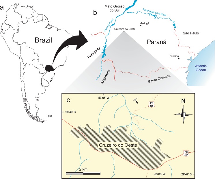
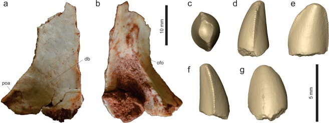
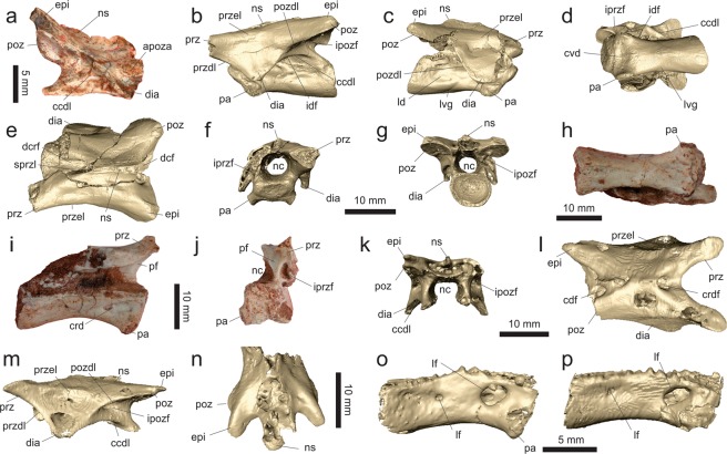
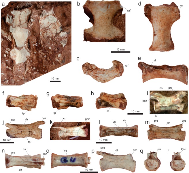
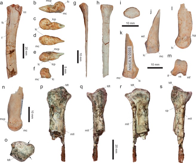
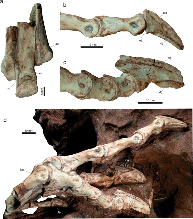
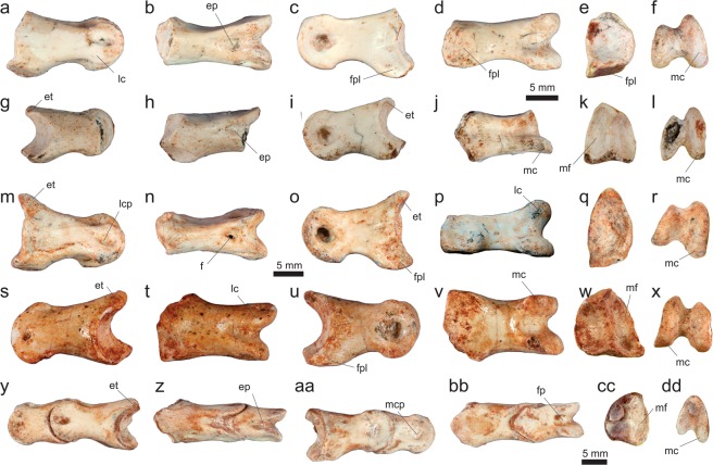
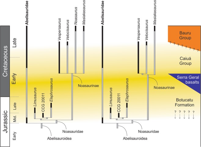
Noasaurines form an enigmatic group of small-bodied predatory theropod dinosaurs known from the Late Cretaceous of Gondwana. They are relatively rare, with notable records in Argentina and Madagascar, and possible remains reported for Brazil, India, and continental Africa. In south-central Brazil, the deposits of the Bauru Basin have yielded a rich tetrapod fauna, which is concentrated in the Bauru Group. The mainly aeolian deposits of the Caiuá Group, on the contrary, bear a scarce fossil record composed only of lizards, turtles, and pterosaurs. Here, we describe the first dinosaur of the Caiuá Group, which also represents the best-preserved theropod of the entire Bauru Basin known to date. The recovered skeletal parts (vertebrae, girdles, limbs, and scarce cranial elements) show that the new taxon was just over 1 m long, with a unique anatomy among theropods. The shafts of its metatarsals II and IV are very lateromedially compressed, as are the blade-like ungual phalanges of the respective digits. This implies that the new taxon could have been functionally monodactyl, with a main central weight-bearing digit, flanked by neighbouring elements positioned very close to digit III or even held free of the ground. Such anatomical adaptation is formerly unrecorded among archosaurs, but has been previously inferred from footprints of the same stratigraphic unit that yielded the new dinosaur. A phylogenetic analysis nests the new taxon within the Noasaurinae clade, which is unresolved because of the multiple alternative positions that Noasaurus leali can acquire in the optimal trees. The exclusion of the latter form results in positioning the new dinosaur as the sister-taxon of the Argentinean Velocisaurus unicus.


Conflict of interest statement

The authors declare no competing interests.

Figures

Figure 1
Figure 1
Location maps for the type-locality of Vespersaurus paranaensis gen. et sp. nov. (a) South America highlighting Paraná state (in black), Brazil. (b) Paraná state showing the location of Cruzeiro do Oeste. (c) Surroundings of the urban area (hatched) of Cruzeiro do Oeste showing the location of the type-locality (hammer). Watercourses marked in blue. PR-487, paved road; PR-180, unpaved road.
Figure 2
Figure 2
Geological context for the type-locality of Vespersaurus paranaensis gen. et sp. nov. (a) Map showing surface exposure of the Rio Paraná and Goio Erê formations in western Paraná and south-western São Paulo (modified from) and the location of sites with dinosaur and mammal (black footprint) and mammal only (white footprint) tracks. (b) Stratigraphic column of the Rio Paraná Formation in the type-locality (indicated in Fig. 1), showing the provenance of the fossils.
Figure 3
Figure 3
Cranial and dental remains of Vespersaurus paranaensis gen. et sp. nov. (a,b) Isolated frontal (MPCO.V 0063b) in dorsal (a) and ventral (b) views. (c–g) Isolated tooth (MPCO.V 0020c) in apical (c), mesial (d), lingual (e), distal (f), and labial (g) views. Anatomical abbreviations: db, dorsal bulbous area; ofo, orbital fossa; poa, postorbital articulation.
Figure 4
Figure 4
Mid-cranial neck vertebrae of Vespersaurus paranaensis gen. et sp. nov. (a) Axis (MPCO.V 0067) in right lateral view; (b–g) cranial cervical vertebra (MPCO.V 0035) in left (b) and right (c) lateral, ventral (d), dorsal (e), cranial (f), and caudal (g) views. (h–j) Cranial cervical vertebra (MPCO.V 0048) in ventral (h), right lateral, with a medial exposure of the left neural arch (i), and cranial (j) views. (l,m) Cranial cervical neural arch (MPCO.V 0034a) in dorsal (l) and left lateral (m) views. (n) Cranial cervical neural arch (MPCO.V 0034b) in dorsal view. (o) Cranial cervical centrum (MPCO.V 0034c) in right lateral view. (p) Cranial cervical centrum (MPCO.V 0034d) in right lateral view. Anatomical abbreviations: apoza, atlantal postzygapophysis articulation facet; ccdl, caudal centrodiapophyseal lamina; dcf, caudodorsal fossa; dcrf, craniodorsal fossa; cvd, cranioventral depression; dia, diapophysisi; epi, epipophysis; idf, infradiapophyseal fossa; ipozf, infrapostzygapophyseal fossa; ipref, infraprezygapophyseal fossa; ld, lateral depression; lf, lateral foramen; lvg, lateroventral groove; nc, neural canal; ns, neural spine; pa, parapophysis; pf, peduncular fossa; poz, postzygapophysis; pozdl, postzygodiapophyseal lamina; prz, prezygapophysis; przdl, prezygodiapophyseal lamina; przel, prezygoepipophyseal lamina; sprzl, spinoprezygapophyseal lamina.
Figure 5
Figure 5
Caudal neck and trunk vertebrae of Vespersaurus paranaensis gen. et sp. nov. (a–e) Caudal neck vertebra (MPCO.V 0017) in right lateral (a), cranial (b), caudal (c), ventral (d), and dorsal (e) views. (f–h) Trunk neural arch (MPCO.V 0066) in lateral (f), cranial (g), and ventral (h) views. (I,j) trunk centrum 0065d10b (holotype) in lateral (i) and ventral (j) views; (km) trunk centrum 0065d10a (holotype) in lateral (k), cranial (l), and caudal (m) views. Anatomical abbreviations, as in Fig. 4, plus: crcdl, cranial centrodiapophyseal lamina; crcpl, cranial centroparapophyseal lamina; dcpo, dorsocaudal pneumatic opening; df1&2, dorsal foramina 1 and 2; ipozl, intrapostzygapophsyeal lamina; lr, lateral ridge; pdl, paradiapophyseal lamina; posf, postspinal fossa; prsf, prespinal fossa; przpl, prezygoparapophyseal; spozl, spinopostzygapophyseal lamina.
Figure 6
Figure 6
Sacral and tail vertebrae of Vespersaurus paranaensis gen. et sp. nov. (a) Sacral vertebrae (MPCO.V 0064a) in ventral view. (b,c) Sacral vertebra (MPCO.V 0065d7) in ventral (b) and cranial (c) views. (d,e) Sacral vertebra (MPCO.V 0065d6, holotype) in ventral (d) and left lateral (e) views. (fh) Tail vertebra (MPCO.V 0065d9, holotype) in dorsal (f), ventral (g) and left lateral (h) views. (i) Tail vertebra (MPCO.V 0065d5, holotype) in right lateral view. (j) Tail vertebra (MPCO.V 0052) in left lateral view. (k) Tail vertebra (MPCO.V 0020) in left lateral view. (lm) Tail vertebra (MPCO.V 0024) in ventral (l) and dorsal (m) views. (nr) Tail vertebra (MPCO.V 0025) in dorsal (n), ventral (o), right lateral (p), cranial (q), and caudal (r) views. Anatomical abbreviations, as in Fig. 4, plus: dlr, dorsolateral ridge; il, ilium; raf, rib articulation facet; sv1-3, sacral vertebrae 1–; tp, transverse process; vg, ventral groove; vr, ventral ridge.
Figure 7
Figure 7
Pectoral girdle and limb elements of Vespersaurus paranaensis gen. et sp. nov. (a–c) Right sacapulocoracoid (MPCO.V 0011) in lateral (a), caudal (b), and medial (c) views. (d) Left sacapulocoracoid (MPCO.V 0006 f) in lateral view. (e–j) Left humerus (MPCO.V 0006d) in proximal (e), cranial (f), medial (g), caudal (h), lateral (i), and distal (j) views. (kp) Left radius (MPCO.V 0006j) in proximal (k), medial (l), caudal (m), lateral (n), cranial (o), and distal (p) views. (q) Right manual digit I (MPCO.V 0006j) in medial view. (r) Right manus (MPCO.V 0006j) in dorsal view. Anatomical abbreviations: acla, acrocoracohumeral ligament attachment; bf, brachial fossa; cbi, M. coracobrachialis insertion; ct, coracoid foramen; ct, coracoid tuber; ctg, groove on coracoid tuber; dbg, dorsal blood groove; dp, descending process; dpc, deltopectoral ridge; drr, distal radial ridge; dsi, M. deltoideous scapularis insertion; entr, entepicondylar ridge; f, foramen; gl, glenoid; gt, greater tubercle; hh, humeral head; it, internal tuberosity; lcs, lateral concave surface; ldi, M. latissimus dorsi insertion; mcIII, metacarpal III; p1-2dI-III, phalanges 1–2 of manual digits I-III; pgd, preglenoid depression; pgf, preglenoid fossa; pgr, preglenoid ridge; rc, radial condyle; scr, scapular ridge; sgd, subglenoid fossa; sgp, supraglenoid pit; sr, stout ridge; ua, ulnar articulation; uc, ulnar condyle; updI-II, ungual phalanges of manual digits I-II; vbg, ventral blood groove.
Figure 8
Figure 8
Pelvic girdle elements of Vespersaurus paranaensis gen. et sp. nov. (a,b) Left ilium (MPCO.V 0065d11, holotype) in lateral (a) and ventral (b) views. (cf) Right pubis (MPCO.V 0042) in dorsal (c), lateral (d), medial (e), and proximal (f) views. (g) Left ischium (MPCO.V 0065d2; holotype) in lateral view. (hl) Left ischium (MPCO.V 0014) in ventral (h), medial (i), proximal (j), lateral (k), and dorsal (l) views. Anatomical abbreviations: acr, acetabular roof; am, acetabular margin; ai, acetabular insisure; ao, M. ambiens origin; bfo, brevis fossa; bs, brevis shelf; ftio, M. flexor tibialis internus origin; fto, M. flexor tibialis musculature origin; ila, iliac articulation; ip, iliac peduncle; isa, ischiadic articulation; ip, ischiadic peduncle; lr, lateral ridge; mbs, “medial brevis shelf”; mc, medial ridge; of, obturator foramen; ofl, obturator flange; or, obturator ridge; pa, pubic apron; paa, postacetabular ala; pp, pubic peduncle; sac, supracetabular crest; sa, symphyseal articulation; sk, socket; sld, semi-lunate depression. Dashed lines represent reconstructed margins.
Figure 9
Figure 9
Tibia, tarsus, and metatarsus of Vespersaurus paranaensis gen. et sp. nov. (a) Right tibial shaft (MPCO.V 0018a-c) in lateral view. (bf) Left metatarsal I (MPCO.V 0057b) in medial (b), lateral (c), ventral (d), dorsal (e), and proximal (f) views. (gi) Right metatarsal II (MPCO.V 0063a) in caudal (g), lateral (h), and proximal (i) views. (jn) Right metatarsal II (MPCO.V 0055) in dorsal (j), ventral (k), lateral (l), distal (m), and medial (n) views. (os) Left third distal tarsal and metatarsals II-III (MPCO.V 0016) in proximal (o), caudal (p), lateral (q), cranial (r), and medial (s) views. Anatomical abbreviations: cc, cnemial crest; cp, caudal projection; ed, extensor depression; f, foramen; fc, fibular crest; lc, lateral condyle; lcp, lateral collateral pit; lf, lateral flange; MII, metatarsal II; mc, medial condyle; mcp, medial collateral pit; mf, medial flange; tdt, third distal tarsal; vs, ventral sulcus. Arrows indicate the contact between metatarsals II and II in proximal view.
Figure 10
Figure 10
Holotype right pes of Vespersaurus paranaensis gen. et sp. nov. (MPCO.V 0065d1). (a) Distal end of metatarsal II-IV in dorsal view. (b) Distal part of digit II in lateral view. (c) Distal part of digit IV in lateral view. (d) Entire foot as preserved in lateral view. Anatomical abbreviations: ed, extensor depression; fd, flexor depression; lcp, lateral collateral pit; mc, lateral crest; plg, proximolateral groove; vg, vertical groove; vlg, ventral lateral groove; vm, ventral margin.
Figure 11
Figure 11
Non-ungual pedal phalanges of Vespersaurus paranaensis gen. et sp. nov. (af) Phalanx 1 of right digit IV (MPCO.V 0056a) in lateral (a), dorsal (b), medial (c), ventral (d), proximal (e), and distal (f) views. (gl) Phalanx 2 of right digit IV (MPCO.V 0056b) in lateral (g), dorsal (h), medial (i), ventral (j), proximal (k), and distal (l) views. (mr) Phalanx 1 of right digit IV (MPCO.V 0060) in lateral (m), dorsal (n), medial (o), ventral (p), proximal (q), and distal (r) views (sx). Phalanx 2 of left digit IV (MPCO.V 0057a) in lateral (s), dorsal (t), medial (u), ventral (v), proximal (w), and distal (x) views. (ydd) Phalanges 2 and 3 of a left digit IV (MPCO.V 0006k) in lateral (y), dorsal (z), medial (aa), ventral (bb), proximal (cc), and distal (dd) views. Anatomical abbreviations: ep, extensor pit; et, extensor tuber; f, foramen; fp, flexor pit; fpl, flexor platform; lc, lateral condyle; lcp, lateral collateral pit; mc, medial condyle; mcp, medial collateral pit; mf, medial facet.
Figure 12
Figure 12
Pedal phalanges of Vespersaurus paranaensis gen. et sp. nov. (af) Phalanx 1 of digit III (MPCO.V 0049) in lateral/medial (a), dorsal (b), lateral/medial (c), ventral (d), proximal (e), and distal (f). (gl) Phalanx 2 of digit III (MPCO.V 0059) in lateral (g), dorsal (h), medial (i), ventral (j), proximal (k), and distal (l). (mq) Ungual phalanx of a right digit IV in medial (m), lateral (n), proximal (o), ventral (p), and dorsal (q). (rv) Ungual phalanx of a left digit I in medial (r), lateral (s), proximal (t), dorsal (u), and ventral (v). Anatomical abbreviations, as in Fig. 11, plus: dlg(m/l), dorsal longitudinal groove (medial/lateral), icg, intercondylar groove; vg, vertical grove; vlg(m/l), ventral longitudinal groove (medial/lateral).
Figure 13
Figure 13
Excerpts (focused on Abelisauroidea) of the Strict Reduced Consensus Trees found in the analyses conducted here (topology to the right excludes N. leali) calibrated into the Jurassic-Cretaceous timescale and a simplified startigraphic chart of the Paraná Basin. Geological correlations and taxon ranges compiled from various sources,,,,,.

References

    1. Sampson SD, Carrano MT, Forster CA. A bizarre predatory dinosaur from the Late Cretaceous of Madagascar. Nature. 2001;409:504–506. doi: 10.1038/35054046. - DOI - PubMed
    1. Carrano MT, Sampson SD, Forster CA. The osteology of Masiakasaurus knopfleri, a small abelisauroid (Dinosauria: Theropoda) from the Late Cretaceous of Madagascar. Journal of Vertebrate Paleontology. 2002;22:510–534. doi: 10.1671/0272-4634(2002)022[0510:TOOMKA]2.0.CO;2. - DOI
    1. Carrano MT, Loewen MA, Sertich JJW. New materials of Masiakasaurus knopfleri Sampson, Carrano and Forster, 2001, and implications for the morphology of the Noasauridae. Smithsonian Contributions to Paleobiology. 2011;95:1–53. doi: 10.5479/si.00810266.95.1. - DOI
    1. Rauhut OWM, Carrano MT. The theropod dinosaur Elaphrosaurus bambergi Janensch, 1920, from the Late Jurassic of Tendaguru, Tanzania. Zoological Journal of the Linnean Society. 2016;178:546–610. doi: 10.1111/zoj.12425. - DOI
    1. Bonaparte JF, Powell JE. A continental assemblage of tetrapods from the Upper Cretaceous beds of El Brete, northwestern Argentina (Sauropoda–Coelurosauria–Carnosauria–Aves) Mémoires de la Société Géologique de France, Nouvelle Série. 1980;139:19–28.

Publication types